A team of molecular biologists has used advanced electron microscopy techniques to unlock the structure of a unique virus that infects bacteria that live under conditions of extreme heat and acidity.
The nonenveloped, rod-shaped virus SIRV2 (
Sulfolobus islandicus rod-shaped virus 2) infects the hyperthermophilic acidophile
Sulfolobus islandicus, a species of archaea that lives in hot springs at 80 degrees Celsius and pH 3. Investigators at the University of Virginia (Charlottesville, USA) wanted to know how the virus managed to safeguard its critical DNA core and whether the virus could be exploited for use as a delivery system for gene therapy in humans.
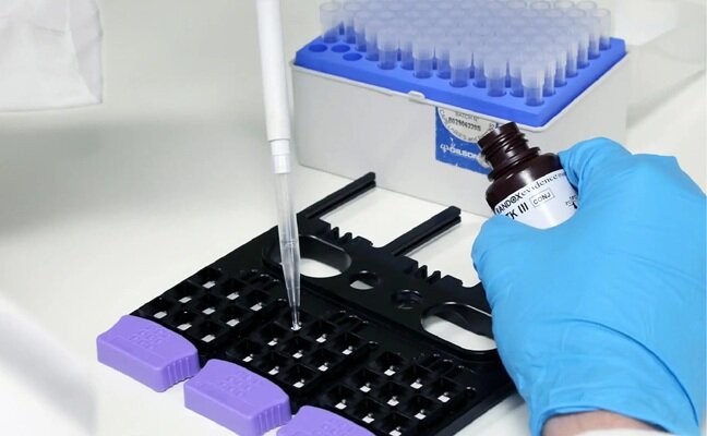
To study the structure of the viral DNA, the investigators turned to the FEI (Hillsboro, OR, USA) Titan Krios electron microscope, which had recently become operational at the University of Virginia. The Titan Krios transmission electron microscope (TEM) was tailored for use in protein and cellular imaging. Its revolutionary cryo-based technology and stability was designed to permit a full range of semi-automated applications, including: electron crystallography, single particle analysis, cryo-electron microscopy, and dual-axis cellular tomography of frozen hydrated cell organelles and cells.
The investigators reported in the May 22, 2015, issue of the journal
Science that they used the Titan Krios to generate a three-dimensional reconstruction of the SIRV2 virion at approximately 0.4 nm resolution. Their study revealed a previously unknown form of virion organization. Although almost half of the capsid protein was unstructured in solution, this unstructured region folded in the virion into a single extended alpha helix that wrapped around the DNA. The DNA was entirely in the A-form, which suggested that there might be a mechanism shared by the virus with bacterial spores for protecting DNA in the most adverse environments.
"Many people have felt that this A-form of DNA is only found in the laboratory under very non-biological conditions, when DNA is dehydrated or dry," said senior author Dr. Edward H. Egelman, professor of biochemistry and molecular genetics at the University of Virginia. "Instead, it appears to be a general mechanism in biology for protecting DNA."
"What is interesting and unusual is being able to see how proteins and DNA can be put together in a way that is absolutely stable under the harshest conditions imaginable," said Dr. Engelman. “We have discovered what appears to be a basic mechanism of resistance—to heat, to desiccation, to ultraviolet radiation. And knowing that, then, we can go in many different directions, including developing ways to package DNA for gene therapy."
Related Links:
University of Virginia

New
Gold Member
Clinical Drug Testing Panel
DOA Urine MultiPlex
POC Helicobacter Pylori Test Kit
Hepy Urease Test
New
Gold Member
Genetic Type 1 Diabetes Risk Test
T1D GRS Array

Gold Member
Collection and Transport System
PurSafe Plus®


")

")
")
")
 in the area surrounding sEcad-high cancer cells (blue, center) (Photo courtesy of Debeb Laboratory)")
")
")
 line (Photo courtesy of Sysmex America)")
")
")
")
")
 system (Deichmann, M. et al., Nat Commun 16, 10306, 2025. DOI: 10.1038/s41467-025-65236-7)")
")
")
")
")
")
")
 will be held at Dubai World Trade Centre from 10-13 February")
")
")
")
")
")
")
")