“Lab-On-A-Disc” Device Paves Way for More Automated Liquid Biopsies
Posted on 05 Feb 2026
Extracellular vesicles (EVs) are tiny particles released by cells into the bloodstream that carry molecular information about a cell’s condition, including whether it is cancerous. However, EVs are highly diverse, scarce, and mixed with many other blood components, making them difficult to isolate and analyze reliably. Existing EV-based liquid biopsy methods are slow, manual, and poorly suited for measuring multiple cancer biomarkers at once. Researchers have now developed an automated system that integrates EV isolation and multiplex protein analysis into a single device, enabling accurate multi-cancer detection from a simple blood sample.
In a study led by Mass General Brigham (Boston, MA, USA), researchers aimed to determine whether EVs could be processed in a fully automated, unbiased, and high-throughput manner suitable for clinical diagnostics. Their goal was to combine plasma separation, EV enrichment, and simultaneous labeling of multiple EV protein markers into one practical device that could be easily adopted in healthcare settings.
")
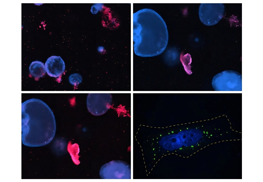
To achieve this, the team developed a centrifugal lab-on-a-disc platform called SpinEx. The device integrates on-disc chromatography to isolate EVs, microbead chambers to capture them, and dedicated compartments to label multiple EV protein targets simultaneously. All steps are performed automatically within the same disc, after which the labeled EVs are quantified using high-throughput flow cytometry.
SpinEx successfully isolated and multiplex-labeled EVs from just 150 microliters of blood, demonstrating fast and fully automated processing. The resulting EV protein profiles accurately distinguished cancer samples from non-cancer samples in an independent validation set. The system also correctly classified five different cancer types based on EV protein patterns.
The technology enables minimally invasive cancer detection using a simple blood draw and a standardized automated workflow. By capturing rich EV protein signatures, SpinEx could support earlier cancer detection, improved tumor-type identification, and longitudinal disease monitoring. Because the platform is compatible with high-throughput testing, it has strong potential for clinical adoption and future large-scale cancer screening programs.
“The most meaningful part of this work is showing that a complex EV workflow can be fully automated without losing analytical depth,” said Hakho Lee, PhD, co-senior author of the paper published in Nature Biomedical Engineering. “EVs have long shown promise for cancer diagnostics, but translation has been limited by labor intensive, fragmented processing steps. SpinEx’s ability to process whole blood and deliver multiplex biomarker results marks a real step toward improving patient care.”
Related Links:
Mass General Brigham