


Raman Technique Visualizes Details Almost to a Trillionth of a Meter
|
By Biotechdaily staff writers Posted on 05 May 2008 |
|
A team of researchers has developed a new type of imaging system that can illuminate tumors in living subjects--obtaining images with a precision of nearly one-trillionth of a meter.
This technique, called Raman spectroscopy, expands the existing range of tools for the field of molecular imaging, according to team leader Sanjiv Sam Gambhir, M.D., Ph.D., professor of radiology at Stanford University School of Medicine (Stanford, CA, USA). He is the senior author of a study describing the application that was published in the March 31, 2008, advance online issue of the journal Proceedings of the [U.S.] National Academy of Sciences (PNAS).
"This is an entirely new way of imaging living subjects, not based on anything previously used,” said Dr. Gambhir, who directs the Molecular Imaging Program at Stanford. He reported that signals from Raman spectroscopy are stronger and longer lived than other available techniques, and the type of particles used in this method can transmit information about multiple types of molecular targets simultaneously. "Usually we can measure one or two things at a time,” he said. "With this, we can now likely see 10, 20, 30 things at once.
Dr. Gambhir reported that he believes this is the first time Raman spectroscopy has been used to image deep within the body, using tiny nanoparticles injected into the body to serve as beacons. When laser light is beamed from a source outside the body, these specialized particles emit signals that can be measured and translated into a visible indicator of their location in the body.
Postdoctoral scholars Shay Keren, Ph.D., and Cristina Zavaleta, Ph.D., co-first authors of the study, found a way to make Raman spectroscopy a medical application. To accomplish this, they used two types of engineered Raman nanoparticles: gold nanoparticles and single-wall carbon nanotubes. First, they injected mice with the some of the nanoparticles. To see the nanoparticles, they used a special microscope that the group had adapted to view anesthetized mice exposed to laser light. The researchers could see that the nanoparticles migrated to the liver, where they were processed for excretion.
To be able to detect molecular events, according to Dr. Zavaleta, they labeled separate batches of spectrally unique Raman nanoparticles with different "tags” (peptides or antibodies) and then injected them into the body simultaneously to see where they went. For example, if each type of particle migrated to a different tumor site, the newly developed Raman microscope would enable the researchers to separate the signals from each batch of particles.
As part of this proof-of-principle work, Dr. Gambhir's team tagged the gold nanoparticles with different bits of proteins that homed in on different tumor molecules. "We could attach pretty much anything,” said Dr. Gambhir. The Raman effect also lasts indefinitely, so the particles do not lose effectiveness as indicators as long as they stay in the body.
Using a microscope they modified to detect Raman nanoparticles, the researchers were able to visualize targets on a scale 1,000 times smaller than what is now obtainable by the most precise fluorescence imaging using quantum dots. When modified for human use, they researchers reported, the technique has the potential to be useful during surgery, for example, in the removal of cancerous tissue. The extreme sensitivity of the imager could enable detection of even the smalltest malignant tissues.
A clinical trial is planned to test the gold nanoparticles in humans for possible use in conjunction with a colonoscopy to indicate early-stage colorectal cancer.
Related Links:
Stanford University School of Medicine
Gold Member
Collection and Transport System
PurSafe Plus®
Anterior Nasal Specimen Collection Swabs
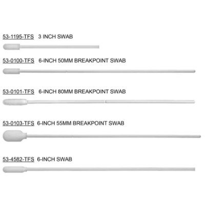
53-1195-TFS, 53-0100-TFS, 53-0101-TFS, 53-4582-TFS
Laboratory Software
ArtelWare
Latest BioResearch News
- Genome Analysis Predicts Likelihood of Neurodisability in Oxygen-Deprived Newborns
- Gene Panel Predicts Disease Progession for Patients with B-cell Lymphoma
- New Method Simplifies Preparation of Tumor Genomic DNA Libraries
- New Tool Developed for Diagnosis of Chronic HBV Infection
- Panel of Genetic Loci Accurately Predicts Risk of Developing Gout
- Disrupted TGFB Signaling Linked to Increased Cancer-Related Bacteria
- Gene Fusion Protein Proposed as Prostate Cancer Biomarker
- NIV Test to Diagnose and Monitor Vascular Complications in Diabetes
- Semen Exosome MicroRNA Proves Biomarker for Prostate Cancer
- Genetic Loci Link Plasma Lipid Levels to CVD Risk
- Newly Identified Gene Network Aids in Early Diagnosis of Autism Spectrum Disorder
- Link Confirmed between Living in Poverty and Developing Diseases
- Genomic Study Identifies Kidney Disease Loci in Type I Diabetes Patients
- Liquid Biopsy More Effective for Analyzing Tumor Drug Resistance Mutations
- New Liquid Biopsy Assay Reveals Host-Pathogen Interactions
- Method Developed for Enriching Trophoblast Population in Samples
Channels
Clinical Chemistry
view channel")
New PSA-Based Prognostic Model Improves Prostate Cancer Risk Assessment
Prostate cancer is the second-leading cause of cancer death among American men, and about one in eight will be diagnosed in their lifetime. Screening relies on blood levels of prostate-specific antigen... Read more
Extracellular Vesicles Linked to Heart Failure Risk in CKD Patients
Chronic kidney disease (CKD) affects more than 1 in 7 Americans and is strongly associated with cardiovascular complications, which account for more than half of deaths among people with CKD.... Read more")
")
Molecular Diagnostics
view channel")
Diagnostic Device Predicts Treatment Response for Brain Tumors Via Blood Test
Glioblastoma is one of the deadliest forms of brain cancer, largely because doctors have no reliable way to determine whether treatments are working in real time. Assessing therapeutic response currently... Read more")
Blood Test Detects Early-Stage Cancers by Measuring Epigenetic Instability
Early-stage cancers are notoriously difficult to detect because molecular changes are subtle and often missed by existing screening tools. Many liquid biopsies rely on measuring absolute DNA methylation... Read more")
“Lab-On-A-Disc” Device Paves Way for More Automated Liquid Biopsies
Extracellular vesicles (EVs) are tiny particles released by cells into the bloodstream that carry molecular information about a cell’s condition, including whether it is cancerous. However, EVs are highly... Read more in the area surrounding sEcad-high cancer cells (blue, center) (Photo courtesy of Debeb Laboratory)")
Blood Test Identifies Inflammatory Breast Cancer Patients at Increased Risk of Brain Metastasis
Brain metastasis is a frequent and devastating complication in patients with inflammatory breast cancer, an aggressive subtype with limited treatment options. Despite its high incidence, the biological... Read moreHematology
view channel")
New Guidelines Aim to Improve AL Amyloidosis Diagnosis
Light chain (AL) amyloidosis is a rare, life-threatening bone marrow disorder in which abnormal amyloid proteins accumulate in organs. Approximately 3,260 people in the United States are diagnosed... Read more")
Fast and Easy Test Could Revolutionize Blood Transfusions
Blood transfusions are a cornerstone of modern medicine, yet red blood cells can deteriorate quietly while sitting in cold storage for weeks. Although blood units have a fixed expiration date, cells from... Read more line (Photo courtesy of Sysmex America)")
Automated Hemostasis System Helps Labs of All Sizes Optimize Workflow
High-volume hemostasis sections must sustain rapid turnaround while managing reruns and reflex testing. Manual tube handling and preanalytical checks can strain staff time and increase opportunities for error.... Read more")
High-Sensitivity Blood Test Improves Assessment of Clotting Risk in Heart Disease Patients
Blood clotting is essential for preventing bleeding, but even small imbalances can lead to serious conditions such as thrombosis or dangerous hemorrhage. In cardiovascular disease, clinicians often struggle... Read moreImmunology
view channelBlood Test Identifies Lung Cancer Patients Who Can Benefit from Immunotherapy Drug
Small cell lung cancer (SCLC) is an aggressive disease with limited treatment options, and even newly approved immunotherapies do not benefit all patients. While immunotherapy can extend survival for some,... Read more")
Whole-Genome Sequencing Approach Identifies Cancer Patients Benefitting From PARP-Inhibitor Treatment
Targeted cancer therapies such as PARP inhibitors can be highly effective, but only for patients whose tumors carry specific DNA repair defects. Identifying these patients accurately remains challenging,... Read more")
Ultrasensitive Liquid Biopsy Demonstrates Efficacy in Predicting Immunotherapy Response
Immunotherapy has transformed cancer treatment, but only a small proportion of patients experience lasting benefit, with response rates often remaining between 10% and 20%. Clinicians currently lack reliable... Read more")
Microbiology
view channel")
Comprehensive Review Identifies Gut Microbiome Signatures Associated With Alzheimer’s Disease
Alzheimer’s disease affects approximately 6.7 million people in the United States and nearly 50 million worldwide, yet early cognitive decline remains difficult to characterize. Increasing evidence suggests... Read moreAI-Powered Platform Enables Rapid Detection of Drug-Resistant C. Auris Pathogens
Infections caused by the pathogenic yeast Candida auris pose a significant threat to hospitalized patients, particularly those with weakened immune systems or those who have invasive medical devices.... Read more")
")
Pathology
view channel system (Deichmann, M. et al., Nat Commun 16, 10306, 2025. DOI: 10.1038/s41467-025-65236-7)")
Engineered Yeast Cells Enable Rapid Testing of Cancer Immunotherapy
Developing new cancer immunotherapies is a slow, costly, and high-risk process, particularly for CAR T cell treatments that must precisely recognize cancer-specific antigens. Small differences in tumor... Read more")
First-Of-Its-Kind Test Identifies Autism Risk at Birth
Autism spectrum disorder is treatable, and extensive research shows that early intervention can significantly improve cognitive, social, and behavioral outcomes. Yet in the United States, the average age... Read more")
")
Technology
view channel")
Robotic Technology Unveiled for Automated Diagnostic Blood Draws
Routine diagnostic blood collection is a high‑volume task that can strain staffing and introduce human‑dependent variability, with downstream implications for sample quality and patient experience.... Read more")
ADLM Launches First-of-Its-Kind Data Science Program for Laboratory Medicine Professionals
Clinical laboratories generate billions of test results each year, creating a treasure trove of data with the potential to support more personalized testing, improve operational efficiency, and enhance patient care.... Read moreAptamer Biosensor Technology to Transform Virus Detection
Rapid and reliable virus detection is essential for controlling outbreaks, from seasonal influenza to global pandemics such as COVID-19. Conventional diagnostic methods, including cell culture, antigen... Read more")
AI Models Could Predict Pre-Eclampsia and Anemia Earlier Using Routine Blood Tests
Pre-eclampsia and anemia are major contributors to maternal and child mortality worldwide, together accounting for more than half a million deaths each year and leaving millions with long-term health complications.... Read moreIndustry
view channelNew Collaboration Brings Automated Mass Spectrometry to Routine Laboratory Testing
Mass spectrometry is a powerful analytical technique that identifies and quantifies molecules based on their mass and electrical charge. Its high selectivity, sensitivity, and accuracy make it indispensable... Read more")
AI-Powered Cervical Cancer Test Set for Major Rollout in Latin America
Noul Co., a Korean company specializing in AI-based blood and cancer diagnostics, announced it will supply its intelligence (AI)-based miLab CER cervical cancer diagnostic solution to Mexico under a multi‑year... Read more")
Diasorin and Fisher Scientific Enter into US Distribution Agreement for Molecular POC Platform
Diasorin (Saluggia, Italy) has entered into an exclusive distribution agreement with Fisher Scientific, part of Thermo Fisher Scientific (Waltham, MA, USA), for the LIAISON NES molecular point-of-care... Read more will be held at Dubai World Trade Centre from 10-13 February")