Quantifying the Size of Holes Antibacterial Compounds Create in Cell Walls
|
By LabMedica International staff writers Posted on 23 Jan 2013 |

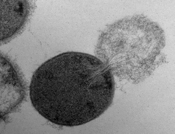
Image: Transmission electron microscopy image of a Streptococcus pyogenes cell experiencing lysis after exposure to the highly active enzyme PlyC (Photo courtesy of Daniel Nelson, UMD).
The emergence of antibiotic-resistant bacteria has initiated a search for alternatives to traditional antibiotics. One potential option is PlyC, a powerful enzyme that kills the bacteria that causes streptococcal toxic shock syndrome and strep throat. PlyC functions by fastening onto the surface of a bacteria cell and chomping a hole in the cell wall large enough for the bacteria’s inner membrane to protrude from the cell, eventually causing the cell to burst and die.
Research has shown that unconventional antimicrobials such as PlyC can effectively kill bacteria. However, essential questions remain about how bacteria respond to the holes that these therapeutics make in their cell wall and what size holes bacteria can withstand before degrading. Solving those problems could improve the efficacy of current antibacterial drugs and begin to develop new ones.
Researchers from the Georgia Institute of Technology (Atlanta, USA) and the University of Maryland (College Park, USA) recently conducted a study to try to answer those questions. The researchers created a biophysical model of the response of a Gram-positive bacterium to the formation of a hole in its cell wall. Then they used experimental measurements to validate the theory, which predicted that a hole in the bacteria cell wall larger than 15–24 nm in diameter would cause the cell to lyse, or burst. These small holes are approximately one-hundredth the diameter of a typical bacterial cell.
“Our model correctly predicted that the membrane and cell contents of Gram-positive bacteria cells explode out of holes in cell walls that exceed a few dozen nanometers. This critical hole size, validated by experiments, is much larger than the holes Gram-positive bacteria use to transport molecules necessary for their survival, which have been estimated to be less than 7 nanometers in diameter,” said Dr. Joshua Weitz, an associate professor in the School of Biology at Georgia Tech.
The study’s findings were published online on January 9, 2013, in the Journal of the Royal Society Interface. Common Gram-positive bacteria that infect humans include Streptococcus, which causes strep throat; Staphylococcus, which causes impetigo; and Clostridium, which causes botulism and tetanus. Gram-negative bacteria include Escherichia, which causes urinary tract infections; Vibrio, which causes cholera; and Neisseria, which causes gonorrhea.
Gram-positive bacteria are different from Gram-negative bacteria in the structure of their cell walls. The cell wall comprises the outer layer of Gram-positive bacteria, whereas the cell wall lies between the inner and outer membrane of Gram-negative bacteria and is therefore protected from direct exposure to the environment.
Georgia Tech biology graduate student Gabriel Mitchell, Georgia Tech physics professor Dr. Kurt Wiesenfeld, and Dr. Weitz developed a biophysical theory of the response of a Gram-positive bacterium to the formation of a hole in its cell wall. The model detailed the effect of pressure, stretching and bending forces on the altering configuration of the cell membrane due to a hole. The force associated with bending and stretching pulls the membrane inward, while the pressure from the inside of the cell pushes the membrane outward through the hole.
“We found that bending forces act to keep the membrane together and push it back inside, but a sufficiently large hole enables the bending forces to be overpowered by the internal pressure forces and the membrane begins to escape out and the cell contents follow,” said Dr. Weitz.
The balance between the bending and pressure forces led to the model prediction that holes 15–24 nm in diameter or larger would cause a bacteria cell to burst. To assess the hypothesis, Dr. Daniel Nelson, an assistant professor at the University of Maryland, utilized transmission electron microscopy images to gauge the size of holes created in lysed Streptococcus pyogenes bacteria cells following PlyC exposure.
Dr. Nelson discovered holes in the lysed bacteria cells that ranged in diameter from 22–180 nm, with a mean diameter of 68 nm. These experimental measurements agreed with the researchers’ theoretic calculation of hole sizes that cause bacterial cell death. According to the researchers, their theoretic model is the first to consider the effects of cell wall thickness on lysis. “Because lysis events occur most often at thinner points in the cell wall, cell wall thickness may play a role in suppressing lysis by serving as a buffer against the formation of large holes,” said Mr. Mitchell.
The combination of research and theory used in this study provided clues into the effect of defects on a cell’s viability and the processes employed by enzymes to interrupt homeostasis and cause bacteria cell death. To additionally determine the processes behind enzyme-induced lysis, the researchers plan to measure membrane dynamics as a function of hole geometry in the future.
Related Links:
Georgia Institute of Technology
University of Maryland
Latest BioResearch News
- Genome Analysis Predicts Likelihood of Neurodisability in Oxygen-Deprived Newborns
- Gene Panel Predicts Disease Progession for Patients with B-cell Lymphoma
- New Method Simplifies Preparation of Tumor Genomic DNA Libraries
- New Tool Developed for Diagnosis of Chronic HBV Infection
- Panel of Genetic Loci Accurately Predicts Risk of Developing Gout
- Disrupted TGFB Signaling Linked to Increased Cancer-Related Bacteria
- Gene Fusion Protein Proposed as Prostate Cancer Biomarker
- NIV Test to Diagnose and Monitor Vascular Complications in Diabetes
- Semen Exosome MicroRNA Proves Biomarker for Prostate Cancer
- Genetic Loci Link Plasma Lipid Levels to CVD Risk
- Newly Identified Gene Network Aids in Early Diagnosis of Autism Spectrum Disorder
- Link Confirmed between Living in Poverty and Developing Diseases
- Genomic Study Identifies Kidney Disease Loci in Type I Diabetes Patients
- Liquid Biopsy More Effective for Analyzing Tumor Drug Resistance Mutations
- New Liquid Biopsy Assay Reveals Host-Pathogen Interactions
- Method Developed for Enriching Trophoblast Population in Samples
Channels
Clinical Chemistry
view channel
New PSA-Based Prognostic Model Improves Prostate Cancer Risk Assessment
Prostate cancer is the second-leading cause of cancer death among American men, and about one in eight will be diagnosed in their lifetime. Screening relies on blood levels of prostate-specific antigen... Read more
Extracellular Vesicles Linked to Heart Failure Risk in CKD Patients
Chronic kidney disease (CKD) affects more than 1 in 7 Americans and is strongly associated with cardiovascular complications, which account for more than half of deaths among people with CKD.... Read moreMolecular Diagnostics
view channel
Diagnostic Device Predicts Treatment Response for Brain Tumors Via Blood Test
Glioblastoma is one of the deadliest forms of brain cancer, largely because doctors have no reliable way to determine whether treatments are working in real time. Assessing therapeutic response currently... Read more
Blood Test Detects Early-Stage Cancers by Measuring Epigenetic Instability
Early-stage cancers are notoriously difficult to detect because molecular changes are subtle and often missed by existing screening tools. Many liquid biopsies rely on measuring absolute DNA methylation... Read more
“Lab-On-A-Disc” Device Paves Way for More Automated Liquid Biopsies
Extracellular vesicles (EVs) are tiny particles released by cells into the bloodstream that carry molecular information about a cell’s condition, including whether it is cancerous. However, EVs are highly... Read more
Blood Test Identifies Inflammatory Breast Cancer Patients at Increased Risk of Brain Metastasis
Brain metastasis is a frequent and devastating complication in patients with inflammatory breast cancer, an aggressive subtype with limited treatment options. Despite its high incidence, the biological... Read moreHematology
view channel
New Guidelines Aim to Improve AL Amyloidosis Diagnosis
Light chain (AL) amyloidosis is a rare, life-threatening bone marrow disorder in which abnormal amyloid proteins accumulate in organs. Approximately 3,260 people in the United States are diagnosed... Read more
Fast and Easy Test Could Revolutionize Blood Transfusions
Blood transfusions are a cornerstone of modern medicine, yet red blood cells can deteriorate quietly while sitting in cold storage for weeks. Although blood units have a fixed expiration date, cells from... Read more
Automated Hemostasis System Helps Labs of All Sizes Optimize Workflow
High-volume hemostasis sections must sustain rapid turnaround while managing reruns and reflex testing. Manual tube handling and preanalytical checks can strain staff time and increase opportunities for error.... Read more
High-Sensitivity Blood Test Improves Assessment of Clotting Risk in Heart Disease Patients
Blood clotting is essential for preventing bleeding, but even small imbalances can lead to serious conditions such as thrombosis or dangerous hemorrhage. In cardiovascular disease, clinicians often struggle... Read moreImmunology
view channelBlood Test Identifies Lung Cancer Patients Who Can Benefit from Immunotherapy Drug
Small cell lung cancer (SCLC) is an aggressive disease with limited treatment options, and even newly approved immunotherapies do not benefit all patients. While immunotherapy can extend survival for some,... Read more
Whole-Genome Sequencing Approach Identifies Cancer Patients Benefitting From PARP-Inhibitor Treatment
Targeted cancer therapies such as PARP inhibitors can be highly effective, but only for patients whose tumors carry specific DNA repair defects. Identifying these patients accurately remains challenging,... Read more
Ultrasensitive Liquid Biopsy Demonstrates Efficacy in Predicting Immunotherapy Response
Immunotherapy has transformed cancer treatment, but only a small proportion of patients experience lasting benefit, with response rates often remaining between 10% and 20%. Clinicians currently lack reliable... Read moreMicrobiology
view channel
Comprehensive Review Identifies Gut Microbiome Signatures Associated With Alzheimer’s Disease
Alzheimer’s disease affects approximately 6.7 million people in the United States and nearly 50 million worldwide, yet early cognitive decline remains difficult to characterize. Increasing evidence suggests... Read moreAI-Powered Platform Enables Rapid Detection of Drug-Resistant C. Auris Pathogens
Infections caused by the pathogenic yeast Candida auris pose a significant threat to hospitalized patients, particularly those with weakened immune systems or those who have invasive medical devices.... Read morePathology
view channel
Engineered Yeast Cells Enable Rapid Testing of Cancer Immunotherapy
Developing new cancer immunotherapies is a slow, costly, and high-risk process, particularly for CAR T cell treatments that must precisely recognize cancer-specific antigens. Small differences in tumor... Read more
First-Of-Its-Kind Test Identifies Autism Risk at Birth
Autism spectrum disorder is treatable, and extensive research shows that early intervention can significantly improve cognitive, social, and behavioral outcomes. Yet in the United States, the average age... Read moreTechnology
view channel
Robotic Technology Unveiled for Automated Diagnostic Blood Draws
Routine diagnostic blood collection is a high‑volume task that can strain staffing and introduce human‑dependent variability, with downstream implications for sample quality and patient experience.... Read more
ADLM Launches First-of-Its-Kind Data Science Program for Laboratory Medicine Professionals
Clinical laboratories generate billions of test results each year, creating a treasure trove of data with the potential to support more personalized testing, improve operational efficiency, and enhance patient care.... Read moreAptamer Biosensor Technology to Transform Virus Detection
Rapid and reliable virus detection is essential for controlling outbreaks, from seasonal influenza to global pandemics such as COVID-19. Conventional diagnostic methods, including cell culture, antigen... Read more
AI Models Could Predict Pre-Eclampsia and Anemia Earlier Using Routine Blood Tests
Pre-eclampsia and anemia are major contributors to maternal and child mortality worldwide, together accounting for more than half a million deaths each year and leaving millions with long-term health complications.... Read moreIndustry
view channelNew Collaboration Brings Automated Mass Spectrometry to Routine Laboratory Testing
Mass spectrometry is a powerful analytical technique that identifies and quantifies molecules based on their mass and electrical charge. Its high selectivity, sensitivity, and accuracy make it indispensable... Read more
AI-Powered Cervical Cancer Test Set for Major Rollout in Latin America
Noul Co., a Korean company specializing in AI-based blood and cancer diagnostics, announced it will supply its intelligence (AI)-based miLab CER cervical cancer diagnostic solution to Mexico under a multi‑year... Read more
Diasorin and Fisher Scientific Enter into US Distribution Agreement for Molecular POC Platform
Diasorin (Saluggia, Italy) has entered into an exclusive distribution agreement with Fisher Scientific, part of Thermo Fisher Scientific (Waltham, MA, USA), for the LIAISON NES molecular point-of-care... Read more


