


AI Image Analysis Module Detects Cancers at the Time of Surgery
|
By LabMedica International staff writers Posted on 26 May 2022 |
|
")
A new image analysis module based on deep learning allows neurosurgeons to identify areas of cancer infiltration in patients undergoing primary treatment of a diffuse glioma, providing cancer detection where they really need it and dramatically improving brain tumor surgery.
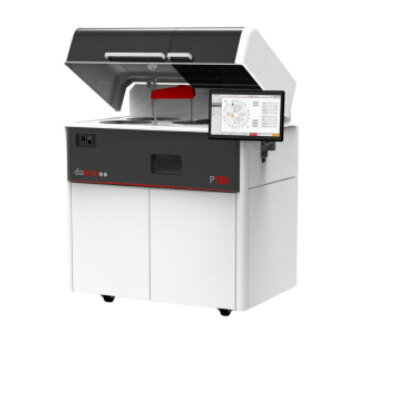
Invenio Imaging Inc.’s (Santa Clara, CA, USA) NIO Laser Imaging System uses Stimulated Raman Histology to image unprocessed tissue specimen without sectioning or staining, enabling histologic evaluation outside the laboratory. It has been used in over 2000 brain tumor procedures across multiple institutions in the US and in Europe. SRH allows three-dimensional imaging of thick specimens using optical sectioning and relies on laser spectroscopy to interrogate the chemical composition of the sample. As such, it does not require physical sectioning, (e.g. with a microtome on frozen or paraffin-embedded tissue) or dye staining, and it allows optical imaging of fresh tissue specimens with minimal tissue preparation.
In contrast to other laser spectroscopy techniques, SRH is unique in that it performs a spectroscopic measurement at each pixel and displays the results as a pseudo-color image, instead of a point spectrum. The NIO Laser Imaging System uses a high numerical aperture objective with 25x magnification and a 0.5mm scan width. Larger areas up to 10mm x 10mm can then be acquired by stitching multiple fields of view in a fully automated process. NIO images are natively digital and can be shared with existing IT infrastructure via a vendor-neutral DICOM interface. The NIO Glioma Reveal image analysis module now adds immediate decision support to the NIO Laser Imaging System by allowing the imaging of multiple samples from the resection cavity. Invenio has received the CE Mark for the NIO Glioma Reveal image analysis module, allowing neurosurgeons in the EU to use it to inform intraoperative decisions.
"By streamlining intraoperative tissue imaging, the NIO Laser Imaging System allows the imaging of multiple samples from the resection cavity. The NIO Glioma Reveal image analysis module now adds immediate decision support", said Chris Freudiger, PhD, co-founder and CTO of Invenio Imaging.
"Glioma Reveal provides cancer detection where we really need it, dramatically improving brain tumor surgery," added Prof. Dr. Jürgen Beck, Chair of Neurosurgery at the University of Freiburg.
"Applying reliable artificial intelligence to digital pathology appears to me, as a surgeon, to be the missing piece in the puzzle of rapid intraoperative histology-based decision-making," said Asst. Prof. Dr. Volker Neuschmelting, Vice-Chair of Neurosurgery at the University of Cologne.
"The NIO Laser Imaging System can also be combined with other important imaging techniques such as 5-ALA fluorescence to further improve brain tumor detection during surgery," explained Prof. Dr. Georg Widhalm, neurosurgeon at the University of Vienna.
Related Links:
Invenio Imaging Inc.


Latest Pathology News
- Engineered Yeast Cells Enable Rapid Testing of Cancer Immunotherapy
- First-Of-Its-Kind Test Identifies Autism Risk at Birth
- AI Algorithms Improve Genetic Mutation Detection in Cancer Diagnostics
- Skin Biopsy Offers New Diagnostic Method for Neurodegenerative Diseases
- Fast Label-Free Method Identifies Aggressive Cancer Cells
- New X-Ray Method Promises Advances in Histology
- Single-Cell Profiling Technique Could Guide Early Cancer Detection
- Intraoperative Tumor Histology to Improve Cancer Surgeries
- Rapid Stool Test Could Help Pinpoint IBD Diagnosis
- AI-Powered Label-Free Optical Imaging Accurately Identifies Thyroid Cancer During Surgery
- Deep Learning–Based Method Improves Cancer Diagnosis
- ADLM Updates Expert Guidance on Urine Drug Testing for Patients in Emergency Departments
- New Age-Based Blood Test Thresholds to Catch Ovarian Cancer Earlier
- Genetics and AI Improve Diagnosis of Aortic Stenosis
- AI Tool Simultaneously Identifies Genetic Mutations and Disease Type
- Rapid Low-Cost Tests Can Prevent Child Deaths from Contaminated Medicinal Syrups
Channels
Clinical Chemistry
view channel")
New PSA-Based Prognostic Model Improves Prostate Cancer Risk Assessment
Prostate cancer is the second-leading cause of cancer death among American men, and about one in eight will be diagnosed in their lifetime. Screening relies on blood levels of prostate-specific antigen... Read more
Extracellular Vesicles Linked to Heart Failure Risk in CKD Patients
Chronic kidney disease (CKD) affects more than 1 in 7 Americans and is strongly associated with cardiovascular complications, which account for more than half of deaths among people with CKD.... Read more")
")
Molecular Diagnostics
view channel")
Diagnostic Device Predicts Treatment Response for Brain Tumors Via Blood Test
Glioblastoma is one of the deadliest forms of brain cancer, largely because doctors have no reliable way to determine whether treatments are working in real time. Assessing therapeutic response currently... Read more")
Blood Test Detects Early-Stage Cancers by Measuring Epigenetic Instability
Early-stage cancers are notoriously difficult to detect because molecular changes are subtle and often missed by existing screening tools. Many liquid biopsies rely on measuring absolute DNA methylation... Read more")
“Lab-On-A-Disc” Device Paves Way for More Automated Liquid Biopsies
Extracellular vesicles (EVs) are tiny particles released by cells into the bloodstream that carry molecular information about a cell’s condition, including whether it is cancerous. However, EVs are highly... Read more in the area surrounding sEcad-high cancer cells (blue, center) (Photo courtesy of Debeb Laboratory)")
Blood Test Identifies Inflammatory Breast Cancer Patients at Increased Risk of Brain Metastasis
Brain metastasis is a frequent and devastating complication in patients with inflammatory breast cancer, an aggressive subtype with limited treatment options. Despite its high incidence, the biological... Read moreHematology
view channel")
New Guidelines Aim to Improve AL Amyloidosis Diagnosis
Light chain (AL) amyloidosis is a rare, life-threatening bone marrow disorder in which abnormal amyloid proteins accumulate in organs. Approximately 3,260 people in the United States are diagnosed... Read more")
Fast and Easy Test Could Revolutionize Blood Transfusions
Blood transfusions are a cornerstone of modern medicine, yet red blood cells can deteriorate quietly while sitting in cold storage for weeks. Although blood units have a fixed expiration date, cells from... Read more line (Photo courtesy of Sysmex America)")
Automated Hemostasis System Helps Labs of All Sizes Optimize Workflow
High-volume hemostasis sections must sustain rapid turnaround while managing reruns and reflex testing. Manual tube handling and preanalytical checks can strain staff time and increase opportunities for error.... Read more")
High-Sensitivity Blood Test Improves Assessment of Clotting Risk in Heart Disease Patients
Blood clotting is essential for preventing bleeding, but even small imbalances can lead to serious conditions such as thrombosis or dangerous hemorrhage. In cardiovascular disease, clinicians often struggle... Read moreImmunology
view channelBlood Test Identifies Lung Cancer Patients Who Can Benefit from Immunotherapy Drug
Small cell lung cancer (SCLC) is an aggressive disease with limited treatment options, and even newly approved immunotherapies do not benefit all patients. While immunotherapy can extend survival for some,... Read more")
Whole-Genome Sequencing Approach Identifies Cancer Patients Benefitting From PARP-Inhibitor Treatment
Targeted cancer therapies such as PARP inhibitors can be highly effective, but only for patients whose tumors carry specific DNA repair defects. Identifying these patients accurately remains challenging,... Read more")
Ultrasensitive Liquid Biopsy Demonstrates Efficacy in Predicting Immunotherapy Response
Immunotherapy has transformed cancer treatment, but only a small proportion of patients experience lasting benefit, with response rates often remaining between 10% and 20%. Clinicians currently lack reliable... Read more")
Microbiology
view channel")
Comprehensive Review Identifies Gut Microbiome Signatures Associated With Alzheimer’s Disease
Alzheimer’s disease affects approximately 6.7 million people in the United States and nearly 50 million worldwide, yet early cognitive decline remains difficult to characterize. Increasing evidence suggests... Read moreAI-Powered Platform Enables Rapid Detection of Drug-Resistant C. Auris Pathogens
Infections caused by the pathogenic yeast Candida auris pose a significant threat to hospitalized patients, particularly those with weakened immune systems or those who have invasive medical devices.... Read more")
")
Pathology
view channel system (Deichmann, M. et al., Nat Commun 16, 10306, 2025. DOI: 10.1038/s41467-025-65236-7)")
Engineered Yeast Cells Enable Rapid Testing of Cancer Immunotherapy
Developing new cancer immunotherapies is a slow, costly, and high-risk process, particularly for CAR T cell treatments that must precisely recognize cancer-specific antigens. Small differences in tumor... Read more")
First-Of-Its-Kind Test Identifies Autism Risk at Birth
Autism spectrum disorder is treatable, and extensive research shows that early intervention can significantly improve cognitive, social, and behavioral outcomes. Yet in the United States, the average age... Read more")
")
Technology
view channel")
Robotic Technology Unveiled for Automated Diagnostic Blood Draws
Routine diagnostic blood collection is a high‑volume task that can strain staffing and introduce human‑dependent variability, with downstream implications for sample quality and patient experience.... Read more")
ADLM Launches First-of-Its-Kind Data Science Program for Laboratory Medicine Professionals
Clinical laboratories generate billions of test results each year, creating a treasure trove of data with the potential to support more personalized testing, improve operational efficiency, and enhance patient care.... Read moreAptamer Biosensor Technology to Transform Virus Detection
Rapid and reliable virus detection is essential for controlling outbreaks, from seasonal influenza to global pandemics such as COVID-19. Conventional diagnostic methods, including cell culture, antigen... Read more")
AI Models Could Predict Pre-Eclampsia and Anemia Earlier Using Routine Blood Tests
Pre-eclampsia and anemia are major contributors to maternal and child mortality worldwide, together accounting for more than half a million deaths each year and leaving millions with long-term health complications.... Read moreIndustry
view channelNew Collaboration Brings Automated Mass Spectrometry to Routine Laboratory Testing
Mass spectrometry is a powerful analytical technique that identifies and quantifies molecules based on their mass and electrical charge. Its high selectivity, sensitivity, and accuracy make it indispensable... Read more")
AI-Powered Cervical Cancer Test Set for Major Rollout in Latin America
Noul Co., a Korean company specializing in AI-based blood and cancer diagnostics, announced it will supply its intelligence (AI)-based miLab CER cervical cancer diagnostic solution to Mexico under a multi‑year... Read more")
Diasorin and Fisher Scientific Enter into US Distribution Agreement for Molecular POC Platform
Diasorin (Saluggia, Italy) has entered into an exclusive distribution agreement with Fisher Scientific, part of Thermo Fisher Scientific (Waltham, MA, USA), for the LIAISON NES molecular point-of-care... Read more will be held at Dubai World Trade Centre from 10-13 February")