Immunochromatographic Strip Test Developed for Rift Valley Fever
|
By LabMedica International staff writers Posted on 03 Oct 2019 |

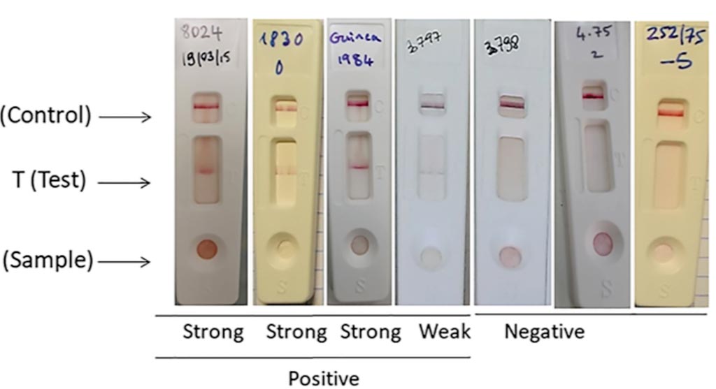
Image: Rift Valley Fever (RVF) lateral flow immunochromatographic (LFT) strip test for the detection of RVF infection using the two selected monoclonal antibodies (Photo courtesy of University of Montpellier).
Rift Valley fever (RVF) is an emerging mosquito-borne disease that affects a wide range of animals and human beings in Africa and the Arabian Peninsula. A rapid and specific test for RVF diagnosis at the site of a suspected outbreak is crucial for the implementation of control measures.
Patients who become ill usually experience fever, generalized weakness, back pain, and dizziness at the onset of the illness. Typically, patients recover within two days to one week after onset of illness. However, a small percentage (8-10%) of people infected with RVFV develops much more severe symptoms. There is a growing concern that RVFV will extend its current range due to the wide variety of mosquito species able to transmit to several mammal hosts.
Scientists at the University of Montpellier (Montpellier, France) and their colleagues developed a first-line lateral flow immunochromatographic strip test (LFT) for the detection of the nucleoprotein (N) of the RVF virus (RVFV). Its diagnostic performance characteristics were evaluated using reference stocks isolates recovered from different hosts and in geographic regions mimicking clinical specimens and from known RVF negative serum samples.
The test strip was constructed on the principles of immunochromatography using colloidal-gold-labeled monoclonal antibodies (Mabs). The team used the two Mabs generated against the N protein of RVFV: the Mab 8E10-4A4 gold conjugate and the Mab 10H3-4E4-3D5. Mab 10H3-4E4-3D5 was immobilized onto a nitrocellulose membrane for the test line zone and rabbit anti-mouse antibodies were immobilized for the control line zone to capture unbound Mab. The test (T) and control (C) lines were checked for the development of color after 10 minutes and again after 30 minutes as it might take longer time for weak positives to form a visual band scored subjectively from negative to strong.
A total of 25 isolated strains mimicking clinical specimens of different geographical origins and 10 clinical samples originating from an ongoing outbreak in 2019 detected RVF positive by a TaqMan RT-PCR technique which is considered as the current reference detection system were also detected positive by the LFT giving a diagnostic sensitivity of 100%. Diagnostic specificity (DSp) was assessed on 160 serum samples known to be negative for RVFV by sero-neutralization test and cELISA but collected in tropical countries where RVF has been circulating to detect possible cross reactions or in other areas where RVF introduction is considered at risk. The test gave a DSp of 98.81% and there was an absence of cross-reactivity with viruses belonging to different genera.
The authors concluded that the specificity and sensitivity of the evaluated test are lower than the ones of molecular-based techniques (LAMP, PCR) but are adequate for specific rapid initial detection of RVF outbreaks or disease surveillance in control programs. The fact no specialized reagents and laboratory equipment are needed, make this assay a valuable, first-line diagnostic tool in resource-poor diagnostic territories for on-site RVFV detection, however the staff require training. The study was published on September 11, 2019, in the journal PLOS NTD.
Related Links:
University of Montpellier
Latest Microbiology News
- Comprehensive Review Identifies Gut Microbiome Signatures Associated With Alzheimer’s Disease
- AI-Powered Platform Enables Rapid Detection of Drug-Resistant C. Auris Pathogens
- New Test Measures How Effectively Antibiotics Kill Bacteria
- New Antimicrobial Stewardship Standards for TB Care to Optimize Diagnostics
- New UTI Diagnosis Method Delivers Antibiotic Resistance Results 24 Hours Earlier
- Breakthroughs in Microbial Analysis to Enhance Disease Prediction
- Blood-Based Diagnostic Method Could Identify Pediatric LRTIs
- Rapid Diagnostic Test Matches Gold Standard for Sepsis Detection
- Rapid POC Tuberculosis Test Provides Results Within 15 Minutes
- Rapid Assay Identifies Bloodstream Infection Pathogens Directly from Patient Samples
- Blood-Based Molecular Signatures to Enable Rapid EPTB Diagnosis
- 15-Minute Blood Test Diagnoses Life-Threatening Infections in Children
- High-Throughput Enteric Panels Detect Multiple GI Bacterial Infections from Single Stool Swab Sample
- Fast Noninvasive Bedside Test Uses Sugar Fingerprint to Detect Fungal Infections
- Rapid Sepsis Diagnostic Device to Enable Personalized Critical Care for ICU Patients
- Microfluidic Platform Assesses Neutrophil Function in Sepsis Patients
Channels
Clinical Chemistry
view channel
New PSA-Based Prognostic Model Improves Prostate Cancer Risk Assessment
Prostate cancer is the second-leading cause of cancer death among American men, and about one in eight will be diagnosed in their lifetime. Screening relies on blood levels of prostate-specific antigen... Read more
Extracellular Vesicles Linked to Heart Failure Risk in CKD Patients
Chronic kidney disease (CKD) affects more than 1 in 7 Americans and is strongly associated with cardiovascular complications, which account for more than half of deaths among people with CKD.... Read moreMolecular Diagnostics
view channel
Diagnostic Device Predicts Treatment Response for Brain Tumors Via Blood Test
Glioblastoma is one of the deadliest forms of brain cancer, largely because doctors have no reliable way to determine whether treatments are working in real time. Assessing therapeutic response currently... Read more
Blood Test Detects Early-Stage Cancers by Measuring Epigenetic Instability
Early-stage cancers are notoriously difficult to detect because molecular changes are subtle and often missed by existing screening tools. Many liquid biopsies rely on measuring absolute DNA methylation... Read more
“Lab-On-A-Disc” Device Paves Way for More Automated Liquid Biopsies
Extracellular vesicles (EVs) are tiny particles released by cells into the bloodstream that carry molecular information about a cell’s condition, including whether it is cancerous. However, EVs are highly... Read more
Blood Test Identifies Inflammatory Breast Cancer Patients at Increased Risk of Brain Metastasis
Brain metastasis is a frequent and devastating complication in patients with inflammatory breast cancer, an aggressive subtype with limited treatment options. Despite its high incidence, the biological... Read moreHematology
view channel
New Guidelines Aim to Improve AL Amyloidosis Diagnosis
Light chain (AL) amyloidosis is a rare, life-threatening bone marrow disorder in which abnormal amyloid proteins accumulate in organs. Approximately 3,260 people in the United States are diagnosed... Read more
Fast and Easy Test Could Revolutionize Blood Transfusions
Blood transfusions are a cornerstone of modern medicine, yet red blood cells can deteriorate quietly while sitting in cold storage for weeks. Although blood units have a fixed expiration date, cells from... Read more
Automated Hemostasis System Helps Labs of All Sizes Optimize Workflow
High-volume hemostasis sections must sustain rapid turnaround while managing reruns and reflex testing. Manual tube handling and preanalytical checks can strain staff time and increase opportunities for error.... Read more
High-Sensitivity Blood Test Improves Assessment of Clotting Risk in Heart Disease Patients
Blood clotting is essential for preventing bleeding, but even small imbalances can lead to serious conditions such as thrombosis or dangerous hemorrhage. In cardiovascular disease, clinicians often struggle... Read moreImmunology
view channelBlood Test Identifies Lung Cancer Patients Who Can Benefit from Immunotherapy Drug
Small cell lung cancer (SCLC) is an aggressive disease with limited treatment options, and even newly approved immunotherapies do not benefit all patients. While immunotherapy can extend survival for some,... Read more
Whole-Genome Sequencing Approach Identifies Cancer Patients Benefitting From PARP-Inhibitor Treatment
Targeted cancer therapies such as PARP inhibitors can be highly effective, but only for patients whose tumors carry specific DNA repair defects. Identifying these patients accurately remains challenging,... Read more
Ultrasensitive Liquid Biopsy Demonstrates Efficacy in Predicting Immunotherapy Response
Immunotherapy has transformed cancer treatment, but only a small proportion of patients experience lasting benefit, with response rates often remaining between 10% and 20%. Clinicians currently lack reliable... Read morePathology
view channel
Engineered Yeast Cells Enable Rapid Testing of Cancer Immunotherapy
Developing new cancer immunotherapies is a slow, costly, and high-risk process, particularly for CAR T cell treatments that must precisely recognize cancer-specific antigens. Small differences in tumor... Read more
First-Of-Its-Kind Test Identifies Autism Risk at Birth
Autism spectrum disorder is treatable, and extensive research shows that early intervention can significantly improve cognitive, social, and behavioral outcomes. Yet in the United States, the average age... Read moreTechnology
view channel
Robotic Technology Unveiled for Automated Diagnostic Blood Draws
Routine diagnostic blood collection is a high‑volume task that can strain staffing and introduce human‑dependent variability, with downstream implications for sample quality and patient experience.... Read more
ADLM Launches First-of-Its-Kind Data Science Program for Laboratory Medicine Professionals
Clinical laboratories generate billions of test results each year, creating a treasure trove of data with the potential to support more personalized testing, improve operational efficiency, and enhance patient care.... Read moreAptamer Biosensor Technology to Transform Virus Detection
Rapid and reliable virus detection is essential for controlling outbreaks, from seasonal influenza to global pandemics such as COVID-19. Conventional diagnostic methods, including cell culture, antigen... Read more
AI Models Could Predict Pre-Eclampsia and Anemia Earlier Using Routine Blood Tests
Pre-eclampsia and anemia are major contributors to maternal and child mortality worldwide, together accounting for more than half a million deaths each year and leaving millions with long-term health complications.... Read moreIndustry
view channelNew Collaboration Brings Automated Mass Spectrometry to Routine Laboratory Testing
Mass spectrometry is a powerful analytical technique that identifies and quantifies molecules based on their mass and electrical charge. Its high selectivity, sensitivity, and accuracy make it indispensable... Read more
AI-Powered Cervical Cancer Test Set for Major Rollout in Latin America
Noul Co., a Korean company specializing in AI-based blood and cancer diagnostics, announced it will supply its intelligence (AI)-based miLab CER cervical cancer diagnostic solution to Mexico under a multi‑year... Read more
Diasorin and Fisher Scientific Enter into US Distribution Agreement for Molecular POC Platform
Diasorin (Saluggia, Italy) has entered into an exclusive distribution agreement with Fisher Scientific, part of Thermo Fisher Scientific (Waltham, MA, USA), for the LIAISON NES molecular point-of-care... Read more


