Newly Identified Stroke Biomarkers Pave Way for Blood Tests to Quickly Diagnose Brain Injuries
|
By LabMedica International staff writers Posted on 05 Mar 2025 |

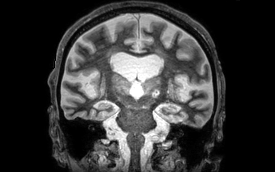
Each year, nearly 800,000 individuals in the U.S. experience a stroke, which occurs when blood flow to specific areas of the brain is insufficient, causing brain cells to die due to a lack of oxygen. While magnetic resonance imaging (MRI) is a valuable diagnostic tool for stroke, delays in treatment can lead to worse outcomes. In 2016, the FDA approved a new therapy for tremor disorders, involving high-intensity focused ultrasound (HIFU) to target and destroy a portion of the thalamus, a brain area often responsible for tremors. A recent study has shown that a molecule known as glial fibrillary acidic protein (GFAP) significantly increases in the blood of patients undergoing HIFU treatment for tremors, which causes damage similar to that of a small stroke. This finding, published in Brain Communications, suggests GFAP could be a promising biomarker for stroke and may eventually lead to blood tests for the rapid diagnosis of brain injuries.
In 2022, researchers at UT Southwestern Medical Center (Dallas, TX, USA) reported a technique that improved HIFU targeting for tremor treatment. Recently, the team noticed that the controlled brain injury caused by this therapy appeared similar to a stroke in brain imaging, with both types of damage sharing features, particularly in how the brain responds to these injuries. The researchers hypothesized that this similarity might help achieve the long-sought goal of diagnosing stroke and brain injuries through blood markers. Previous attempts have faced challenges such as a lack of blood samples taken before a stroke, differences in the locations of brain injuries in stroke patients, uncertainty around the timing of strokes, and variations among patients.
The UT Southwestern team believed that using HIFU as a research tool could help overcome these obstacles. In the study, 30 patients with tremor-dominant Parkinson’s disease or essential tremor, another movement disorder, received HIFU treatment. Blood samples were collected before the procedure and at one hour and 48 hours post-treatment. The researchers then measured concentrations of five molecular markers previously identified as potentially useful for diagnosing brain injuries: GFAP, neurofilament light chain, amyloid-beta 40, amyloid-beta 42, and phosphorylated tau 181 (pTau-181). Forty-eight hours following HIFU treatment, all markers except pTau-181 showed significant increases, with GFAP rising the most—more than four times its pre-treatment levels on average.
These results suggest that GFAP could serve as a reliable marker for stroke and other brain injuries. The researchers plan to further investigate GFAP levels at various time points following HIFU treatment to assess its potential as a diagnostic marker for brain injuries. Additionally, they are studying other molecules that might indicate brain injuries even earlier than GFAP. The team has also begun collecting blood from emergency stroke patients to determine whether GFAP levels are elevated in this group.
“This is the first study to use HIFU as a controlled model to evaluate brain injury biomarker dynamics,” said Bhavya R. Shah, M.D., Associate Professor of Radiology and Neurological Surgery at UT Southwestern as well as in the Advanced Imaging Research Center. “The ability to pair a timed pre- and post-HIFU measurement with precise lesion delivery is unprecedented and offers extraordinary potential for validating blood biomarkers of brain injury in a way that has not been done before.”
Latest Molecular Diagnostics News
- Diagnostic Device Predicts Treatment Response for Brain Tumors Via Blood Test
- Blood Test Detects Early-Stage Cancers by Measuring Epigenetic Instability
- Two-in-One DNA Analysis Improves Diagnostic Accuracy While Saving Time and Costs
- “Lab-On-A-Disc” Device Paves Way for More Automated Liquid Biopsies
- New Tool Maps Chromosome Shifts in Cancer Cells to Predict Tumor Evolution
- Blood Test Identifies Inflammatory Breast Cancer Patients at Increased Risk of Brain Metastasis
- Newly-Identified Parkinson’s Biomarkers to Enable Early Diagnosis Via Blood Tests
- New Blood Test Could Detect Pancreatic Cancer at More Treatable Stage
- Liquid Biopsy Could Replace Surgical Biopsy for Diagnosing Primary Central Nervous Lymphoma
- New Tool Reveals Hidden Metabolic Weakness in Blood Cancers
- World's First Blood Test Distinguishes Between Benign and Cancerous Lung Nodules
- Rapid Test Uses Mobile Phone to Identify Severe Imported Malaria Within Minutes
- Gut Microbiome Signatures Predict Long-Term Outcomes in Acute Pancreatitis
- Blood Test Promises Faster Answers for Deadly Fungal Infections
- Blood Test Could Detect Infection Exposure History
- Urine-Based MRD Test Tracks Response to Bladder Cancer Surgery
Channels
Clinical Chemistry
view channel
New PSA-Based Prognostic Model Improves Prostate Cancer Risk Assessment
Prostate cancer is the second-leading cause of cancer death among American men, and about one in eight will be diagnosed in their lifetime. Screening relies on blood levels of prostate-specific antigen... Read more
Extracellular Vesicles Linked to Heart Failure Risk in CKD Patients
Chronic kidney disease (CKD) affects more than 1 in 7 Americans and is strongly associated with cardiovascular complications, which account for more than half of deaths among people with CKD.... Read moreHematology
view channel
New Guidelines Aim to Improve AL Amyloidosis Diagnosis
Light chain (AL) amyloidosis is a rare, life-threatening bone marrow disorder in which abnormal amyloid proteins accumulate in organs. Approximately 3,260 people in the United States are diagnosed... Read more
Fast and Easy Test Could Revolutionize Blood Transfusions
Blood transfusions are a cornerstone of modern medicine, yet red blood cells can deteriorate quietly while sitting in cold storage for weeks. Although blood units have a fixed expiration date, cells from... Read more
Automated Hemostasis System Helps Labs of All Sizes Optimize Workflow
High-volume hemostasis sections must sustain rapid turnaround while managing reruns and reflex testing. Manual tube handling and preanalytical checks can strain staff time and increase opportunities for error.... Read more
High-Sensitivity Blood Test Improves Assessment of Clotting Risk in Heart Disease Patients
Blood clotting is essential for preventing bleeding, but even small imbalances can lead to serious conditions such as thrombosis or dangerous hemorrhage. In cardiovascular disease, clinicians often struggle... Read moreImmunology
view channelBlood Test Identifies Lung Cancer Patients Who Can Benefit from Immunotherapy Drug
Small cell lung cancer (SCLC) is an aggressive disease with limited treatment options, and even newly approved immunotherapies do not benefit all patients. While immunotherapy can extend survival for some,... Read more
Whole-Genome Sequencing Approach Identifies Cancer Patients Benefitting From PARP-Inhibitor Treatment
Targeted cancer therapies such as PARP inhibitors can be highly effective, but only for patients whose tumors carry specific DNA repair defects. Identifying these patients accurately remains challenging,... Read more
Ultrasensitive Liquid Biopsy Demonstrates Efficacy in Predicting Immunotherapy Response
Immunotherapy has transformed cancer treatment, but only a small proportion of patients experience lasting benefit, with response rates often remaining between 10% and 20%. Clinicians currently lack reliable... Read moreMicrobiology
view channel
Comprehensive Review Identifies Gut Microbiome Signatures Associated With Alzheimer’s Disease
Alzheimer’s disease affects approximately 6.7 million people in the United States and nearly 50 million worldwide, yet early cognitive decline remains difficult to characterize. Increasing evidence suggests... Read moreAI-Powered Platform Enables Rapid Detection of Drug-Resistant C. Auris Pathogens
Infections caused by the pathogenic yeast Candida auris pose a significant threat to hospitalized patients, particularly those with weakened immune systems or those who have invasive medical devices.... Read morePathology
view channel
Engineered Yeast Cells Enable Rapid Testing of Cancer Immunotherapy
Developing new cancer immunotherapies is a slow, costly, and high-risk process, particularly for CAR T cell treatments that must precisely recognize cancer-specific antigens. Small differences in tumor... Read more
First-Of-Its-Kind Test Identifies Autism Risk at Birth
Autism spectrum disorder is treatable, and extensive research shows that early intervention can significantly improve cognitive, social, and behavioral outcomes. Yet in the United States, the average age... Read moreTechnology
view channel
Robotic Technology Unveiled for Automated Diagnostic Blood Draws
Routine diagnostic blood collection is a high‑volume task that can strain staffing and introduce human‑dependent variability, with downstream implications for sample quality and patient experience.... Read more
ADLM Launches First-of-Its-Kind Data Science Program for Laboratory Medicine Professionals
Clinical laboratories generate billions of test results each year, creating a treasure trove of data with the potential to support more personalized testing, improve operational efficiency, and enhance patient care.... Read moreAptamer Biosensor Technology to Transform Virus Detection
Rapid and reliable virus detection is essential for controlling outbreaks, from seasonal influenza to global pandemics such as COVID-19. Conventional diagnostic methods, including cell culture, antigen... Read more
AI Models Could Predict Pre-Eclampsia and Anemia Earlier Using Routine Blood Tests
Pre-eclampsia and anemia are major contributors to maternal and child mortality worldwide, together accounting for more than half a million deaths each year and leaving millions with long-term health complications.... Read moreIndustry
view channelNew Collaboration Brings Automated Mass Spectrometry to Routine Laboratory Testing
Mass spectrometry is a powerful analytical technique that identifies and quantifies molecules based on their mass and electrical charge. Its high selectivity, sensitivity, and accuracy make it indispensable... Read more
AI-Powered Cervical Cancer Test Set for Major Rollout in Latin America
Noul Co., a Korean company specializing in AI-based blood and cancer diagnostics, announced it will supply its intelligence (AI)-based miLab CER cervical cancer diagnostic solution to Mexico under a multi‑year... Read more
Diasorin and Fisher Scientific Enter into US Distribution Agreement for Molecular POC Platform
Diasorin (Saluggia, Italy) has entered into an exclusive distribution agreement with Fisher Scientific, part of Thermo Fisher Scientific (Waltham, MA, USA), for the LIAISON NES molecular point-of-care... Read more


