Automated Malaria Diagnosis Enhanced by Deep Neural Networks
|
By LabMedica International staff writers Posted on 14 Aug 2020 |


|
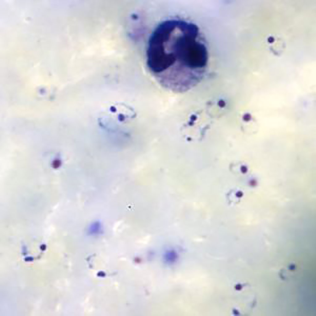
Ring-form trophozoites of Plasmodium falciparum and a white blood cell in a thick blood film (Photo courtesy of Medical Care Development International).
Plasmodium falciparum malaria remains one of the greatest global health burdens with over 228 million cases globally in 2018. In that year there were approximately 405,000 deaths due to malaria worldwide, with the African region accounting for 93% of these deaths, mostly among children.
Although there are a range of techniques that have been developed for the diagnosis of malaria, conventional light microscopy on Giemsa‐stained thick and thin blood films remains the gold standard. Techniques such as polymerase chain reaction, flow cytometric assay and fluorescence‐dye based approaches lack a universally standardized methodology, present high costs, and require quality control improvement.
A team of scientists from University College London (London, UK) leveraged routine clinical‐microscopy labels from their quality‐controlled malaria clinics, to train a Deep Malaria Convolutional Neural Network classifier (DeepMCNN) for automated malaria diagnosis. The DeepMCNN system also provides total Malaria Parasite (MP) and White Blood Cell (WBC) counts allowing parasitaemia estimation in MP/μL. Malaria parasites were detected and counted using human‐expert operated microscopy following Giemsa staining of thick and thin blood films. The criterion for declaring a participant to be malaria parasite‐free was no detectable parasites in 100 high‐power (100×) fields in thick films.
The investigators captured images using an upright bright-field BX63 microscope (Olympus, Tokyo, Japan) fitted with a 100×/1.4 NA objective lens, a motorized x‐y sample positioning stage (Prior Scientific, Cambridge, UK) and a color camera to capture images of Giemsa‐stained, thick blood smears. These smears prepared in their clinics tested the use of deep learning‐based object detection methods to identify both P. falciparum parasites and white‐blood‐cell (WBC) nuclei in the digitized extended depth of field (EDoF) thick blood films images.
The team reported that the prospective validation of the DeepMCNN achieved sensitivity/specificity of 0.92/0.90 against expert‐level malaria diagnosis. The PPV/NPV performance was 0.92/0.90, which is clinically usable in their holoendemic settings in a densely populated metropolis.
The authors concluded that their open data and easily deployable DeepMCNN provide a clinically relevant platform, where other healthcare providers could harness their readily available patient level diagnostic labels, to tailor and further improve the accuracy of the DeepMCNN classifier for their clinical pathway settings. The study was published in the August 2020 issue of the American Journal of Hematology.
Related Links:
University College London
Olympus
Prior Scientific






Although there are a range of techniques that have been developed for the diagnosis of malaria, conventional light microscopy on Giemsa‐stained thick and thin blood films remains the gold standard. Techniques such as polymerase chain reaction, flow cytometric assay and fluorescence‐dye based approaches lack a universally standardized methodology, present high costs, and require quality control improvement.
A team of scientists from University College London (London, UK) leveraged routine clinical‐microscopy labels from their quality‐controlled malaria clinics, to train a Deep Malaria Convolutional Neural Network classifier (DeepMCNN) for automated malaria diagnosis. The DeepMCNN system also provides total Malaria Parasite (MP) and White Blood Cell (WBC) counts allowing parasitaemia estimation in MP/μL. Malaria parasites were detected and counted using human‐expert operated microscopy following Giemsa staining of thick and thin blood films. The criterion for declaring a participant to be malaria parasite‐free was no detectable parasites in 100 high‐power (100×) fields in thick films.
The investigators captured images using an upright bright-field BX63 microscope (Olympus, Tokyo, Japan) fitted with a 100×/1.4 NA objective lens, a motorized x‐y sample positioning stage (Prior Scientific, Cambridge, UK) and a color camera to capture images of Giemsa‐stained, thick blood smears. These smears prepared in their clinics tested the use of deep learning‐based object detection methods to identify both P. falciparum parasites and white‐blood‐cell (WBC) nuclei in the digitized extended depth of field (EDoF) thick blood films images.
The team reported that the prospective validation of the DeepMCNN achieved sensitivity/specificity of 0.92/0.90 against expert‐level malaria diagnosis. The PPV/NPV performance was 0.92/0.90, which is clinically usable in their holoendemic settings in a densely populated metropolis.
The authors concluded that their open data and easily deployable DeepMCNN provide a clinically relevant platform, where other healthcare providers could harness their readily available patient level diagnostic labels, to tailor and further improve the accuracy of the DeepMCNN classifier for their clinical pathway settings. The study was published in the August 2020 issue of the American Journal of Hematology.
Related Links:
University College London
Olympus
Prior Scientific

Platinum Member
Automated Coagulation Analyzer
Hemolumi H6
Gold Member
Platelet Function Analyzer
PL-12
HPV Test
Allplex HPV28 Detection
Fully Automated Urinalysis System
DxU 1800 Fully Automated Urinalysis System
Latest Molecular Diagnostics News
- Noninvasive Cervical Test Detects Endometrial Cancer for Triage
- AI Tool Classifies Pathogenic Variants in Severe Childhood Epilepsy
- Molecular Test Detects Barrett’s Esophagus to Guide Endoscopy Referral
- Blood-Based Metagenomic Test Detects Wide Range of Pathogens from Single Sample
- New RNA Detection Tool Shows Promise for HIV and Other Viral Testing
- Tissue-Specific Vesicle Isolation Expands Molecular Insights Into Liver Disease
- Whole Genome Sequencing Solution Targets Unsolved Rare Disease Cases
- Validated Test Predicts Recurrence Risk and Radiation Benefit in Early-Stage Breast Cancer
- Study Shows How Genome Reference Bias Can Mislead Preventive Testing
- Genomic Screening Expands Detection of Treatable Conditions in Newborns
- Age-Based Genetic Testing May Miss Most Inherited Cancer Risk Variants
- Pooled Sputum Testing Offers Lower-Cost Molecular Diagnosis for Tuberculosis
- Expanded Sequencing Panel Supports Myeloid Malignancy Variant Profiling
- Tumor Markers Help Predict Response to Less Intensive HER2+ Chemotherapy
- New NGS Suite Expands Clonality and MRD Research in Lymphoid Malignancies
- Tumor-Informed ctDNA Test Shows Utility Across Gastrointestinal and Gynecologic Cancers
Channels
Clinical Chemistry
view channel
Multimodal Clinical Data Models Predict Cachexia Risk Across Cancers
Cancer cachexia is a severe wasting syndrome that affects roughly half of patients with cancer and cannot be reversed simply by increasing food intake. It can reduce treatment tolerance, impair quality... Read more
Study Evaluates Alzheimer’s Blood Biomarker Across Racial and Ethnic Groups
Alzheimer’s disease trials increasingly depend on identifying cognitively unimpaired people who already have elevated brain amyloid. Blood biomarkers may help reduce reliance on positron emission tomography... Read more
Hematology
view channel
New Myelofibrosis Guidelines Recommend Expanded Genetic Testing at Diagnosis
Myelofibrosis is a rare, life-threatening blood cancer affecting up to six per 100,000 people. The disease causes scarring in the bone marrow, limiting the production of healthy blood cells and potentially... Read more
Handheld Point-of-Care Test Aims to Enable Rapid Coagulation Assessment in Trauma
Severe blood loss and inadequate tissue oxygenation are leading causes of preventable death after traumatic injury. Blood products are essential in trauma care, but cold-chain storage, short shelf life,... Read more

Immunology
view channel
FDA Approval Adds Automated Testing Option for Parvovirus B19 Serology
Human Parvovirus B19 is a highly contagious erythrovirus transmitted mainly through respiratory droplets and close contact. Diagnosis relies on IgM antibodies to detect active infection and IgG antibodies... Read more
Blood-Based Immune Aging Map Could Help Spot Disease Risk Earlier
Chronological age does not always reflect immune health or future disease risk. Two adults of the same age can have immune systems that age very differently, with some remaining resilient while others... Read more
Microbiology
view channel
Rapid Host-Response Test Aims to Predict Corticosteroid Benefit in Sepsis
Sepsis and severe infections are difficult to treat because patients with similar clinical presentations may have very different underlying immune responses. Corticosteroids are commonly used in severe... Read more
Oral and Lung Microbiome Changes Linked to Severe Long Covid Symptoms
Long Covid can involve persistent respiratory, cognitive, and gastrointestinal symptoms after acute infection, creating a difficult diagnostic and management challenge. Because COVID-19 initially affects... Read more

Pathology
view channel
Molecular Testing Refines Barrett’s Esophagus Progression Risk Beyond Biopsy Findings
Barrett’s esophagus is a precancerous condition associated with increased risk of esophageal adenocarcinoma. Routine endoscopic biopsy findings guide surveillance and therapy, but histopathology may not... Read more
Updated Digital Pathology Platform Expands AI Integration for Cancer Diagnosis
Digital pathology is reshaping how laboratories manage case review, image analysis, and collaboration. As diagnostic workflows become more complex, pathology teams need platforms that can support digital... Read more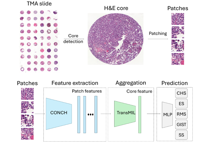
Technology
view channel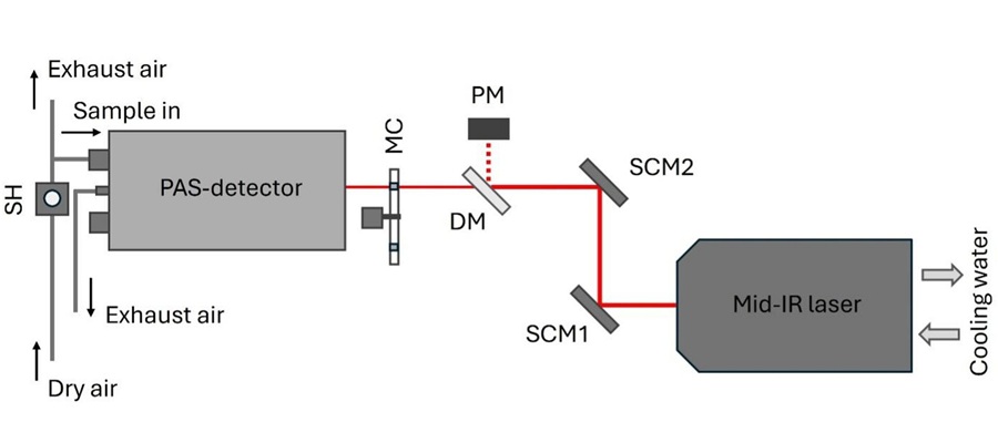
Laser-Based Swab Analysis Shows Promise for Detecting Disease-Linked Odor Patterns
Disease-related changes in volatile organic compounds can alter body odor, producing measurable patterns in exhaled breath and bodily fluids. Current analytical methods can be complex, time-consuming,... Read more
Laser-Enhanced Assay Boosts Sensitivity for Colorectal Cancer Biomarker Detection
Colorectal cancer is the third most commonly diagnosed cancer and the second leading cause of cancer-related death worldwide. Early detection remains critical, but cancer biomarkers can produce only faint... Read more

Industry
view channel
Global TB Summit Connects Diagnostic Testing With Prevention and Clinical Care
Tuberculosis remains a major global infectious disease challenge despite declining deaths. In 2024, an estimated 10.7 million people worldwide developed the disease and nearly 1.2 million died from it.... Read more