New X-Ray Crystallography Study Confirms Structure of Empty Cowpea mosaic virus Particles
|
By LabMedica International staff writers Posted on 10 Apr 2016 |
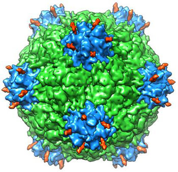
Image: A study shows that a hollowed-out version of Cowpea mosaic virus could be useful in human therapies (Photo courtesy of the Scripps Research Institute).
An X-ray crystallography study confirmed that empty Cowpea mosaic virus (CPMV) particles (eVLPs) were structurally similar to the intact virus and showed that they could be used for drug transport and other biomedical applications.
CPMV is a plant virus of the Comovirus group. Its genome consists of two molecules of positive-sense RNA, which are separately encapsulated. The virus particles are 28 nanometers in diameter and contain 60 copies each of a Large (L) and Small (S) coat protein. The structure is well characterized to atomic resolution, and the viral particles are thermostable. CPMV displays a number of features that can be exploited for nanoscale biomaterial fabrication. Its genetic, biological, and physical properties are well characterized, and it can be isolated readily from plants. There are many stable mutants already prepared that allow specific modification of the capsid surface. It is possible to attach a number of different chemicals to the virus surface and to construct multilayer arrays of such nanoparticles on solid surfaces. This gives the natural or genetically engineered nanoparticles a range of properties which could be useful in nanotechnological applications such as biosensors, catalysis and nanoelectronic devices.
Empty CPMV particles (eVLPs) can be modified to entrap drugs or other molecules while the outside surface can be coated with peptides that direct the particles to a specific class of target cells.
Investigators at The Scripps Research Institute (La Jolla, CA, USA) reported in the March 24, 2016, online edition of the journal Structure that they had used X-ray crystallography at 2.3 angstrom resolution to determine the crystal structure of CPMV eVLPs and then compared it to previously reported cryo-electron microscopy (cryo-EM) reports of eVLPs and virion crystal structures.
The new study revealed that although the X-ray and cryo-EM structures of eVLPs were mostly similar, there existed significant differences at the C-terminus of the small (S) subunit. The intact C-terminus of the S subunit plays a critical role in enabling the efficient assembly of CPMV virions and eVLPs, but undergoes proteolysis after particle formation. In addition, the results of mass spectrometry-based proteomics analysis of coat protein subunits from CPMV eVLPs and virions showed that the C-termini of S subunits underwent proteolytic cleavages at multiple sites instead of a single cleavage site as previously observed.
"By studying the structure of the viral particles, we can get important information for transforming this plant virus into a useful therapeutic," said senior author Dr. Vijay Reddy, an associate professor at The Scripps Research Institute. "The eVLP is no longer a virus; it is just a protein capsule."
Related Links:
The Scripps Research Institute
Latest BioResearch News
- Genome Analysis Predicts Likelihood of Neurodisability in Oxygen-Deprived Newborns
- Gene Panel Predicts Disease Progession for Patients with B-cell Lymphoma
- New Method Simplifies Preparation of Tumor Genomic DNA Libraries
- New Tool Developed for Diagnosis of Chronic HBV Infection
- Panel of Genetic Loci Accurately Predicts Risk of Developing Gout
- Disrupted TGFB Signaling Linked to Increased Cancer-Related Bacteria
- Gene Fusion Protein Proposed as Prostate Cancer Biomarker
- NIV Test to Diagnose and Monitor Vascular Complications in Diabetes
- Semen Exosome MicroRNA Proves Biomarker for Prostate Cancer
- Genetic Loci Link Plasma Lipid Levels to CVD Risk
- Newly Identified Gene Network Aids in Early Diagnosis of Autism Spectrum Disorder
- Link Confirmed between Living in Poverty and Developing Diseases
- Genomic Study Identifies Kidney Disease Loci in Type I Diabetes Patients
- Liquid Biopsy More Effective for Analyzing Tumor Drug Resistance Mutations
- New Liquid Biopsy Assay Reveals Host-Pathogen Interactions
- Method Developed for Enriching Trophoblast Population in Samples
Channels
Clinical Chemistry
view channel
Existing Hospital Analyzers Can Identify Fake Liquid Medical Products
Counterfeit and substandard medicines remain a serious global health threat, with World Health Organization estimates suggesting that 10.5% of medicines in low- and middle-income countries are either fake... Read more
Rapid Blood Testing Method Aids Safer Decision-Making in Drug-Related Emergencies
Acute recreational drug toxicity is a frequent reason for emergency department visits, yet clinicians rarely have access to confirmatory toxicology results in real time. Instead, treatment decisions are... Read moreMolecular Diagnostics
view channel
New Extraction Kit Enables Consistent, Scalable cfDNA Isolation from Multiple Biofluids
Circulating cell-free DNA (cfDNA) found in plasma, serum, urine, and cerebrospinal fluid is typically present at low concentrations and is often highly fragmented, making efficient recovery challenging... Read more
AI-Powered Liquid Biopsy Classifies Pediatric Brain Tumors with High Accuracy
Liquid biopsies offer a noninvasive way to study cancer by analyzing circulating tumor DNA in body fluids. However, in pediatric brain tumors, the small amount of ctDNA in cerebrospinal fluid has limited... Read moreHematology
view channel
Rapid Cartridge-Based Test Aims to Expand Access to Hemoglobin Disorder Diagnosis
Sickle cell disease and beta thalassemia are hemoglobin disorders that often require referral to specialized laboratories for definitive diagnosis, delaying results for patients and clinicians.... Read more
New Guidelines Aim to Improve AL Amyloidosis Diagnosis
Light chain (AL) amyloidosis is a rare, life-threatening bone marrow disorder in which abnormal amyloid proteins accumulate in organs. Approximately 3,260 people in the United States are diagnosed... Read moreImmunology
view channel
New Biomarker Predicts Chemotherapy Response in Triple-Negative Breast Cancer
Triple-negative breast cancer is an aggressive form of breast cancer in which patients often show widely varying responses to chemotherapy. Predicting who will benefit from treatment remains challenging,... Read moreBlood Test Identifies Lung Cancer Patients Who Can Benefit from Immunotherapy Drug
Small cell lung cancer (SCLC) is an aggressive disease with limited treatment options, and even newly approved immunotherapies do not benefit all patients. While immunotherapy can extend survival for some,... Read more
Whole-Genome Sequencing Approach Identifies Cancer Patients Benefitting From PARP-Inhibitor Treatment
Targeted cancer therapies such as PARP inhibitors can be highly effective, but only for patients whose tumors carry specific DNA repair defects. Identifying these patients accurately remains challenging,... Read more
Ultrasensitive Liquid Biopsy Demonstrates Efficacy in Predicting Immunotherapy Response
Immunotherapy has transformed cancer treatment, but only a small proportion of patients experience lasting benefit, with response rates often remaining between 10% and 20%. Clinicians currently lack reliable... Read moreMicrobiology
view channel
Rapid Test Promises Faster Answers for Drug-Resistant Infections
Drug-resistant pathogens continue to pose a growing threat in healthcare facilities, where delayed detection can impede outbreak control and increase mortality. Candida auris is notoriously difficult to... Read more
CRISPR-Based Technology Neutralizes Antibiotic-Resistant Bacteria
Antibiotic resistance has accelerated into a global health crisis, with projections estimating more than 10 million deaths per year by 2050 as drug-resistant “superbugs” continue to spread.... Read more
Comprehensive Review Identifies Gut Microbiome Signatures Associated With Alzheimer’s Disease
Alzheimer’s disease affects approximately 6.7 million people in the United States and nearly 50 million worldwide, yet early cognitive decline remains difficult to characterize. Increasing evidence suggests... Read morePathology
view channel
Single Sample Classifier Predicts Cancer-Associated Fibroblast Subtypes in Patient Samples
Pancreatic ductal adenocarcinoma (PDAC) remains one of the deadliest cancers, in part because of its dense tumor microenvironment that influences how tumors grow and respond to treatment.... Read more
New AI-Driven Platform Standardizes Tuberculosis Smear Microscopy Workflow
Sputum smear microscopy remains central to tuberculosis treatment monitoring and follow-up, particularly in high‑burden settings where serial testing is routine. Yet consistent, repeatable bacillary assessment... Read more
AI Tool Uses Blood Biomarkers to Predict Transplant Complications Before Symptoms Appear
Stem cell and bone marrow transplants can be lifesaving, but serious complications may arise months after patients leave the hospital. One of the most dangerous is chronic graft-versus-host disease, in... Read moreTechnology
view channel
Blood Test “Clocks” Predict Start of Alzheimer’s Symptoms
More than 7 million Americans live with Alzheimer’s disease, and related health and long-term care costs are projected to reach nearly USD 400 billion in 2025. The disease has no cure, and symptoms often... Read more
AI-Powered Biomarker Predicts Liver Cancer Risk
Liver cancer, or hepatocellular carcinoma, causes more than 800,000 deaths worldwide each year and often goes undetected until late stages. Even after treatment, recurrence rates reach 70% to 80%, contributing... Read more
Robotic Technology Unveiled for Automated Diagnostic Blood Draws
Routine diagnostic blood collection is a high‑volume task that can strain staffing and introduce human‑dependent variability, with downstream implications for sample quality and patient experience.... Read more
ADLM Launches First-of-Its-Kind Data Science Program for Laboratory Medicine Professionals
Clinical laboratories generate billions of test results each year, creating a treasure trove of data with the potential to support more personalized testing, improve operational efficiency, and enhance patient care.... Read moreIndustry
view channel
QuidelOrtho Collaborates with Lifotronic to Expand Global Immunoassay Portfolio
QuidelOrtho (San Diego, CA, USA) has entered a long-term strategic supply agreement with Lifotronic Technology (Shenzhen, China) to expand its global immunoassay portfolio and accelerate customer access... Read more


