Molecular Mechanism Regulating Regeneration of Spinal Nerves Identified
|
By LabMedica International staff writers Posted on 29 Mar 2016 |


|
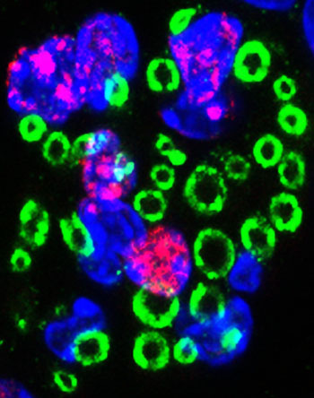
Image: A confocal micrograph of the lesion core following spinal cord injury. Nuclear EdU (red) shows the presence of newly differentiated cells which produce Schwann cell myelin (green). These peripheral-like Schwann cells remyelinate central axons in the injured spinal cord and are important for spontaneous repair and functional recovery after spinal cord injury (Photo courtesy of King\'s College London).
The protein neuregulin-1 was found to regulate the repair mechanism that attempts to restore lost myelin following spinal cord injury.
Following traumatic spinal cord injury, acute demyelination of spinal axons is followed by a period of spontaneous remyelination. However, this endogenous repair response is incomplete and may account for the chronic loss of function demonstrated by surviving axons. Spontaneous remyelination is largely mediated by Schwann cells, where demyelinated central axons, particularly in the dorsal columns, become associated with peripheral myelin. The molecular control mechanism, functional role, and origin of these central remyelinating Schwann cells are currently unknown.
The growth factor neuregulin-1 (Nrg1, encoded by the NRG1 gene) is a key signaling factor controlling myelination in the peripheral nervous system, via signaling through ErbB tyrosine kinase receptors. The neuregulins are a family of four structurally related proteins that are part of the EGF (epidermal growth factor) family of proteins, which have been shown to have diverse functions in the development of the nervous system.
Investigators at King's College London (United Kingdom) and the University of Oxford (United Kingdom) examined whether Nrg1 was required for Schwann cell-mediated remyelination of central dorsal column axons and whether removal of Nrg1 would influence the degree of spontaneous remyelination and functional recovery following spinal cord injury.
They reported in the March 17, 2016, online edition of the journal Brain that Nrg1 signaling mediated an endogenous regenerative event in which Schwann cells remyelinated denuded central axons after traumatic spinal cord injury and that Nrg1 was an important mediator of spontaneous functional repair after spinal cord injury. In mice lacking the NRG1 gene, spontaneous myelin repair was completely prevented and spinal nerve fibers remained demyelinated. Furthermore, mice without NRG1 showed worse outcomes after spinal cord injury compared to mice with the gene intact, particularly in walking, balance, and coordinated movements.
Senior author Dr. Elizabeth Bradbury, professor of regenerative medicine and neuroplasticity at King's College London, said, "Spinal cord injury could happen to anyone, at any time. In an instant your life could change and you could lose all feeling and function below the level of the injury. Existing treatments are largely ineffective, so there is a pressing need for new regenerative therapies to repair tissue damage and restore function after spinal cord injury."
"These new findings advance our understanding of the molecular mechanisms which may orchestrate the body's remarkable capacity for natural repair," said Dr. Bradbury.
"By enhancing this spontaneous response, we may be able to significantly improve spinal cord function after injury. Our research also has wider implications for other disorders of the central nervous system which share this demyelinating pathology, such as multiple sclerosis."
Related Links:
King's College London
University of Oxford


.jpg)





Following traumatic spinal cord injury, acute demyelination of spinal axons is followed by a period of spontaneous remyelination. However, this endogenous repair response is incomplete and may account for the chronic loss of function demonstrated by surviving axons. Spontaneous remyelination is largely mediated by Schwann cells, where demyelinated central axons, particularly in the dorsal columns, become associated with peripheral myelin. The molecular control mechanism, functional role, and origin of these central remyelinating Schwann cells are currently unknown.
The growth factor neuregulin-1 (Nrg1, encoded by the NRG1 gene) is a key signaling factor controlling myelination in the peripheral nervous system, via signaling through ErbB tyrosine kinase receptors. The neuregulins are a family of four structurally related proteins that are part of the EGF (epidermal growth factor) family of proteins, which have been shown to have diverse functions in the development of the nervous system.
Investigators at King's College London (United Kingdom) and the University of Oxford (United Kingdom) examined whether Nrg1 was required for Schwann cell-mediated remyelination of central dorsal column axons and whether removal of Nrg1 would influence the degree of spontaneous remyelination and functional recovery following spinal cord injury.
They reported in the March 17, 2016, online edition of the journal Brain that Nrg1 signaling mediated an endogenous regenerative event in which Schwann cells remyelinated denuded central axons after traumatic spinal cord injury and that Nrg1 was an important mediator of spontaneous functional repair after spinal cord injury. In mice lacking the NRG1 gene, spontaneous myelin repair was completely prevented and spinal nerve fibers remained demyelinated. Furthermore, mice without NRG1 showed worse outcomes after spinal cord injury compared to mice with the gene intact, particularly in walking, balance, and coordinated movements.
Senior author Dr. Elizabeth Bradbury, professor of regenerative medicine and neuroplasticity at King's College London, said, "Spinal cord injury could happen to anyone, at any time. In an instant your life could change and you could lose all feeling and function below the level of the injury. Existing treatments are largely ineffective, so there is a pressing need for new regenerative therapies to repair tissue damage and restore function after spinal cord injury."
"These new findings advance our understanding of the molecular mechanisms which may orchestrate the body's remarkable capacity for natural repair," said Dr. Bradbury.
"By enhancing this spontaneous response, we may be able to significantly improve spinal cord function after injury. Our research also has wider implications for other disorders of the central nervous system which share this demyelinating pathology, such as multiple sclerosis."
Related Links:
King's College London
University of Oxford

Platinum Member
Automated Coagulation Analyzer
Hemolumi H6
Gold Member
Clinical Chemistry Assay
Sorbitol Dehydrogenase (SDH)
New
Silver Member
Vitamin D Assay
EZ Vitamin D Assay
New
Gastrointestinal Panel
Xpert® GI Panel
Latest BioResearch News
- Genetic Study Links Inherited Blindness to Gene Tied to Rare Metabolic Disorder
- Population-Scale Proteomics Study Identifies New Genetic-Protein Links
- Circulating Tumor DNA Helps Identify Targets for Personalized Immunotherapy
- Researchers Classify 108 Lysosomal Disorders in Updated Diagnostic Framework
- New Autoantibody Target Identified in Neuromyelitis Optica Spectrum Disorder
- New Immune Target Could Support More Precise High Blood Pressure Treatment
- New Gene-Disease Link May Help Diagnose Rare Neurodevelopmental Disorders
- Researchers Identify Shared Molecular Networks Behind Fatigue-Related Illnesses
- Blood Metabolite Signature Predicts ALS Progression and Points to Treatment Strategy
- Whole-Blood Extracellular Vesicle Analysis Captures Molecular Signals Missed by Plasma Testing
- Age-Related Genomic Differences Could Refine Treatment Decisions in Lung Cancer
- Computational Tool Identifies Central Asthma Genes for Target Discovery
- Molecular Pathway Reveals Driver of Triple-Negative Breast Cancer Spread
- D-Serine May Predict Immune Checkpoint Therapy Resistance in Gastric Cancer
- New Genetic Cause Identified for Neurodevelopmental Disorder
- New Genetic Discovery Could Support Precision Diabetes Care
Channels
Clinical Chemistry
view channel
Hemoglobin Levels May Help Signal Long-Term Dementia Risk
Dementia is a growing health challenge in aging societies, with limited treatment options and no cure. Identifying long-term risk remains difficult, particularly when specialist scans or emerging biomarkers... Read more
Liquid Biopsy Platform Shows Promise for Early Gynecologic Cancer Detection
Gynecologic cancers can be difficult to detect early, particularly when symptoms overlap with benign conditions. Earlier cancer detection remains a diagnostic need in ovarian cancer, described as one of... Read more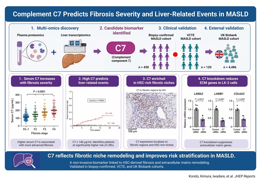
Multi-Omics Study Identifies Blood Marker for MASLD Progression
Metabolic dysfunction-associated steatotic liver disease (MASLD) is a major cause of liver-related morbidity and mortality worldwide. As MASLD progresses, fibrosis and liver-related events, including hepatocellular... Read more
Global Survey Finds Pediatric Troponin Testing Lacks Consistent Reference Intervals
Cardiac troponin is released into the blood when heart muscle is damaged and is widely used as a rapid marker of cardiac injury in adults. In children, interpreting troponin results remains difficult because... Read moreMolecular Diagnostics
view channel
Validated Test Predicts Recurrence Risk and Radiation Benefit in Early-Stage Breast Cancer
For women with early-stage invasive breast cancer who undergo breast-conserving surgery, clinicians must assess both the risk of locoregional recurrence and the potential benefit of adjuvant radiation therapy.... Read more
Genomic Screening Expands Detection of Treatable Conditions in Newborns
Conventional newborn screening can miss conditions that lack biochemical biomarkers or present atypically. Initial hearing screens may also fail to detect hearing loss that is later identified through... Read more
Hematology
view channel
New Donor Genetic Marker May Help Predict Stem Cell Transplant Success
Donor selection for hematopoietic stem cell transplantation plays a major role in relapse risk and survival for patients with blood cancers and other blood disorders. Despite advances in genotyping, uncertainty... Read more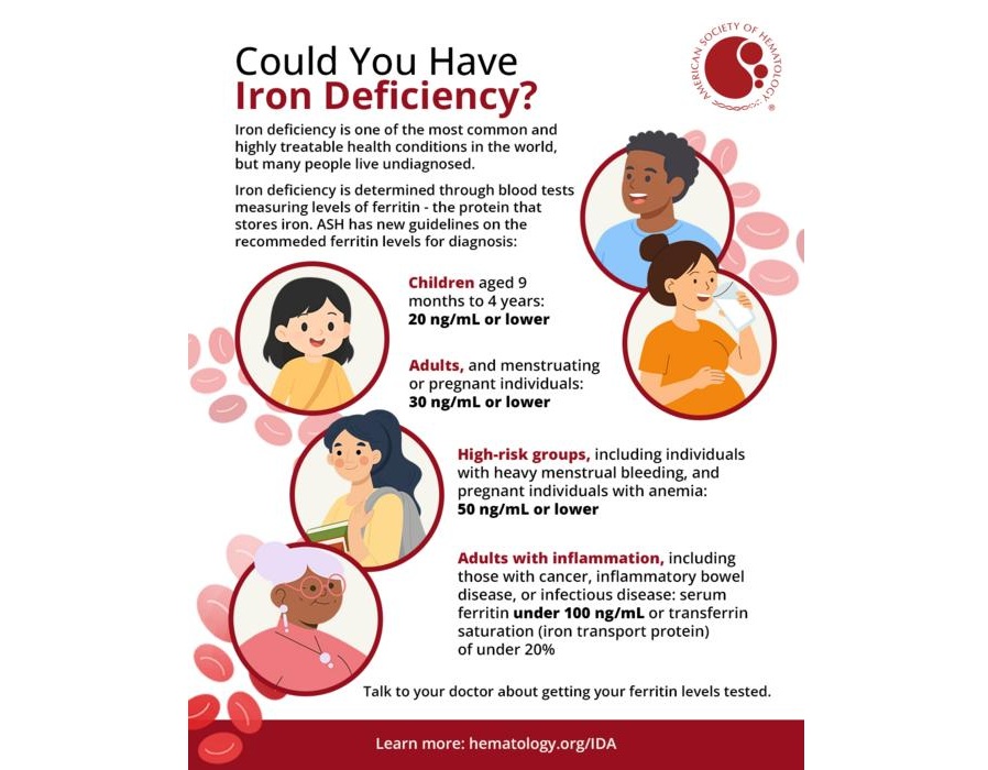
Updated Ferritin Thresholds Improve Detection of Iron Deficiency
Iron deficiency is one of the most common health conditions worldwide, yet its nonspecific symptoms can delay diagnosis for months. Variation in testing practices and ferritin thresholds may contribute... Read more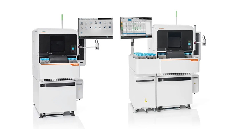
Immunology
view channel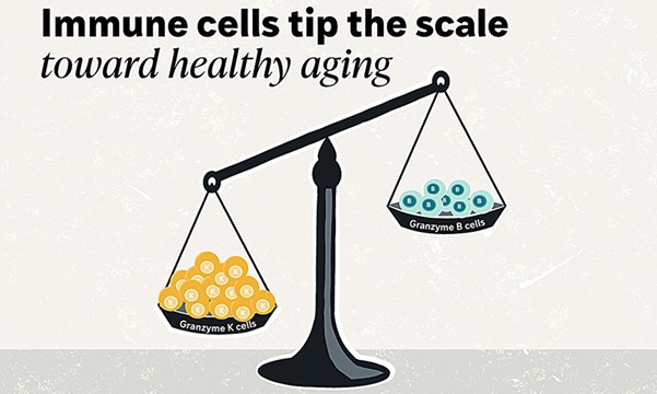
Blood-Based Immune Aging Map Could Help Spot Disease Risk Earlier
Chronological age does not always reflect immune health or future disease risk. Two adults of the same age can have immune systems that age very differently, with some remaining resilient while others... Read more
Blood-Based Immune Signal Could Reveal Lung Cancer Risk Before Tumors Develop
Lung cancer remains difficult to detect before symptoms appear, when advanced disease is harder to treat. In the U.K., about 32,800 people die from lung cancer each year, and only 11.1% of those diagnosed... Read more
Microbiology
view channel
Rapid Urine Test Aids Diagnosis of Invasive Aspergillosis
Invasive aspergillosis is an uncommon mold infection in the general population but can pose serious risks for people with weakened immune defenses. Diagnosis can be difficult because existing approaches... Read more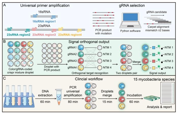
Nanodroplet CRISPR Technology Supports Rapid, Multiplexed Mycobacterial Identification
Mycobacterial infections are difficult to diagnose because closely related species can have different clinical and therapeutic implications. Nontuberculous mycobacteria (NTM) are increasingly recognized... Read more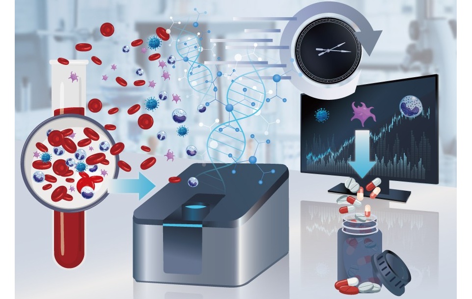
Genomic Workflow Identifies Fungal Pathogens Before Blood Cultures Turn Positive
Fungal bloodstream infections pose a major threat to hospitalized patients. Candida species cause most invasive fungal infections worldwide and are among the leading causes of hospital-acquired bloodstream... Read more
FDA-Cleared Multiplex PCR Test Detects 13 Respiratory Pathogens in a Single Sample
Respiratory tract infections can be difficult to distinguish at presentation because many cause overlapping, nonspecific symptoms and are initially grouped as influenza-like illnesses. Causes span a range... Read morePathology
view channel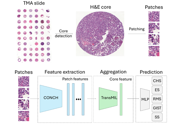
AI Histopathology Tool Distinguishes Ewing Sarcoma for Diagnosis
Ewing sarcoma is a rare malignant tumor that mainly affects children, adolescents, and young adults. It usually arises in bone but can also develop in soft tissue, where its microscopic appearance may... Read more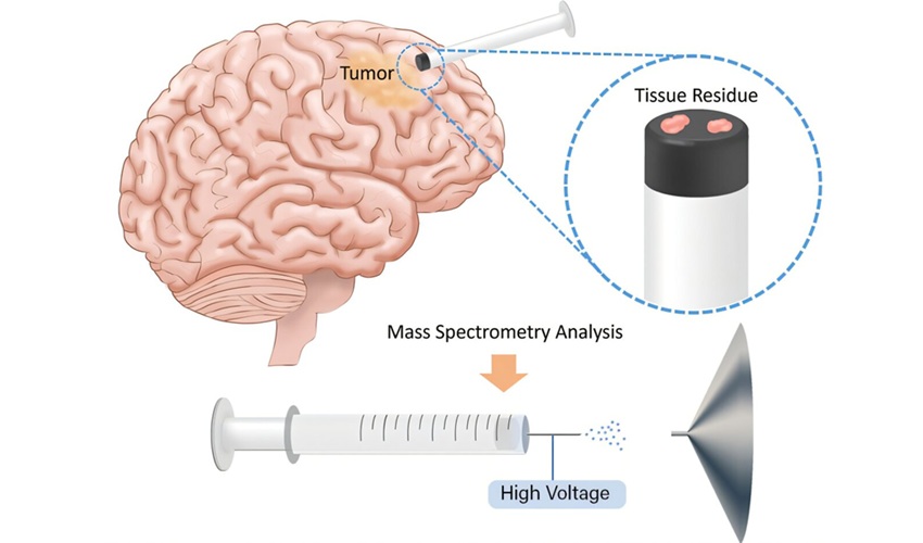
Rapid Mass Spectrometry Test May Aid Glioma Margin Decisions
Glioma brain tumors are highly infiltrative and can extend into nearby healthy brain tissue, making tumor margins difficult to define during surgery. Residual tumor cells may contribute to recurrence and... Read more
Genomic Classifier Predicts Benefit From Adding Hormone Therapy to Salvage Prostate Radiation
Men who have undergone prostatectomy for prostate cancer may later develop a detectable or rising prostate-specific antigen, prompting salvage radiation therapy. A key challenge is determining who is most... Read more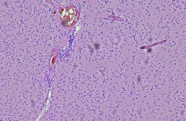
Technology
view channel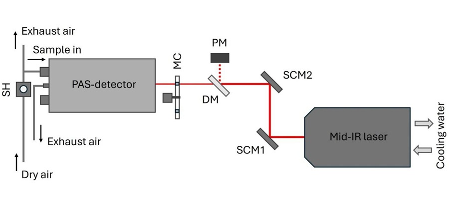
Laser-Based Swab Analysis Shows Promise for Detecting Disease-Linked Odor Patterns
Disease-related changes in volatile organic compounds can alter body odor, producing measurable patterns in exhaled breath and bodily fluids. Current analytical methods can be complex, time-consuming,... Read more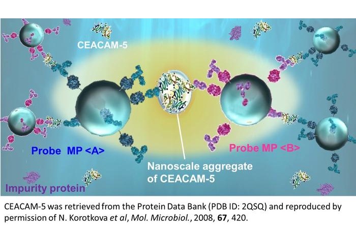
Laser-Enhanced Assay Boosts Sensitivity for Colorectal Cancer Biomarker Detection
Colorectal cancer is the third most commonly diagnosed cancer and the second leading cause of cancer-related death worldwide. Early detection remains critical, but cancer biomarkers can produce only faint... Read more

Industry
view channel
Multi-Cancer Screening Blood Test Gains FDA Breakthrough Designation
Natera Inc.’s (Austin, TX, USA) multi-cancer early detection (MCED) assay has received Breakthrough Device designation (BDD) from the U.S. Food and Drug Administration (FDA). The assay analyzes cell-free... Read more
European Approval Expands Liquid Biopsy Use in Advanced Breast Cancer Testing
Estrogen receptor-positive (ER+), HER2-negative advanced breast cancer may require molecular testing to guide treatment selection, with blood-based assays offering a way to identify tumor mutations that... Read more