AI Tool Enhances Interpretation of Tissue Samples by Pathologists
Posted on 09 Jul 2025
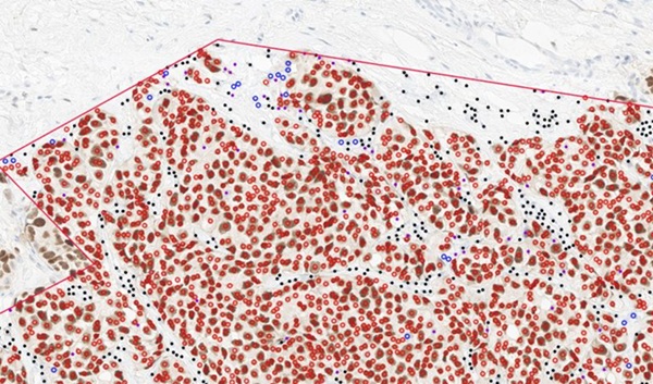
Malignant melanoma, a form of skin cancer, is diagnosed by pathologists based on tissue samples. A crucial aspect of this process is estimating the presence of tumor-infiltrating lymphocytes (TILs), immune cells that significantly influence the body's response to cancer. However, TIL assessments can vary depending on the pathologist, which compromises medical safety and can lead to inaccuracies in determining the severity of the disease. This variability is critical as TILs play a role in prognosis and treatment decisions, with a high presence being considered favorable. Now, an artificial intelligence (AI)-based tool that quantifies TILs in tissue samples has been found to improve the consistency and accuracy of these assessments. The tool provides more reliable data, which is vital for determining treatment options.
The AI tool, developed through a collaboration between Karolinska Institutet (Stockholm, Sweden) and Yale University (New Haven, CT, USA), was designed to support pathologists in quantifying TILs. The solution aims to reduce variability in assessments and help make more accurate predictions about a patient's disease prognosis. The researchers used a set of digital images of stained tissue sections from patients with malignant melanoma to develop and test the tool. This AI support was incorporated into the workflow of experienced pathologists and other professionals with some experience in assessing pathological images. The tool works by analyzing the tissue samples and providing a quantification of the TILs, offering a clearer picture of the disease's progression.
")
The study tested the AI-assisted assessments by comparing them to those made by pathologists working without AI support. The research involved 98 participants who evaluated 60 tissue samples. The results showed that AI-supported assessments were superior in terms of reproducibility, with high consistency across different assessors. Additionally, the AI-enhanced assessments led to more accurate prognoses, as the results closely aligned with the retrospective 'correct answer'. These findings, published in JAMA Network Open, suggest that AI can be a powerful tool for clinical pathology, particularly in melanoma diagnosis. The researchers plan to conduct further studies to refine the AI tool and explore its potential for broader clinical use.
"Understanding the severity of a patient's disease based on tissue samples is important, among other things, for determining how aggressively it should be treated," said Balazs Acs, associate professor at the Department of Oncology-Pathology at Karolinska Institutet. “We now have an AI-based tool that can quantify the TIL biomarker, which could help with treatment decisions in the future. However, more studies are needed before this AI tool can be used in clinical practice, but the results so far are promising and suggest that it could be a very useful tool in clinical pathology.”