Magnetic Needle Performs Less Invasive, More Precise Surgery and Diagnostic Tests
|
By LabMedica International staff writers Posted on 25 Apr 2022 |


|

A tiny, untethered needle can enter the body through an incision no larger than a pin prick to perform biopsies, suture wounds, and even deliver cancer-fighting chemotherapy directly to tumors. Controlled by externally applied magnetics forces—no attached, guiding wires, or human or robotic hands—these miniscule tools promise a future of more precise, safer, and far less invasive surgery, experts say. However, as devices get smaller, so does their response to the magnetic forces that cause them to move and steer their course. Now, researchers have a solution: a surgical needle equipped with small magnets inside that, when stimulated by the externally applied forces, slip from one end of the needle to another, tapping against a rigid plate and supplying ample force to penetrate tissue.
In a study, a team of researchers at the Johns Hopkins Whiting School of Engineering (Baltimore, MD, USA) have demonstrated for the first time that untethered magnetic needles can be forceful enough to accomplish surgery. The researchers call their device the "Pulse Actuated Collisions for Tissue-penetrating Needle," or MPACT-Needle. For their experiments, the team connected a slender thread of suturing material to the needle, using a joystick connected to a computer to deliver commands that enabled the MPACT-Needle to perform surgical suturing on a sample rabbit cornea.
The team's next step is to develop better, more accurate motion-control algorithms equipped with imaging modalities to precisely control the movement of the device, making surgeries and procedures safer. The researchers believe that if the technology becomes successful, their new magnetic needles would make it possible to access hard-to-reach, delicate areas of the body, such as the bile duct, to deliver drugs directly to tumors, extract biopsy samples, or suture a wound rapidly and effectively to stem internal bleeding.
"These extremely small tools have the potential to revolutionize medicine, but as anyone who has played with magnets knows, size is important," said Axel Krieger, an assistant professor of mechanical engineering at Johns Hopkins Whiting School of Engineering. "The smaller the surgical tool, the less invasive the surgery, but also the weaker the device's response to magnetic force. One of the biggest challenges facing us is how to make sure that these mini-tools can be moved with enough force to penetrate tissue and do the job they are there to do."
"We proved that these tiny magnetic needles can have strong enough forces to perform delicate surgery with limited invasiveness," said study leader Onder Erin, a postdoctoral fellow in mechanical engineering at Johns Hopkins. "I can envision a time when our device will also be used to perform biopsies, and even deliver therapeutics and chemotherapy directly to tumors."
Related Links:
Johns Hopkins Whiting School of Engineering







Latest Technology News
- New AI-Powered System Aims to Streamline Laboratory and Manufacturing Workflows
- Laser-Based Swab Analysis Shows Promise for Detecting Disease-Linked Odor Patterns
- Laser-Enhanced Assay Boosts Sensitivity for Colorectal Cancer Biomarker Detection
- New Automation Workflow Streamlines NGS Library Preparation for High-Volume Labs
- ADLM Calls for CLIA Updates to Support Safe AI Use in Laboratory Medicine
- Multimodal AI Framework Aims to Guide Cancer Immunotherapy Decisions
- New Multipurpose Centrifuge Combines High Capacity with Sustainable Cooling
- Bacterial Vesicle Expression System Streamlines Production of Cancer Diagnostic Proteins
- Autonomous Robotic System Receives FDA Authorization for Blood Collection
- Interoperable Data Platform Standardizes Multi-Cancer Blood Test Results
- Training Device Improves Accuracy of Pooled Molecular Diagnostics
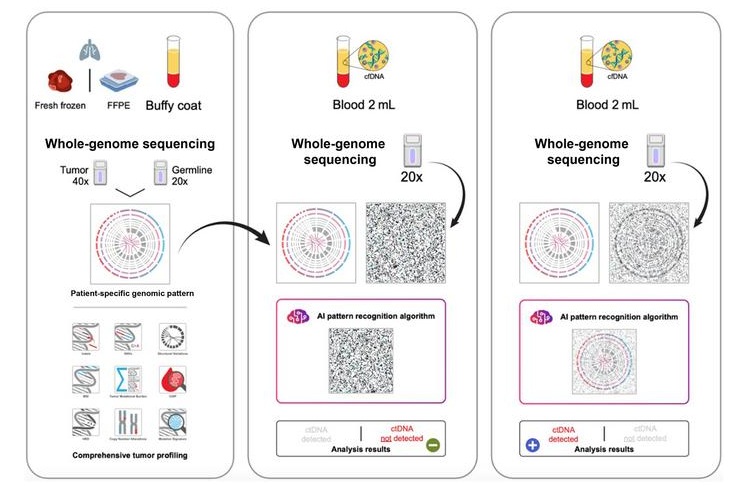
- New CE-Certified Software Advances Whole-Genome Cancer Testing
- National Rare Disease Registry Standardizes Genetic and Clinical Data for Coordinated Care
- AI Platform Links Biomarker Results to Cancer Clinical Trials and Guidelines
- Agentic AI Platform Supports Genomic Decision-Making in Oncology
- Algorithm Panel Aids Liver Fibrosis Assessment and Liver Cancer Surveillance
Channels
Clinical Chemistry
view channel
Point-of-Care Blood Biomarker Test Detects Brain Injury Missed by CT
Traumatic brain injury remains difficult to assess in emergency settings because some patients continue to have symptoms despite normal computed tomography findings. Approximately 2.8 million people in the U.... Read more
Daily Blood Testing May Help Guide Earlier Antibiotic Stopping in Sepsis
Sepsis is a life-threatening condition caused by the body’s overwhelming response to infection and accounts for thousands of deaths each year. In intensive care, concern about stopping treatment too early... Read more

Molecular Diagnostics
view channel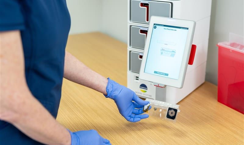
Molecular Vaginitis Panel Gains FDA Clearance and CLIA Waiver for Point-of-Care Use
Vaginitis affects most women at least once in their lifetime and can cause painful, disruptive symptoms. Common causes include bacterial vaginosis, vulvovaginal candidiasis, and trichomoniasis, reflecting... Read more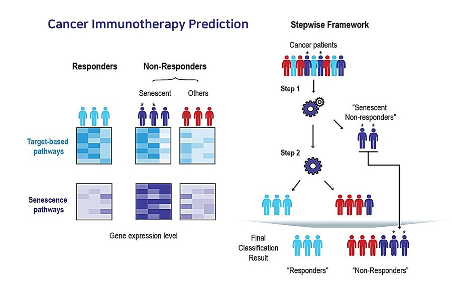
AI Tool Predicts Immunotherapy Response Using Tumor Senescence Signals
Patients receiving the same cancer immunotherapy can have markedly different outcomes, with some showing little or no response. Identifying these patients earlier could reduce the costs of ineffective... Read more
Hematology
view channel
New Myelofibrosis Guidelines Recommend Expanded Genetic Testing at Diagnosis
Myelofibrosis is a rare, life-threatening blood cancer affecting up to six per 100,000 people. The disease causes scarring in the bone marrow, limiting the production of healthy blood cells and potentially... Read more
Handheld Point-of-Care Test Aims to Enable Rapid Coagulation Assessment in Trauma
Severe blood loss and inadequate tissue oxygenation are leading causes of preventable death after traumatic injury. Blood products are essential in trauma care, but cold-chain storage, short shelf life,... Read more

Immunology
view channel
Antibody Profiling Predicts Inflammatory Bowel Disease Years Before Diagnosis
Inflammatory bowel disease (IBD), which includes Crohn’s disease and ulcerative colitis, involves abnormal immune responses that cause persistent inflammation and damage in the digestive system.... Read more
Immune Profiling Initiative Aims to Improve Bone Marrow Analysis in Leukemia
Acute myeloid leukemia (AML) is an aggressive blood cancer in which assessing disease burden after therapy can help guide clinical evaluation. Patients with measurable residual disease (MRD) after treatment... Read more
Immune Profiling Identifies Early Maternal Autoantibodies Linked to Preterm Birth Risk
A typical pregnancy lasts about 40 weeks, but labor begins prematurely in roughly one in 10 pregnancies. Preterm birth is the leading cause of newborn death worldwide, yet its underlying causes and which... Read more
Longitudinal Blood Testing Reveals Changes in Allergen Sensitization During Childhood
Food allergy is a major childhood health concern, and distinguishing persistent risk from transient sensitization can guide clinical decisions. Specific IgE antibodies may be detectable even when a child... Read moreMicrobiology
view channel
Molecular Panel Gains WHO Emergency Use Listing to Support Ebola Outbreak Response
The ongoing Ebola Bundibugyo outbreak in the Democratic Republic of the Congo and neighboring countries has become one of the largest outbreaks of this Ebola virus species recorded to date.... Read more
Rapid Host-Response Test Aims to Predict Corticosteroid Benefit in Sepsis
Sepsis and severe infections are difficult to treat because patients with similar clinical presentations may have very different underlying immune responses. Corticosteroids are commonly used in severe... Read more

Pathology
view channel
Digital Pathology Test Stratifies Recurrence Risk in Early Breast Cancer
In hormone receptor-positive, human epidermal growth factor receptor 2 (HER2)-negative early breast cancer, accurate recurrence-risk assessment helps guide treatment decisions. However, individual risks... Read more
AI Pathology Tool Predicts DNA Repair Deficiency in High-Grade Serous Ovarian Cancer
High-grade serous tubo-ovarian carcinoma is an aggressive cancer arising in the ovaries or fallopian tubes. Some tumors exhibit homologous recombination deficiency (HRD), a feature reflecting impaired DNA repair.... Read more

Technology
view channel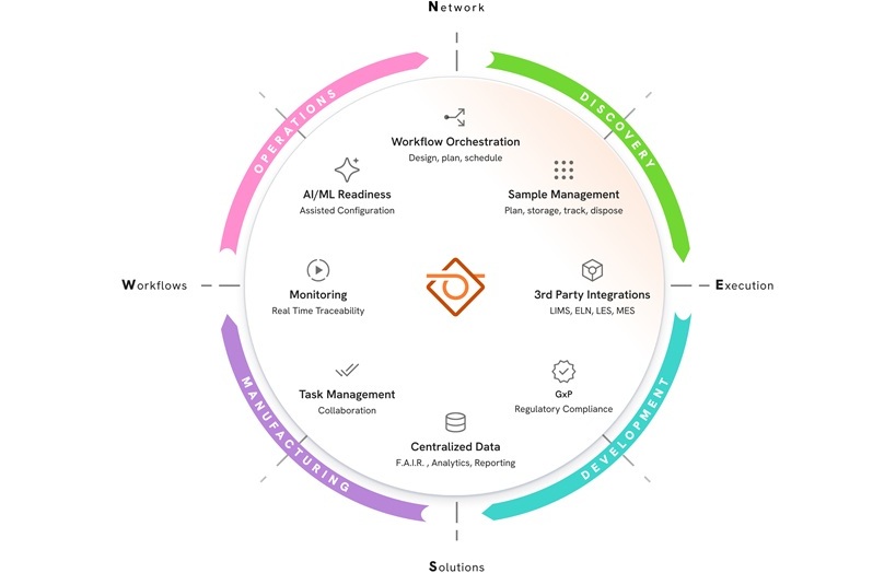
New AI-Powered System Aims to Streamline Laboratory and Manufacturing Workflows
Scientific laboratories supporting drug development, biotechnology, and materials research increasingly rely on coordinated digital workflows to move data, samples, and activities across teams.... Read more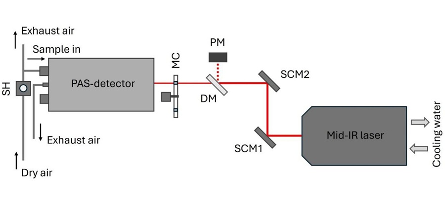
Laser-Based Swab Analysis Shows Promise for Detecting Disease-Linked Odor Patterns
Disease-related changes in volatile organic compounds can alter body odor, producing measurable patterns in exhaled breath and bodily fluids. Current analytical methods can be complex, time-consuming,... Read more

Industry
view channel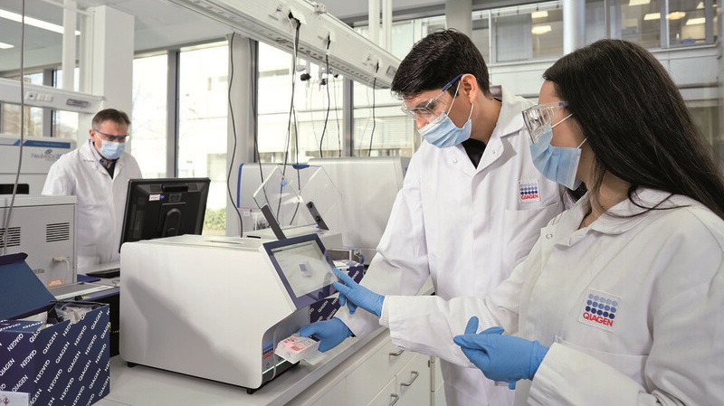
Qiagen Agrees to Go Private Through EQT-Led Acquisition
QIAGEN N.V. (Venlo, the Netherlands) and EQT Private Equity (Stockholm, Sweden) have agreed to an all-cash public takeover offer for all outstanding QIAGEN shares at EUR 46.50 per share.... Read more
Hologic and Inpeco Partner to Advance Molecular Laboratory Automation in Europe
Hologic, Inc. (Marlborough, MA, USA) and Inpeco S.A. (Novazzano, Switzerland) have signed a co-marketing partnership in Europe for Inpeco’s recently launched FlexLab X automation solution and Hologic’s... Read more