






Non‐Invasive Urine Markers Differentiate Renal Cancer from Oncocytoma
|
By LabMedica International staff writers Posted on 25 May 2021 |


|
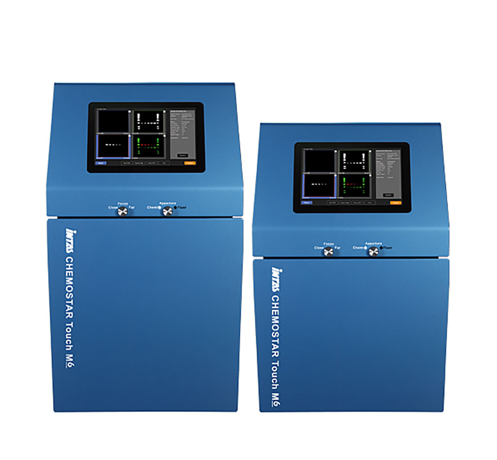
Image: The CHEMOSTAR Touch ECL & Fluorescence Imager (Photo courtesy of INTAS)
Clear cell renal cell carcinoma, (ccRCC), is a type of kidney cancer. Clear cell renal cell carcinoma is also called conventional renal cell carcinoma. Clear cell renal cell carcinoma is named after how the tumor looks under the microscope.
Renal oncocytoma is a benign (noncancerous) growth of the kidney. They generally do not cause any signs or symptoms and are often discovered incidentally while a person is undergoing diagnostic imaging for other conditions. Some people with renal oncocytoma will have abdominal or flank pain; blood in the urine; and/or an abdominal mass.
Urologists at the University Hospital Cologne (Cologne, Germany) have shown that that Vim3 is overexpressed in tissue samples of renal oncocytomas and Mxi‐2 in clear cell renal carcinoma (ccRCC). They collected urine samples from patients suspected of kidney cancer between 2015 and 2018. The final cohort consisted of 350 patients, with pathohistological proof of an oncocytoma in 20 patients or a RCC (50 chromophobe; 40 papillary); 200 clear cell RCC (ccRCC), as well as 40 negative controls.
The medical scientists performed Western blots on urine samples which were analyzed with INTAS Chemostar (Göttingen, Germany). ELISAs were performed using incubated 3B4 Vimentin antibody and for Mxi‐2 and analyzed on the FLUOstar Omega plate reader (BMG LABTECH, Ortenberg, Germany). The team also carried out lateral flow assays; and quantitative real‐time polymerase chain reaction (qRT‐PCR).
The investigators reported that there was a significant increase of Vim3 in urines from patients with oncocytoma and was detectable with ELISA compared to all other subtypes of RCCs. Mxi‐2 was predominantly overexpressed in ccRCCs. Lateral flow assay of Vim3 and Mxi‐2 shows two bands in the case of oncocytoma and ccRCC indicating the specificity of this test. For small renal masses (SRMs), an overexpression of miR‐15a/Mxi2 was detectable in urine samples from ccRCC and chromoRCC patients. In contrast to that, miR‐498/Vim3 were predominantly overexpressed in oncocytoma patients.
The authors concluded that using the highly specific, non‐invasive urine markers, Vim3 and Mxi‐2, they were able to fill the gap and reduce the number of overtreated patients as well as the number of unnecessary surgeries. Furthermore, even the detection as well as pre‐surgical differentiation in small kidney cancers is possible with the measurement of the two predicted miRs as well as the proteins. Both proteins (Vim3 and Mxi‐2) were detectable in patients’ urines and can be used for the non‐invasive differentiation of kidney cancers. The study was published on May 7, 2021 in the Journal of Clinical Laboratory Analysis.
Related Links:
University Hospital Cologne
INTAS
BMG LABTECH






Renal oncocytoma is a benign (noncancerous) growth of the kidney. They generally do not cause any signs or symptoms and are often discovered incidentally while a person is undergoing diagnostic imaging for other conditions. Some people with renal oncocytoma will have abdominal or flank pain; blood in the urine; and/or an abdominal mass.
Urologists at the University Hospital Cologne (Cologne, Germany) have shown that that Vim3 is overexpressed in tissue samples of renal oncocytomas and Mxi‐2 in clear cell renal carcinoma (ccRCC). They collected urine samples from patients suspected of kidney cancer between 2015 and 2018. The final cohort consisted of 350 patients, with pathohistological proof of an oncocytoma in 20 patients or a RCC (50 chromophobe; 40 papillary); 200 clear cell RCC (ccRCC), as well as 40 negative controls.
The medical scientists performed Western blots on urine samples which were analyzed with INTAS Chemostar (Göttingen, Germany). ELISAs were performed using incubated 3B4 Vimentin antibody and for Mxi‐2 and analyzed on the FLUOstar Omega plate reader (BMG LABTECH, Ortenberg, Germany). The team also carried out lateral flow assays; and quantitative real‐time polymerase chain reaction (qRT‐PCR).
The investigators reported that there was a significant increase of Vim3 in urines from patients with oncocytoma and was detectable with ELISA compared to all other subtypes of RCCs. Mxi‐2 was predominantly overexpressed in ccRCCs. Lateral flow assay of Vim3 and Mxi‐2 shows two bands in the case of oncocytoma and ccRCC indicating the specificity of this test. For small renal masses (SRMs), an overexpression of miR‐15a/Mxi2 was detectable in urine samples from ccRCC and chromoRCC patients. In contrast to that, miR‐498/Vim3 were predominantly overexpressed in oncocytoma patients.
The authors concluded that using the highly specific, non‐invasive urine markers, Vim3 and Mxi‐2, they were able to fill the gap and reduce the number of overtreated patients as well as the number of unnecessary surgeries. Furthermore, even the detection as well as pre‐surgical differentiation in small kidney cancers is possible with the measurement of the two predicted miRs as well as the proteins. Both proteins (Vim3 and Mxi‐2) were detectable in patients’ urines and can be used for the non‐invasive differentiation of kidney cancers. The study was published on May 7, 2021 in the Journal of Clinical Laboratory Analysis.
Related Links:
University Hospital Cologne
INTAS
BMG LABTECH

Platinum Member
Automated Coagulation Analyzer
Hemolumi H6
Gold Member
Quantitative POC Immunoassay Analyzer
EASY READER+
New
Nucleic Acid Purification Instrument
QIAsymphony Connect
Automated Clinical Chemistry Analyzer
Envoy 500+
Latest Clinical Chem. News
- Blood Antibody Linked to Lower Depression and PTSD Symptoms After Brain Injury
- New Reference Material Aims to Improve Comparability of Angiotensin II Testing
- Childhood Blood Biomarkers May Reveal Early Heart, Kidney, and Metabolic Risks
- Blood Biomarker Study Reveals Early Signals of Future Cognitive Difficulties
- International Consensus Guidance Outlines General Population Screening for Early-Stage Type 1 Diabetes
- Electrical Fingerprint of Extracellular Vesicles Could Detect Pancreatic Cancer
- Algorithm Uses Blood Biomarkers to Improve Alzheimer’s Trial Screening
- Blood Biomarker May Track Bone Metastases in Medullary Thyroid Cancer
- Blood Biomarkers Help Assess Growth Risks During Steroid Treatment in Duchenne Muscular Dystrophy
- New Blood Biomarker Predicts May Predict Preeclampsia and Fetal Growth Restriction
- Biomarker Score Helps Differentiate Takotsubo Syndrome from Myocardial Infarction
- New Biomarker Recommendations Aim to Personalize Stroke Rehabilitation
- Validated Biomarker Blood Test Detects Early Lung Cancer and Supports Risk Assessment
- Machine Learning Model Uses Routine Lab Tests to Support Heart Failure Classification
- NIH-Funded Study Targets Saliva-Based Long COVID Diagnosis
- Multiprotein Plasma Panel Identifies Remission in Vasculitis
Channels
Molecular Diagnostics
view channel
Blood-Based DNA Analysis Method Detects Early-Stage Solid Cancers
Early detection of stage I solid cancers remains a significant unmet clinical need. Liquid biopsy approaches that can distinguish tumor-derived signals from background cell-free DNA are therefore drawing... Read more
Combined Blood and Genetic Tests Estimate Timing of Alzheimer’s Symptom Onset
Predicting when Alzheimer’s disease will manifest remains a major clinical challenge, particularly before cognitive decline is evident. Blood biomarkers and genetic risk markers can flag elevated risk,... Read more

Hematology
view channel
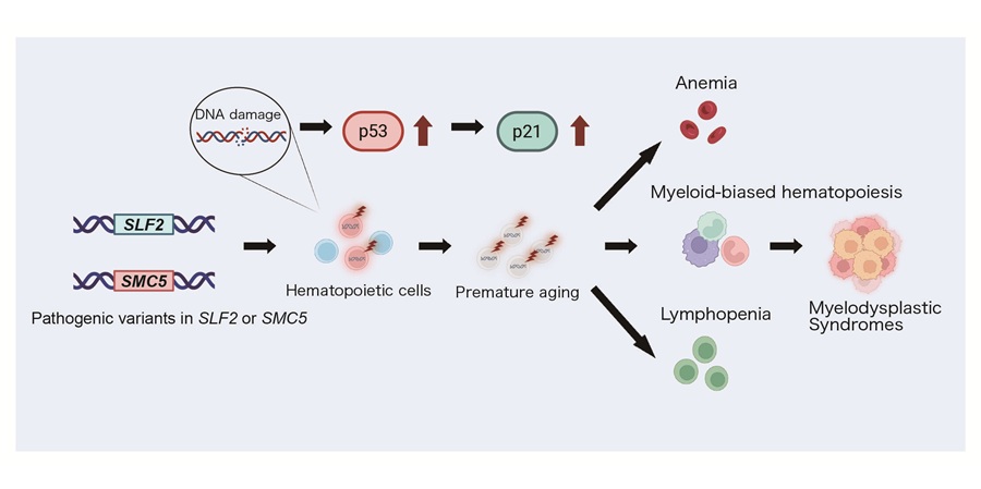
Hidden Molecular Mechanism Behind HIT Blood-Clotting Disorder Revealed
Immune-mediated thrombosis following heparin therapy can be sudden and severe, creating urgent diagnostic challenges. In heparin-induced thrombocytopenia (HIT), antibodies against platelet factor 4 (PF4)... Read more
Borderline Anemia May Carry Higher Risk Than Current Thresholds Suggest
Hemoglobin concentration is among the most frequently ordered blood tests in primary and hospital care. In older adults, both high and low values have been linked to adverse outcomes, including cognitive... Read more
Immunology
view channel
New Cellular Map May Help Predict Crohn’s Disease Course in Children
Crohn’s disease in children is a common and debilitating form of inflammatory bowel disease, marked by chronic intestinal inflammation and limited pediatric-specific treatments. Clinicians must decide... Read more
Temporal Immune Profiling Reveals How Sepsis States Change Over Time
Sepsis is a life-threatening condition in which the immune response to infection becomes dysregulated, leading to rapid organ failure and death. Although antimicrobials and organ support remain standard... Read more
Study Identifies Immune Cells That Drive Harmful Autoantibody Responses in COVID-19
Autoantibodies that mistakenly attack the body’s own tissues have been linked to severe COVID-19, Long COVID, and increased risk of autoimmune disease. However, the origins of these autoantibodies during... Read more
Microbiology
view channel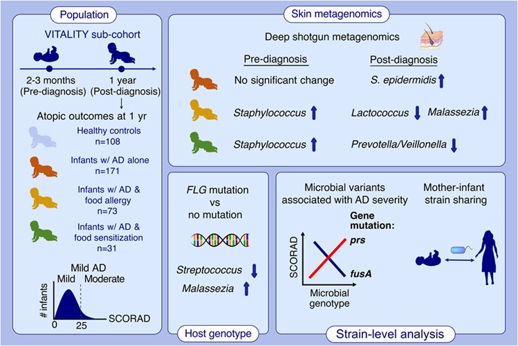
Infant Skin Microbiome Show Links to Later Eczema and Food Allergies
Eczema, or atopic dermatitis, and food allergies often begin in infancy and are among the earliest stages of the atopic march. Atopic dermatitis alone can affect up to 20% of children, yet objective indicators... Read more
Diagnostic Stewardship Guide Targets Testing Overuse and Hospital Cost Pressures
Hospitals face persistent pressure to improve diagnostic accuracy while reducing unnecessary testing and costs. Misordered or overused assays can complicate infection metrics, trigger penalties, and strain... Read more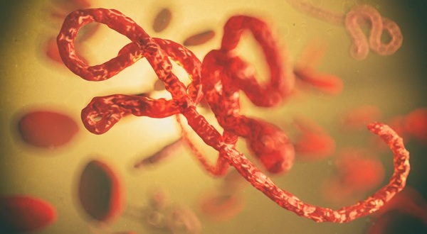
Research Strengthens Bundibugyo Virus Outbreak Readiness with Faster Diagnostics
Bundibugyo virus (BDBV), a species of ebolavirus, causes severe hemorrhagic disease and can be difficult to diagnose rapidly during outbreaks. Recent regulatory changes have further complicated swift deployment... Read more
Pathology
view channel
AI Spatial Analysis of Routine Slides Helps Predict Pancreatic Cancer Recurrence
Predicting which patients with pancreatic cancer will recur after treatment and surgery remains challenging. Pathology evaluation typically measures residual tumor burden, but recent work found that tumor... Read more
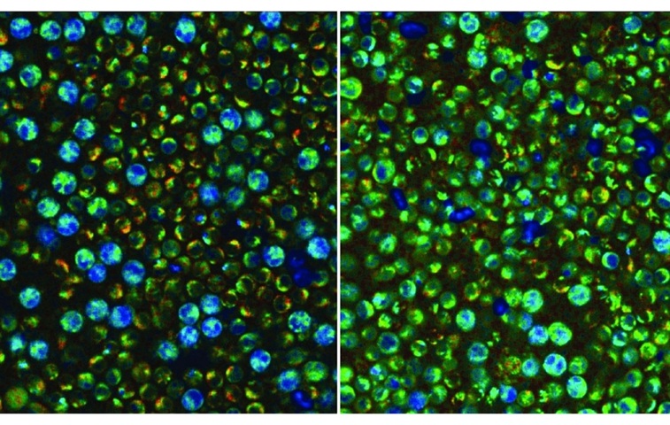
Hybrid Computational Imaging Method Improves Digital Pathology Resolution
Digital pathology depends on high-resolution whole-slide imaging to capture diagnostically relevant cellular and tissue features, yet conventional methods often force trade-offs between speed, cost, and detail.... Read more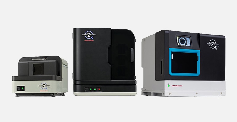
Technology
view channel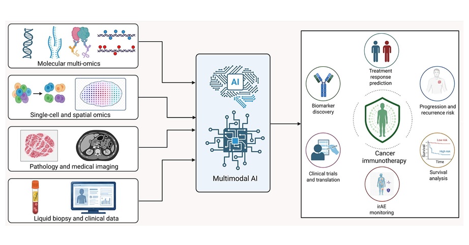
Multimodal AI Framework Aims to Guide Cancer Immunotherapy Decisions
Cancer immunotherapy has reshaped oncology, but heterogeneous responses and immune-related toxicities continue to complicate routine decision-making. Standard biomarkers, including programmed death-ligand... Read more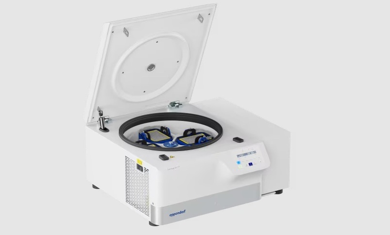
New Multipurpose Centrifuge Combines High Capacity with Sustainable Cooling
Laboratories often need centrifugation that accommodates multiple vessel formats while maintaining controlled temperatures to protect sensitive samples. Intuitive controls and repeatable operation can... Read more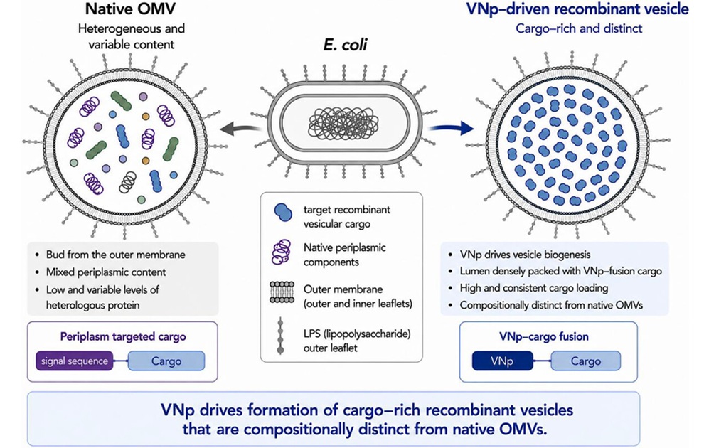
Bacterial Vesicle Expression System Streamlines Production of Cancer Diagnostic Proteins
Recombinant proteins are central to many cancer diagnostics and therapies, but numerous targets remain difficult and costly to produce because they are unstable, toxic to microbial hosts, or require precise folding.... Read more
Industry
view channel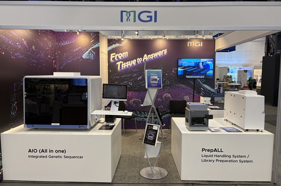
MGI Launches Automated Multiplex Tissue Staining and Imaging System at ECP 2026
MGI Tech Co., Ltd. (Shenzhen, China) introduced VisiOmics (PMIF-20RS), a fully automated multiplex immunofluorescence staining and imaging system, at the 38th European Congress of Pathology (ECP 2026)... Read more
.jpeg "Image Credit: Adobe Stock")