Sensitive Assays Enable Early Detection of Prion Infection
|
By LabMedica International staff writers Posted on 04 Feb 2019 |

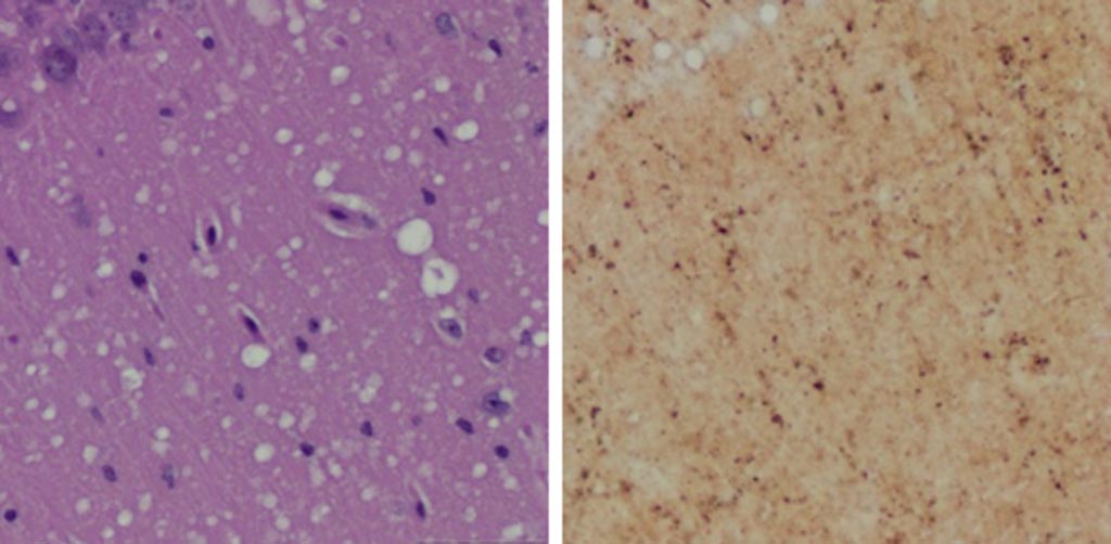
Image: A Microscopic examination of brain tissues of prion-infected animals. (Left) Staining shows spongiform degeneration. (Right) Staining shows intense misfolded prion protein (Photo courtesy of Case Western Reserve University).
Researchers working with rodent models have demonstrated the potential for developing a skin test for the early diagnosis of prion diseases in humans.
Prions are proteinaceous, infectious particles that completely lack any genetic material. These particles are transmissible pathogens, which cause neurodegenerative disorders in humans and animals. Prions show strikingly different biochemical and biophysical properties from other pathogens, such as fungi, bacteria, and viruses, as well as differing host-pathogen interactions. Prions are unusually resistant to many conventional chemical and physical treatments to reduce infectivity, such as intensive ultraviolet radiation, heat, and nuclease treatment.
Furthermore, prion infection induces no humoral or innate immune responses in the host. Prions are also peculiar in the way they multiply, which involves protein-protein interactions followed by conformational conversion. The normal form of prion protein is called PrPC, while the infectious form is called PrPSc – the C refers to cellular PrP (prion protein), while the Sc refers to scrapie, the prototypic prion disease, occurring in sheep. While PrPC is structurally well defined, PrPSc is polydisperse and impossible to define.
A definitive pre-mortem diagnosis of a prion disease, such as Creutzfeldt-Jakob disease (CJD) in humans, bovine spongiform encephalopathy (BSE) in cattle, or chronic wasting disease (CWD) in elk and deer depends on brain biopsy for prion detection, and no validated alternative preclinical diagnostic tests have been reported to date.
To improve this situation, investigators at Case Western Reserve University (Cleveland, OH, USA) sought to determine the feasibility of using a noninvasive skin test for preclinical diagnosis. This idea was based on previous findings, which showed that autopsy skin samples from human prion disease patients exhibited prion seeding and infectivity.
To test the hypothesis, the investigators examined skin PrPSc in hamsters and humanized transgenic (Tg) mice at different time points after intracerebral prion inoculation using the highly sensitive RT-QuIC and sPMCA assays.
The real-time quaking induced conversion (RT-QuIC) assay uses recombinant prion protein to which potentially infectious tissue homogenate is added. If the tissue has prion seeding activity, it induces aggregation in recombinant protein, which can be monitored by the fluorophore Thioflavin T (ThT). For aggregation to occur, intermittent double-orbital shaking at 42 degrees Celsius is required over the assay duration of up to 68 hours. ThT fluorescence is acquired every 15 minutes to report on aggregation status.
The serial protein misfolding cyclic amplification (sPMCA) technique initially incubates a small amount of abnormal prion with an excess of normal protein, so that some conversion takes place. The growing chain of misfolded protein is then blasted with ultrasound, breaking it down into smaller chains and so rapidly increasing the amount of abnormal protein available to cause conversions. By repeating the cycle, the mass of normal protein is rapidly changed into misfolded PrPSc prions. One round of PMCA cycling results in a 2500-fold increase in sensitivity of detection over western blotting, whereas two and seven rounds of successive PMCA cycling result in six million- and three billion-fold increases in sensitivity of detection over western blotting. Thus, PMCA is capable of detecting as little as a single molecule of oligomeric infectious PrPSc.
The investigators reported in the January 16, 2019, online edition of the journal Nature Communications that sPMCA detected skin PrPSc as early as two weeks post inoculation (wpi) in hamsters and four wpi in Tg40h mice. The RT-QuIC assay revealed earliest skin prion-seeding activity at three wpi in hamsters and 20 wpi in Tg40h mice. Unlike prion-inoculated animals, mock-inoculated animals showed detectable skin/brain PrPSc only after long cohabitation periods with scrapie-infected animals.
“Currently a definitive diagnosis of Creutzfeldt-Jakob disease is dependent on the examination of diseased brain tissue obtained at biopsy or autopsy. It has been impossible to detect at the early preclinical stage,” said senior author Dr. Wenquan Zou, associate professor of pathology at Case Western Reserve University. “Since the skin is readily accessible and skin biopsy is minimally invasive, detection of skin prions will be very useful for monitoring disease progression and assessing therapeutic efficacy during clinical trials or treatments when prion therapy becomes available in the future.”
“Sensitive, minimally invasive detection of various misfolded proteins in skin, such as tau in Alzheimer’s disease and alpha-synuclein in Parkinson’s disease, could be highly valuable for disease diagnosis and monitoring of disease progression and efficacy of treatments,” said Dr. Zou. “It is possible that the skin will ultimately serve as a mirror for us to monitor these misfolded proteins that accumulate and damage the brain in patients with these conditions.”
Related Links:
Case Western Reserve University
Latest BioResearch News
- Genome Analysis Predicts Likelihood of Neurodisability in Oxygen-Deprived Newborns
- Gene Panel Predicts Disease Progession for Patients with B-cell Lymphoma
- New Method Simplifies Preparation of Tumor Genomic DNA Libraries
- New Tool Developed for Diagnosis of Chronic HBV Infection
- Panel of Genetic Loci Accurately Predicts Risk of Developing Gout
- Disrupted TGFB Signaling Linked to Increased Cancer-Related Bacteria
- Gene Fusion Protein Proposed as Prostate Cancer Biomarker
- NIV Test to Diagnose and Monitor Vascular Complications in Diabetes
- Semen Exosome MicroRNA Proves Biomarker for Prostate Cancer
- Genetic Loci Link Plasma Lipid Levels to CVD Risk
- Newly Identified Gene Network Aids in Early Diagnosis of Autism Spectrum Disorder
- Link Confirmed between Living in Poverty and Developing Diseases
- Genomic Study Identifies Kidney Disease Loci in Type I Diabetes Patients
- Liquid Biopsy More Effective for Analyzing Tumor Drug Resistance Mutations
- New Liquid Biopsy Assay Reveals Host-Pathogen Interactions
- Method Developed for Enriching Trophoblast Population in Samples
Channels
Clinical Chemistry
view channel
New PSA-Based Prognostic Model Improves Prostate Cancer Risk Assessment
Prostate cancer is the second-leading cause of cancer death among American men, and about one in eight will be diagnosed in their lifetime. Screening relies on blood levels of prostate-specific antigen... Read more
Extracellular Vesicles Linked to Heart Failure Risk in CKD Patients
Chronic kidney disease (CKD) affects more than 1 in 7 Americans and is strongly associated with cardiovascular complications, which account for more than half of deaths among people with CKD.... Read moreMolecular Diagnostics
view channel
Diagnostic Device Predicts Treatment Response for Brain Tumors Via Blood Test
Glioblastoma is one of the deadliest forms of brain cancer, largely because doctors have no reliable way to determine whether treatments are working in real time. Assessing therapeutic response currently... Read more
Blood Test Detects Early-Stage Cancers by Measuring Epigenetic Instability
Early-stage cancers are notoriously difficult to detect because molecular changes are subtle and often missed by existing screening tools. Many liquid biopsies rely on measuring absolute DNA methylation... Read more
“Lab-On-A-Disc” Device Paves Way for More Automated Liquid Biopsies
Extracellular vesicles (EVs) are tiny particles released by cells into the bloodstream that carry molecular information about a cell’s condition, including whether it is cancerous. However, EVs are highly... Read more
Blood Test Identifies Inflammatory Breast Cancer Patients at Increased Risk of Brain Metastasis
Brain metastasis is a frequent and devastating complication in patients with inflammatory breast cancer, an aggressive subtype with limited treatment options. Despite its high incidence, the biological... Read moreHematology
view channel
New Guidelines Aim to Improve AL Amyloidosis Diagnosis
Light chain (AL) amyloidosis is a rare, life-threatening bone marrow disorder in which abnormal amyloid proteins accumulate in organs. Approximately 3,260 people in the United States are diagnosed... Read more
Fast and Easy Test Could Revolutionize Blood Transfusions
Blood transfusions are a cornerstone of modern medicine, yet red blood cells can deteriorate quietly while sitting in cold storage for weeks. Although blood units have a fixed expiration date, cells from... Read more
Automated Hemostasis System Helps Labs of All Sizes Optimize Workflow
High-volume hemostasis sections must sustain rapid turnaround while managing reruns and reflex testing. Manual tube handling and preanalytical checks can strain staff time and increase opportunities for error.... Read more
High-Sensitivity Blood Test Improves Assessment of Clotting Risk in Heart Disease Patients
Blood clotting is essential for preventing bleeding, but even small imbalances can lead to serious conditions such as thrombosis or dangerous hemorrhage. In cardiovascular disease, clinicians often struggle... Read moreImmunology
view channelBlood Test Identifies Lung Cancer Patients Who Can Benefit from Immunotherapy Drug
Small cell lung cancer (SCLC) is an aggressive disease with limited treatment options, and even newly approved immunotherapies do not benefit all patients. While immunotherapy can extend survival for some,... Read more
Whole-Genome Sequencing Approach Identifies Cancer Patients Benefitting From PARP-Inhibitor Treatment
Targeted cancer therapies such as PARP inhibitors can be highly effective, but only for patients whose tumors carry specific DNA repair defects. Identifying these patients accurately remains challenging,... Read more
Ultrasensitive Liquid Biopsy Demonstrates Efficacy in Predicting Immunotherapy Response
Immunotherapy has transformed cancer treatment, but only a small proportion of patients experience lasting benefit, with response rates often remaining between 10% and 20%. Clinicians currently lack reliable... Read moreMicrobiology
view channel
Comprehensive Review Identifies Gut Microbiome Signatures Associated With Alzheimer’s Disease
Alzheimer’s disease affects approximately 6.7 million people in the United States and nearly 50 million worldwide, yet early cognitive decline remains difficult to characterize. Increasing evidence suggests... Read moreAI-Powered Platform Enables Rapid Detection of Drug-Resistant C. Auris Pathogens
Infections caused by the pathogenic yeast Candida auris pose a significant threat to hospitalized patients, particularly those with weakened immune systems or those who have invasive medical devices.... Read morePathology
view channel
Engineered Yeast Cells Enable Rapid Testing of Cancer Immunotherapy
Developing new cancer immunotherapies is a slow, costly, and high-risk process, particularly for CAR T cell treatments that must precisely recognize cancer-specific antigens. Small differences in tumor... Read more
First-Of-Its-Kind Test Identifies Autism Risk at Birth
Autism spectrum disorder is treatable, and extensive research shows that early intervention can significantly improve cognitive, social, and behavioral outcomes. Yet in the United States, the average age... Read moreTechnology
view channel
Robotic Technology Unveiled for Automated Diagnostic Blood Draws
Routine diagnostic blood collection is a high‑volume task that can strain staffing and introduce human‑dependent variability, with downstream implications for sample quality and patient experience.... Read more
ADLM Launches First-of-Its-Kind Data Science Program for Laboratory Medicine Professionals
Clinical laboratories generate billions of test results each year, creating a treasure trove of data with the potential to support more personalized testing, improve operational efficiency, and enhance patient care.... Read moreAptamer Biosensor Technology to Transform Virus Detection
Rapid and reliable virus detection is essential for controlling outbreaks, from seasonal influenza to global pandemics such as COVID-19. Conventional diagnostic methods, including cell culture, antigen... Read more
AI Models Could Predict Pre-Eclampsia and Anemia Earlier Using Routine Blood Tests
Pre-eclampsia and anemia are major contributors to maternal and child mortality worldwide, together accounting for more than half a million deaths each year and leaving millions with long-term health complications.... Read moreIndustry
view channelNew Collaboration Brings Automated Mass Spectrometry to Routine Laboratory Testing
Mass spectrometry is a powerful analytical technique that identifies and quantifies molecules based on their mass and electrical charge. Its high selectivity, sensitivity, and accuracy make it indispensable... Read more
AI-Powered Cervical Cancer Test Set for Major Rollout in Latin America
Noul Co., a Korean company specializing in AI-based blood and cancer diagnostics, announced it will supply its intelligence (AI)-based miLab CER cervical cancer diagnostic solution to Mexico under a multi‑year... Read more
Diasorin and Fisher Scientific Enter into US Distribution Agreement for Molecular POC Platform
Diasorin (Saluggia, Italy) has entered into an exclusive distribution agreement with Fisher Scientific, part of Thermo Fisher Scientific (Waltham, MA, USA), for the LIAISON NES molecular point-of-care... Read more


