Manipulating MicroRNA Levels May Return Cancer Cells to Normalcy
|
By LabMedica International staff writers Posted on 07 Sep 2015 |


|
A possible approach for inducing cancer cells to revert to a precancerous state is based on the protein PLEKHA7 (Pleckstrin homology domain-containing family A member 7), which regulates the levels of select microRNAs (miRNAs) to suppress expression of cell transforming factors.
Investigators at the Mayo Clinic (Jacksonville, FL, USA) had been trying to explain why two proteins, E-cadherin and p120 catenin (catenin [cadherin-associated protein], delta 1 or p120) sometimes seemed to suppress cancer formation and at other times seemed to promote it.
Cadherins (named for “calcium-dependent adhesion”) are a class of type-1 transmembrane proteins. They play important roles in cell adhesion, ensuring that cells within tissues are bound together. They are dependent on calcium (Ca2+) ions to function, hence their name. Loss of E-cadherin function or expression has been implicated in cancer progression and metastasis. E-cadherin downregulation decreases the strength of cellular adhesion within a tissue, resulting in an increase in cellular motility. This in turn may allow cancer cells to cross the basement membrane and invade surrounding tissues.
The gene for p120 encodes a member of the Armadillo protein family, which function in adhesion between cells and signal transduction.
The investigators reported in the August 24, 2015, online edition of the journal Nature Cell Biology that PLEKHA7 recruited the so-called "microprocessor complex" (association of DROSHA and DGCR8 proteins) to a growth-inhibiting site (apical zonula adherens) in epithelial cells instead of sites at basolateral areas of cell–cell contact. If the microprocessor complex was recruited to a basolateral area instead of to the apical zonula adherens, miRNA regulation was disrupted, causing tumor growth. Restoring normal miRNA levels in tumor cells reversed that aberrant cell growth.
"We believe that loss of the apical PLEKHA7-microprocessor complex is an early and somewhat universal event in cancer," said senior author Dr. Panos Anastasiadis, chairman of the department of cancer biology at the Mayo Clinic. "In the vast majority of human tumor samples we examined, this apical structure is absent, although E-cadherin and p120 are still present. This produces the equivalent of a speeding car that has a lot of gas (the bad p120) and no brakes (the PLEKHA7-microprocessor complex). By administering the affected miRNAs in cancer cells to restore their normal levels, we should be able to reestablish the brakes and restore normal cell function. Initial experiments in some aggressive types of cancer are indeed very promising."
Related Links:
Mayo Clinic







Investigators at the Mayo Clinic (Jacksonville, FL, USA) had been trying to explain why two proteins, E-cadherin and p120 catenin (catenin [cadherin-associated protein], delta 1 or p120) sometimes seemed to suppress cancer formation and at other times seemed to promote it.
Cadherins (named for “calcium-dependent adhesion”) are a class of type-1 transmembrane proteins. They play important roles in cell adhesion, ensuring that cells within tissues are bound together. They are dependent on calcium (Ca2+) ions to function, hence their name. Loss of E-cadherin function or expression has been implicated in cancer progression and metastasis. E-cadherin downregulation decreases the strength of cellular adhesion within a tissue, resulting in an increase in cellular motility. This in turn may allow cancer cells to cross the basement membrane and invade surrounding tissues.
The gene for p120 encodes a member of the Armadillo protein family, which function in adhesion between cells and signal transduction.
The investigators reported in the August 24, 2015, online edition of the journal Nature Cell Biology that PLEKHA7 recruited the so-called "microprocessor complex" (association of DROSHA and DGCR8 proteins) to a growth-inhibiting site (apical zonula adherens) in epithelial cells instead of sites at basolateral areas of cell–cell contact. If the microprocessor complex was recruited to a basolateral area instead of to the apical zonula adherens, miRNA regulation was disrupted, causing tumor growth. Restoring normal miRNA levels in tumor cells reversed that aberrant cell growth.
"We believe that loss of the apical PLEKHA7-microprocessor complex is an early and somewhat universal event in cancer," said senior author Dr. Panos Anastasiadis, chairman of the department of cancer biology at the Mayo Clinic. "In the vast majority of human tumor samples we examined, this apical structure is absent, although E-cadherin and p120 are still present. This produces the equivalent of a speeding car that has a lot of gas (the bad p120) and no brakes (the PLEKHA7-microprocessor complex). By administering the affected miRNAs in cancer cells to restore their normal levels, we should be able to reestablish the brakes and restore normal cell function. Initial experiments in some aggressive types of cancer are indeed very promising."
Related Links:
Mayo Clinic

Platinum Member
Automated Coagulation Analyzer
Hemolumi H6
HPV Test
Allplex HPV28 Detection
Gold Member
NEW PRODUCT : SILICONE WASHING MACHINE TRAY COVER WITH VICOLAB SILICONE NET VICOLAB®
REGISTRED 682.9
Latest BioResearch News
- Single-Cell Profiling Reveals Chemotherapy Resistance Signals in Ovarian Cancer
- Genomic Tool Supports Analysis of Repeat Expansions Linked to Inherited Disorders
- CSF Biomarker Meprin-Beta Increases in Alzheimer’s Disease
- Cancer DNA Trapping Mechanism Reveals Potential Target for Future Therapies
- Blood Antibody Signals May Offer Clues to the Origins of Childhood Cancer
- Genetic Study Links Inherited Blindness to Gene Tied to Rare Metabolic Disorder
- Population-Scale Proteomics Study Identifies New Genetic-Protein Links
- Circulating Tumor DNA Helps Identify Targets for Personalized Immunotherapy
- Researchers Classify 108 Lysosomal Disorders in Updated Diagnostic Framework
- New Autoantibody Target Identified in Neuromyelitis Optica Spectrum Disorder
- New Immune Target Could Support More Precise High Blood Pressure Treatment
- New Gene-Disease Link May Help Diagnose Rare Neurodevelopmental Disorders
- Researchers Identify Shared Molecular Networks Behind Fatigue-Related Illnesses
- Blood Metabolite Signature Predicts ALS Progression and Points to Treatment Strategy
- Whole-Blood Extracellular Vesicle Analysis Captures Molecular Signals Missed by Plasma Testing
- Age-Related Genomic Differences Could Refine Treatment Decisions in Lung Cancer
Channels
Clinical Chemistry
view channel
Point-of-Care Blood Biomarker Test Detects Brain Injury Missed by CT
Traumatic brain injury remains difficult to assess in emergency settings because some patients continue to have symptoms despite normal computed tomography findings. Approximately 2.8 million people in the U.... Read more
Daily Blood Testing May Help Guide Earlier Antibiotic Stopping in Sepsis
Sepsis is a life-threatening condition caused by the body’s overwhelming response to infection and accounts for thousands of deaths each year. In intensive care, concern about stopping treatment too early... Read more
Molecular Diagnostics
view channel
Molecular Vaginitis Panel Gains FDA Clearance and CLIA Waiver for Point-of-Care Use
Vaginitis affects most women at least once in their lifetime and can cause painful, disruptive symptoms. Common causes include bacterial vaginosis, vulvovaginal candidiasis, and trichomoniasis, reflecting... Read more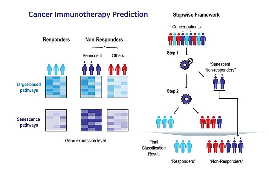
AI Tool Predicts Immunotherapy Response Using Tumor Senescence Signals
Patients receiving the same cancer immunotherapy can have markedly different outcomes, with some showing little or no response. Identifying these patients earlier could reduce the costs of ineffective... Read more
Hematology
view channel
New Myelofibrosis Guidelines Recommend Expanded Genetic Testing at Diagnosis
Myelofibrosis is a rare, life-threatening blood cancer affecting up to six per 100,000 people. The disease causes scarring in the bone marrow, limiting the production of healthy blood cells and potentially... Read more
Handheld Point-of-Care Test Aims to Enable Rapid Coagulation Assessment in Trauma
Severe blood loss and inadequate tissue oxygenation are leading causes of preventable death after traumatic injury. Blood products are essential in trauma care, but cold-chain storage, short shelf life,... Read more

Immunology
view channel
Antibody Profiling Predicts Inflammatory Bowel Disease Years Before Diagnosis
Inflammatory bowel disease (IBD), which includes Crohn’s disease and ulcerative colitis, involves abnormal immune responses that cause persistent inflammation and damage in the digestive system.... Read more
Immune Profiling Initiative Aims to Improve Bone Marrow Analysis in Leukemia
Acute myeloid leukemia (AML) is an aggressive blood cancer in which assessing disease burden after therapy can help guide clinical evaluation. Patients with measurable residual disease (MRD) after treatment... Read more
Immune Profiling Identifies Early Maternal Autoantibodies Linked to Preterm Birth Risk
A typical pregnancy lasts about 40 weeks, but labor begins prematurely in roughly one in 10 pregnancies. Preterm birth is the leading cause of newborn death worldwide, yet its underlying causes and which... Read more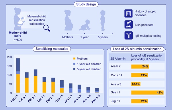
Longitudinal Blood Testing Reveals Changes in Allergen Sensitization During Childhood
Food allergy is a major childhood health concern, and distinguishing persistent risk from transient sensitization can guide clinical decisions. Specific IgE antibodies may be detectable even when a child... Read moreMicrobiology
view channel
Molecular Panel Gains WHO Emergency Use Listing to Support Ebola Outbreak Response
The ongoing Ebola Bundibugyo outbreak in the Democratic Republic of the Congo and neighboring countries has become one of the largest outbreaks of this Ebola virus species recorded to date.... Read more
Rapid Host-Response Test Aims to Predict Corticosteroid Benefit in Sepsis
Sepsis and severe infections are difficult to treat because patients with similar clinical presentations may have very different underlying immune responses. Corticosteroids are commonly used in severe... Read more

Pathology
view channel
Digital Pathology Test Stratifies Recurrence Risk in Early Breast Cancer
In hormone receptor-positive, human epidermal growth factor receptor 2 (HER2)-negative early breast cancer, accurate recurrence-risk assessment helps guide treatment decisions. However, individual risks... Read more
AI Pathology Tool Predicts DNA Repair Deficiency in High-Grade Serous Ovarian Cancer
High-grade serous tubo-ovarian carcinoma is an aggressive cancer arising in the ovaries or fallopian tubes. Some tumors exhibit homologous recombination deficiency (HRD), a feature reflecting impaired DNA repair.... Read more

Technology
view channel
New AI-Powered System Aims to Streamline Laboratory and Manufacturing Workflows
Scientific laboratories supporting drug development, biotechnology, and materials research increasingly rely on coordinated digital workflows to move data, samples, and activities across teams.... Read more
Laser-Based Swab Analysis Shows Promise for Detecting Disease-Linked Odor Patterns
Disease-related changes in volatile organic compounds can alter body odor, producing measurable patterns in exhaled breath and bodily fluids. Current analytical methods can be complex, time-consuming,... Read more

Industry
view channel
Hologic and Inpeco Partner to Advance Molecular Laboratory Automation in Europe
Hologic, Inc. (Marlborough, MA, USA) and Inpeco S.A. (Novazzano, Switzerland) have signed a co-marketing partnership in Europe for Inpeco’s recently launched FlexLab X automation solution and Hologic’s... Read more
Collaboration to Expand Cervical Cancer Screening Access With At-Home HPV Collection
Cervical cancer is largely preventable with regular screening and early detection of human papillomavirus (HPV), the leading cause of the disease. Screening gaps persist, in part because traditional sample... Read more
Alzheimer’s Progression Blood Test Gains U.S. Patent Ahead of Planned Launch
Alzheimer’s disease is the most common cause of dementia, accounting for an estimated 60–70% of cases worldwide. Its progressive effects on memory, thinking, behavior, and daily functioning underscore... Read more