X-Ray Crystallography Reveals the Two Faces of Flavivirus Nonstructural Protein
|
By LabMedica International staff writers Posted on 17 Feb 2014 |
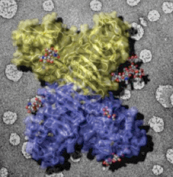
Image: The external face of the Flavivirus NS1 protein (sugars in grey balls) is exposed on infected cell surfaces where it can interact with the immune system. This face is also exposed in secreted NS1 particles present in patient sera. The background image shows artificial membranes coated with the NS1 protein (Photo courtesy of the University of Michigan).
High-resolution X-ray crystallography and electron microscopy have revealed that the Flavivirus NS1 (nonstructural protein 1) has two distinct faces, one that interacts with the interior of the infected host cell and the other that is exposed to antiviral elements in the host's immune system.
Flaviviruses are responsible for several severe diseases of humans including dengue fever, West Nile fever, tick-borne encephalitis, and yellow fever. All the flaviviruses produce nonstructural protein 1 (NS1), which functions in genome replication as an intracellular dimer and in immune system evasion as a secreted hexamer. In general, nonstructural proteins are encoded by the viral genome and are produced in the organisms they infect, but are not packaged into the virus particles. Some of these proteins may play roles within the infected cell during virus replication or act in regulation of virus replication or virus assembly.
Investigators at the University of Michigan (Ann Arbor, USA) and colleagues at Purdue University (Lafayette, IN, USA) chose X-ray crystallography as an approach to developing a better understanding of how NS1 functions.
The investigators isolated and crystallized NS1 from Dengue virus and West Nile virus. They then created a three-dimensional atomic structure map of the protein crystals based on data obtained from X-ray crystallography carried out at the Advanced Photon Source at the Argonne National Laboratory (Illinois, USA). Subsequently, electron microscopy was used to elucidate how NS1 associated with membranes of infected cells.
The investigators reported the crystal structures for full-length, glycosylated NS1 from West Nile and dengue viruses in the February 6, 2014, online edition of the journal Science. Their results revealed that the NS1 hexamer in crystal structures was similar to a solution hexamer visualized by single-particle electron microscopy. Recombinant NS1 bound to lipid bilayers and remodeled large liposomes into lipoprotein nanoparticles. The NS1 structures revealed distinct domains for membrane association of the dimer and interactions with the immune system, and will form a basis for elucidating the molecular mechanism of NS1 function.
"Isolating the protein in order to study it has been a challenge for researchers," said senior author Dr. Janet Smith, professor of biological chemistry at the University of Michigan. "Once we discovered how to do that, it crystallized beautifully. Seeing the design of this key protein provides a target for a potential vaccine or even a therapeutic drug."
"The two faces of NS1 define the regions responsible for its two major functions," said Dr. Smith. "This understanding will guide future research into dissecting and targeting these regions in disease treatment or prevention. We are now collaborating with the Purdue virologists to understand exactly how the two faces of NS1 help the virus survive and thrive in patients. These studies are the next steps toward a vaccine or an antiviral drug."
Related Links:
University of Michigan
Purdue University
Argonne National Laboratory
Latest BioResearch News
- Genome Analysis Predicts Likelihood of Neurodisability in Oxygen-Deprived Newborns
- Gene Panel Predicts Disease Progession for Patients with B-cell Lymphoma
- New Method Simplifies Preparation of Tumor Genomic DNA Libraries
- New Tool Developed for Diagnosis of Chronic HBV Infection
- Panel of Genetic Loci Accurately Predicts Risk of Developing Gout
- Disrupted TGFB Signaling Linked to Increased Cancer-Related Bacteria
- Gene Fusion Protein Proposed as Prostate Cancer Biomarker
- NIV Test to Diagnose and Monitor Vascular Complications in Diabetes
- Semen Exosome MicroRNA Proves Biomarker for Prostate Cancer
- Genetic Loci Link Plasma Lipid Levels to CVD Risk
- Newly Identified Gene Network Aids in Early Diagnosis of Autism Spectrum Disorder
- Link Confirmed between Living in Poverty and Developing Diseases
- Genomic Study Identifies Kidney Disease Loci in Type I Diabetes Patients
- Liquid Biopsy More Effective for Analyzing Tumor Drug Resistance Mutations
- New Liquid Biopsy Assay Reveals Host-Pathogen Interactions
- Method Developed for Enriching Trophoblast Population in Samples
Channels
Clinical Chemistry
view channel
Simple Blood Test Offers New Path to Alzheimer’s Assessment in Primary Care
Timely evaluation of cognitive symptoms in primary care is often limited by restricted access to specialized diagnostics and invasive confirmatory procedures. Clinicians need accessible tools to determine... Read more
Existing Hospital Analyzers Can Identify Fake Liquid Medical Products
Counterfeit and substandard medicines remain a serious global health threat, with World Health Organization estimates suggesting that 10.5% of medicines in low- and middle-income countries are either fake... Read moreMolecular Diagnostics
view channel
Changes In Lymphatic Vessels Can Aid Early Identification of Aggressive Oral Cancer
Oral cancers are the most common malignant tumors in the head and neck region and cause more than 188,000 deaths worldwide each year. Unlike many other cancers, even small, early-stage oral tumors can... Read more
Molecular Monitoring Approach Helps Bladder Cancer Patients Avoid Surgery
Muscle-invasive bladder cancer is typically treated with chemotherapy followed by radical cystectomy, the complete removal of the bladder. While often effective, the surgery significantly affects quality... Read more
Genetic Tests to Speed Diagnosis of Lymphatic Disorders
Defects in the lymphatic system affect approximately one in every 3,500 newborns and can lead to severe complications, including organ failure, breathing difficulties, and life-threatening infections.... Read more
New Extraction Kit Enables Consistent, Scalable cfDNA Isolation from Multiple Biofluids
Circulating cell-free DNA (cfDNA) found in plasma, serum, urine, and cerebrospinal fluid is typically present at low concentrations and is often highly fragmented, making efficient recovery challenging... Read moreHematology
view channel
Rapid Cartridge-Based Test Aims to Expand Access to Hemoglobin Disorder Diagnosis
Sickle cell disease and beta thalassemia are hemoglobin disorders that often require referral to specialized laboratories for definitive diagnosis, delaying results for patients and clinicians.... Read more
New Guidelines Aim to Improve AL Amyloidosis Diagnosis
Light chain (AL) amyloidosis is a rare, life-threatening bone marrow disorder in which abnormal amyloid proteins accumulate in organs. Approximately 3,260 people in the United States are diagnosed... Read moreImmunology
view channel
New Biomarker Predicts Chemotherapy Response in Triple-Negative Breast Cancer
Triple-negative breast cancer is an aggressive form of breast cancer in which patients often show widely varying responses to chemotherapy. Predicting who will benefit from treatment remains challenging,... Read moreBlood Test Identifies Lung Cancer Patients Who Can Benefit from Immunotherapy Drug
Small cell lung cancer (SCLC) is an aggressive disease with limited treatment options, and even newly approved immunotherapies do not benefit all patients. While immunotherapy can extend survival for some,... Read more
Whole-Genome Sequencing Approach Identifies Cancer Patients Benefitting From PARP-Inhibitor Treatment
Targeted cancer therapies such as PARP inhibitors can be highly effective, but only for patients whose tumors carry specific DNA repair defects. Identifying these patients accurately remains challenging,... Read more
Ultrasensitive Liquid Biopsy Demonstrates Efficacy in Predicting Immunotherapy Response
Immunotherapy has transformed cancer treatment, but only a small proportion of patients experience lasting benefit, with response rates often remaining between 10% and 20%. Clinicians currently lack reliable... Read moreMicrobiology
view channel
Three-Test Panel Launched for Detection of Liver Fluke Infections
Parasitic liver fluke infections remain endemic in parts of Asia, where transmission commonly occurs through consumption of raw freshwater fish or aquatic plants. Chronic infection is a well-established... Read more
Rapid Test Promises Faster Answers for Drug-Resistant Infections
Drug-resistant pathogens continue to pose a growing threat in healthcare facilities, where delayed detection can impede outbreak control and increase mortality. Candida auris is notoriously difficult to... Read more
CRISPR-Based Technology Neutralizes Antibiotic-Resistant Bacteria
Antibiotic resistance has accelerated into a global health crisis, with projections estimating more than 10 million deaths per year by 2050 as drug-resistant “superbugs” continue to spread.... Read more
Comprehensive Review Identifies Gut Microbiome Signatures Associated With Alzheimer’s Disease
Alzheimer’s disease affects approximately 6.7 million people in the United States and nearly 50 million worldwide, yet early cognitive decline remains difficult to characterize. Increasing evidence suggests... Read morePathology
view channel
Single Sample Classifier Predicts Cancer-Associated Fibroblast Subtypes in Patient Samples
Pancreatic ductal adenocarcinoma (PDAC) remains one of the deadliest cancers, in part because of its dense tumor microenvironment that influences how tumors grow and respond to treatment.... Read more
New AI-Driven Platform Standardizes Tuberculosis Smear Microscopy Workflow
Sputum smear microscopy remains central to tuberculosis treatment monitoring and follow-up, particularly in high‑burden settings where serial testing is routine. Yet consistent, repeatable bacillary assessment... Read more
AI Tool Uses Blood Biomarkers to Predict Transplant Complications Before Symptoms Appear
Stem cell and bone marrow transplants can be lifesaving, but serious complications may arise months after patients leave the hospital. One of the most dangerous is chronic graft-versus-host disease, in... Read moreTechnology
view channel
Blood Test “Clocks” Predict Start of Alzheimer’s Symptoms
More than 7 million Americans live with Alzheimer’s disease, and related health and long-term care costs are projected to reach nearly USD 400 billion in 2025. The disease has no cure, and symptoms often... Read more
AI-Powered Biomarker Predicts Liver Cancer Risk
Liver cancer, or hepatocellular carcinoma, causes more than 800,000 deaths worldwide each year and often goes undetected until late stages. Even after treatment, recurrence rates reach 70% to 80%, contributing... Read more
Robotic Technology Unveiled for Automated Diagnostic Blood Draws
Routine diagnostic blood collection is a high‑volume task that can strain staffing and introduce human‑dependent variability, with downstream implications for sample quality and patient experience.... Read more
ADLM Launches First-of-Its-Kind Data Science Program for Laboratory Medicine Professionals
Clinical laboratories generate billions of test results each year, creating a treasure trove of data with the potential to support more personalized testing, improve operational efficiency, and enhance patient care.... Read moreIndustry
view channel
QuidelOrtho Collaborates with Lifotronic to Expand Global Immunoassay Portfolio
QuidelOrtho (San Diego, CA, USA) has entered a long-term strategic supply agreement with Lifotronic Technology (Shenzhen, China) to expand its global immunoassay portfolio and accelerate customer access... Read more


