


Benchtop Technology Designed for Marking, Selecting Cells
|
By LabMedica International staff writers Posted on 22 Jan 2013 |
|
.")
Image: The most common jellyfish protein used in lab research glows green when illuminated with UV light (Photo courtesy of NA/TSRI).
Chemists have found a simpler way to perform one of the most essential chores in molecular biology. Their new method allows scientists to add a marker to specific cells, so that these cells may be easily located and/or chosen from a larger cell population.
The technique, which is described in January 2012 in the chemistry journal Angewandte Chemie International Edition, makes use of the tight binding of two proteins that are inexpensively available but are not found in human or other mammalian cells. Therefore, it has advantages over existing cell-marking techniques.
“This new technique is cheap, easy, and sensitive,” said the Scripps Research Institute (TSRI; La Jolla, CA, USA) Prof. Richard A. Lerner, who is the senior author of the new report. “The method should be useful in a variety of applications that require separating out certain types of cells.”
The best-known cell marker currently used is green fluorescent protein (GFP), a protein derived from a jellyfish that emits a characteristic green light when illuminated by specific light wavelengths. When scientists need to add a new gene to cells, for example, to produce a therapeutic protein, they frequently construct a genetic sequence that also includes the GFP gene. Consequently, the cells that effectively produce the new protein will also generate GFP, whose fluorescence allows these cells to be identified and even sorted out from a larger population.
However, fluorescence-based cell sorting is comparatively costly and cumbersome. Alternative cell-marking techniques use marker molecules to which metals or antibodies will bind tightly, but these tend to have annoying side effects on the cells that they mark. Prof. Lerner’s team, led by first author Yingjie Peng, a postdoctoral fellow, set out to devise a better approach.
The new method exploits a special characteristic of chitinase enzymes, which evolved degrade chitin—a rugged, sugar-derived substance found, for example, in squid beaks, crab shells, and the cell walls of fungi. In addition to a main chitin-breaking domain, chitinases have another active structure, a chitin-binding domain (ChBD). “It makes a super-strong bond with chitin,” said Dr. Peng. Recently, scientists have begun to use this high-affinity binding of ChBD and chitin as a marker system, typically for selecting ChBD-tagged proteins in a lab dish. The new method uses ChBD to mark and select cells.
A new gene, in the method, can be added to cells within a larger DNA vector that also includes the genetic sequences for ChBD and GFP. The ChBD molecule will be produced in such a way that it ends up being held on the outer surface of its host cell’s plasma membrane—and the GFP molecule will sit just inside the membrane. The GFP acts as a visual beacon, while the ChBD serves as a handy gripping point for cell selection.
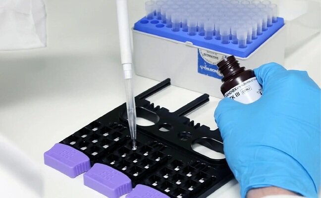
The scientists, after exposing a culture of test cells to this research ChBD-containing vector, were able to visualize, by way of the GFP tags, which cells were expressing them, and was able to pick them out easily, with high sensitivity, utilizing magnetic beads coated with chitin. “This is a relatively easy benchtop method,” Dr. Peng said. Significantly, these selected cells could generate progeny cells that appeared healthy.
Because the ChBD marker, in the vector, is produced in a manner that secures it to a cell’s membrane, it also can serve as a vital application for selecting just the membrane fraction of a sample of cellular material. Dr. Peng and his colleagues showed this using chitin beads to rapidly isolate a pure fraction of membrane material from ChBD-marked test cells. Cellulase enzymes, which degrade the common plant compound cellulose, also have a high-affinity cellulase-binding domain, which can be employed in the same way as the ChBD.
The scientists expect that the new cell-marking method will help to streamline another major molecular biology technique, which was developed by the Lerner laboratory in parallel with the group of Sir Gregory Winter at the Laboratory of Molecular Biology (Cambridge, UK). This technique allows scientists to produce very large and diverse libraries of antibody arms, and to sift through them, or “pan”—in the way miners pan for gold nuggets—for those that might be of use, for example in therapies. ChBD-based markers should be beneficial in boosting the effectiveness of this panning process, according to Dr. Peng.
The investigators are also assessing the potential use of ChBD-based cell marking in living animals, for example, to monitor the outcome of selected cell types throughout an animal’s lifecycle.
Related Links:
Scripps Research Institute


 (3) (1).png)
The technique, which is described in January 2012 in the chemistry journal Angewandte Chemie International Edition, makes use of the tight binding of two proteins that are inexpensively available but are not found in human or other mammalian cells. Therefore, it has advantages over existing cell-marking techniques.
“This new technique is cheap, easy, and sensitive,” said the Scripps Research Institute (TSRI; La Jolla, CA, USA) Prof. Richard A. Lerner, who is the senior author of the new report. “The method should be useful in a variety of applications that require separating out certain types of cells.”
The best-known cell marker currently used is green fluorescent protein (GFP), a protein derived from a jellyfish that emits a characteristic green light when illuminated by specific light wavelengths. When scientists need to add a new gene to cells, for example, to produce a therapeutic protein, they frequently construct a genetic sequence that also includes the GFP gene. Consequently, the cells that effectively produce the new protein will also generate GFP, whose fluorescence allows these cells to be identified and even sorted out from a larger population.
However, fluorescence-based cell sorting is comparatively costly and cumbersome. Alternative cell-marking techniques use marker molecules to which metals or antibodies will bind tightly, but these tend to have annoying side effects on the cells that they mark. Prof. Lerner’s team, led by first author Yingjie Peng, a postdoctoral fellow, set out to devise a better approach.
The new method exploits a special characteristic of chitinase enzymes, which evolved degrade chitin—a rugged, sugar-derived substance found, for example, in squid beaks, crab shells, and the cell walls of fungi. In addition to a main chitin-breaking domain, chitinases have another active structure, a chitin-binding domain (ChBD). “It makes a super-strong bond with chitin,” said Dr. Peng. Recently, scientists have begun to use this high-affinity binding of ChBD and chitin as a marker system, typically for selecting ChBD-tagged proteins in a lab dish. The new method uses ChBD to mark and select cells.
A new gene, in the method, can be added to cells within a larger DNA vector that also includes the genetic sequences for ChBD and GFP. The ChBD molecule will be produced in such a way that it ends up being held on the outer surface of its host cell’s plasma membrane—and the GFP molecule will sit just inside the membrane. The GFP acts as a visual beacon, while the ChBD serves as a handy gripping point for cell selection.
The scientists, after exposing a culture of test cells to this research ChBD-containing vector, were able to visualize, by way of the GFP tags, which cells were expressing them, and was able to pick them out easily, with high sensitivity, utilizing magnetic beads coated with chitin. “This is a relatively easy benchtop method,” Dr. Peng said. Significantly, these selected cells could generate progeny cells that appeared healthy.
Because the ChBD marker, in the vector, is produced in a manner that secures it to a cell’s membrane, it also can serve as a vital application for selecting just the membrane fraction of a sample of cellular material. Dr. Peng and his colleagues showed this using chitin beads to rapidly isolate a pure fraction of membrane material from ChBD-marked test cells. Cellulase enzymes, which degrade the common plant compound cellulose, also have a high-affinity cellulase-binding domain, which can be employed in the same way as the ChBD.
The scientists expect that the new cell-marking method will help to streamline another major molecular biology technique, which was developed by the Lerner laboratory in parallel with the group of Sir Gregory Winter at the Laboratory of Molecular Biology (Cambridge, UK). This technique allows scientists to produce very large and diverse libraries of antibody arms, and to sift through them, or “pan”—in the way miners pan for gold nuggets—for those that might be of use, for example in therapies. ChBD-based markers should be beneficial in boosting the effectiveness of this panning process, according to Dr. Peng.
The investigators are also assessing the potential use of ChBD-based cell marking in living animals, for example, to monitor the outcome of selected cell types throughout an animal’s lifecycle.
Related Links:
Scripps Research Institute
New
Gold Member
Clinical Drug Testing Panel
DOA Urine MultiPlex
New
Gold Member
Genetic Type 1 Diabetes Risk Test
T1D GRS Array
Pipette
Accumax Smart Series
Latest BioResearch News
- Genome Analysis Predicts Likelihood of Neurodisability in Oxygen-Deprived Newborns
- Gene Panel Predicts Disease Progession for Patients with B-cell Lymphoma
- New Method Simplifies Preparation of Tumor Genomic DNA Libraries
- New Tool Developed for Diagnosis of Chronic HBV Infection
- Panel of Genetic Loci Accurately Predicts Risk of Developing Gout
- Disrupted TGFB Signaling Linked to Increased Cancer-Related Bacteria
- Gene Fusion Protein Proposed as Prostate Cancer Biomarker
- NIV Test to Diagnose and Monitor Vascular Complications in Diabetes
- Semen Exosome MicroRNA Proves Biomarker for Prostate Cancer
- Genetic Loci Link Plasma Lipid Levels to CVD Risk
- Newly Identified Gene Network Aids in Early Diagnosis of Autism Spectrum Disorder
- Link Confirmed between Living in Poverty and Developing Diseases
- Genomic Study Identifies Kidney Disease Loci in Type I Diabetes Patients
- Liquid Biopsy More Effective for Analyzing Tumor Drug Resistance Mutations
- New Liquid Biopsy Assay Reveals Host-Pathogen Interactions
- Method Developed for Enriching Trophoblast Population in Samples
Channels
Clinical Chemistry
view channel")
New PSA-Based Prognostic Model Improves Prostate Cancer Risk Assessment
Prostate cancer is the second-leading cause of cancer death among American men, and about one in eight will be diagnosed in their lifetime. Screening relies on blood levels of prostate-specific antigen... Read more
Extracellular Vesicles Linked to Heart Failure Risk in CKD Patients
Chronic kidney disease (CKD) affects more than 1 in 7 Americans and is strongly associated with cardiovascular complications, which account for more than half of deaths among people with CKD.... Read more")
")
Molecular Diagnostics
view channel")
Diagnostic Device Predicts Treatment Response for Brain Tumors Via Blood Test
Glioblastoma is one of the deadliest forms of brain cancer, largely because doctors have no reliable way to determine whether treatments are working in real time. Assessing therapeutic response currently... Read more")
Blood Test Detects Early-Stage Cancers by Measuring Epigenetic Instability
Early-stage cancers are notoriously difficult to detect because molecular changes are subtle and often missed by existing screening tools. Many liquid biopsies rely on measuring absolute DNA methylation... Read more")
“Lab-On-A-Disc” Device Paves Way for More Automated Liquid Biopsies
Extracellular vesicles (EVs) are tiny particles released by cells into the bloodstream that carry molecular information about a cell’s condition, including whether it is cancerous. However, EVs are highly... Read more in the area surrounding sEcad-high cancer cells (blue, center) (Photo courtesy of Debeb Laboratory)")
Blood Test Identifies Inflammatory Breast Cancer Patients at Increased Risk of Brain Metastasis
Brain metastasis is a frequent and devastating complication in patients with inflammatory breast cancer, an aggressive subtype with limited treatment options. Despite its high incidence, the biological... Read moreHematology
view channel")
New Guidelines Aim to Improve AL Amyloidosis Diagnosis
Light chain (AL) amyloidosis is a rare, life-threatening bone marrow disorder in which abnormal amyloid proteins accumulate in organs. Approximately 3,260 people in the United States are diagnosed... Read more")
Fast and Easy Test Could Revolutionize Blood Transfusions
Blood transfusions are a cornerstone of modern medicine, yet red blood cells can deteriorate quietly while sitting in cold storage for weeks. Although blood units have a fixed expiration date, cells from... Read more line (Photo courtesy of Sysmex America)")
Automated Hemostasis System Helps Labs of All Sizes Optimize Workflow
High-volume hemostasis sections must sustain rapid turnaround while managing reruns and reflex testing. Manual tube handling and preanalytical checks can strain staff time and increase opportunities for error.... Read more")
High-Sensitivity Blood Test Improves Assessment of Clotting Risk in Heart Disease Patients
Blood clotting is essential for preventing bleeding, but even small imbalances can lead to serious conditions such as thrombosis or dangerous hemorrhage. In cardiovascular disease, clinicians often struggle... Read moreImmunology
view channelBlood Test Identifies Lung Cancer Patients Who Can Benefit from Immunotherapy Drug
Small cell lung cancer (SCLC) is an aggressive disease with limited treatment options, and even newly approved immunotherapies do not benefit all patients. While immunotherapy can extend survival for some,... Read more")
Whole-Genome Sequencing Approach Identifies Cancer Patients Benefitting From PARP-Inhibitor Treatment
Targeted cancer therapies such as PARP inhibitors can be highly effective, but only for patients whose tumors carry specific DNA repair defects. Identifying these patients accurately remains challenging,... Read more")
Ultrasensitive Liquid Biopsy Demonstrates Efficacy in Predicting Immunotherapy Response
Immunotherapy has transformed cancer treatment, but only a small proportion of patients experience lasting benefit, with response rates often remaining between 10% and 20%. Clinicians currently lack reliable... Read more")
Microbiology
view channel")
Comprehensive Review Identifies Gut Microbiome Signatures Associated With Alzheimer’s Disease
Alzheimer’s disease affects approximately 6.7 million people in the United States and nearly 50 million worldwide, yet early cognitive decline remains difficult to characterize. Increasing evidence suggests... Read moreAI-Powered Platform Enables Rapid Detection of Drug-Resistant C. Auris Pathogens
Infections caused by the pathogenic yeast Candida auris pose a significant threat to hospitalized patients, particularly those with weakened immune systems or those who have invasive medical devices.... Read more")
")
Pathology
view channel system (Deichmann, M. et al., Nat Commun 16, 10306, 2025. DOI: 10.1038/s41467-025-65236-7)")
Engineered Yeast Cells Enable Rapid Testing of Cancer Immunotherapy
Developing new cancer immunotherapies is a slow, costly, and high-risk process, particularly for CAR T cell treatments that must precisely recognize cancer-specific antigens. Small differences in tumor... Read more")
First-Of-Its-Kind Test Identifies Autism Risk at Birth
Autism spectrum disorder is treatable, and extensive research shows that early intervention can significantly improve cognitive, social, and behavioral outcomes. Yet in the United States, the average age... Read more")
")
Technology
view channel")
Robotic Technology Unveiled for Automated Diagnostic Blood Draws
Routine diagnostic blood collection is a high‑volume task that can strain staffing and introduce human‑dependent variability, with downstream implications for sample quality and patient experience.... Read more")
ADLM Launches First-of-Its-Kind Data Science Program for Laboratory Medicine Professionals
Clinical laboratories generate billions of test results each year, creating a treasure trove of data with the potential to support more personalized testing, improve operational efficiency, and enhance patient care.... Read moreAptamer Biosensor Technology to Transform Virus Detection
Rapid and reliable virus detection is essential for controlling outbreaks, from seasonal influenza to global pandemics such as COVID-19. Conventional diagnostic methods, including cell culture, antigen... Read more")
AI Models Could Predict Pre-Eclampsia and Anemia Earlier Using Routine Blood Tests
Pre-eclampsia and anemia are major contributors to maternal and child mortality worldwide, together accounting for more than half a million deaths each year and leaving millions with long-term health complications.... Read moreIndustry
view channelNew Collaboration Brings Automated Mass Spectrometry to Routine Laboratory Testing
Mass spectrometry is a powerful analytical technique that identifies and quantifies molecules based on their mass and electrical charge. Its high selectivity, sensitivity, and accuracy make it indispensable... Read more")
AI-Powered Cervical Cancer Test Set for Major Rollout in Latin America
Noul Co., a Korean company specializing in AI-based blood and cancer diagnostics, announced it will supply its intelligence (AI)-based miLab CER cervical cancer diagnostic solution to Mexico under a multi‑year... Read more")
Diasorin and Fisher Scientific Enter into US Distribution Agreement for Molecular POC Platform
Diasorin (Saluggia, Italy) has entered into an exclusive distribution agreement with Fisher Scientific, part of Thermo Fisher Scientific (Waltham, MA, USA), for the LIAISON NES molecular point-of-care... Read more will be held at Dubai World Trade Centre from 10-13 February")