MAIT Cell Activation Dynamics Associated with COVID-19 Disease Severity
|
By LabMedica International staff writers Posted on 13 Oct 2020 |

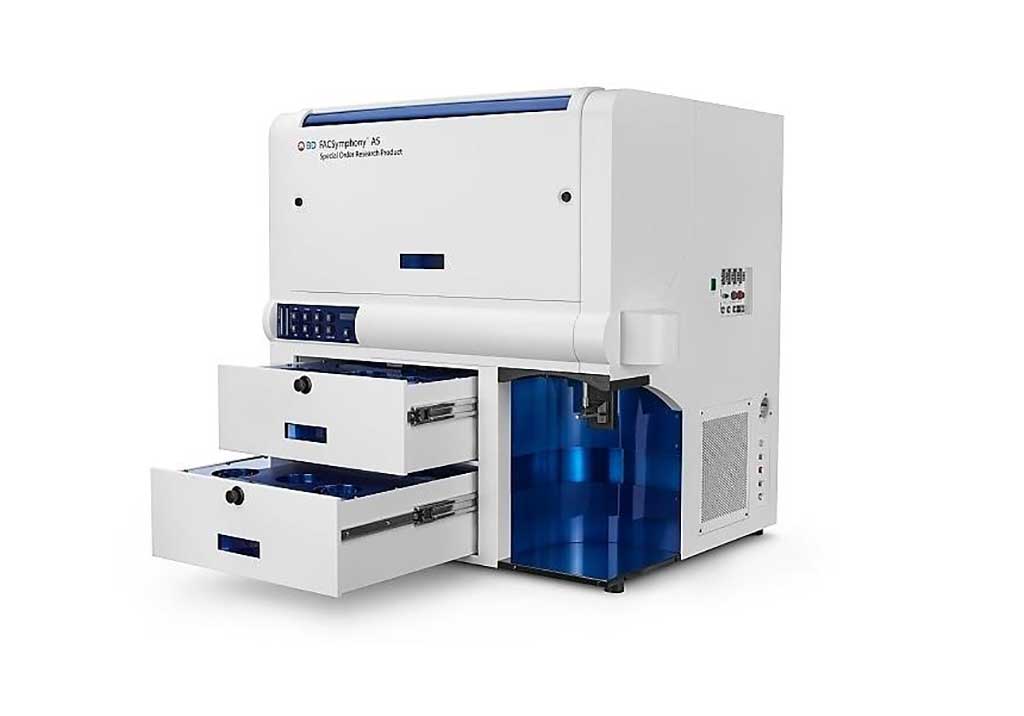
Image: The BD FACSymphony A5 flow cytometer (Photo courtesy of BD Biosciences).
The balance between protective versus pathological immune responses in COVID-19 has been a concern since the onset of the pandemic. SARS-CoV-2 infection can lead to acute respiratory distress syndrome (ARDS), a condition characterized by aggressive inflammatory responses in the lower airways.
Severe COVID-19 is not only due to direct effects of the virus, but also in part to a misdirected host response with complex immune dysregulation of both innate and adaptive immune and inflammatory components. Emerging evidence indicates that mucosa-associated invariant T (MAIT) cells are innate-like sensors of viral infection.
Infectious Disease specialists at the Karolinska University Hospital (Stockholm, Sweden) recruited 69 SARS-CoV-2-infected patients 18 to 78 years old with acute COVID-19 disease admitted to the hospital, or followed up in convalescent phase. The team examined blood samples from 24 patients admitted to the Karolinska University Hospital with COVID-19 disease and compared them to blood samples from 14 healthy controls and 45 individuals who had recovered from COVID-19.
Absolute counts in whole blood were assessed by flow cytometry using BD Multitest 6-color TBNK reagents in association with BD Trucount tubes (BD Biosciences, San Jose, CA, USA). Sera were evaluated for soluble factors using proximity extension assay technology (Olink AB, Uppsala, Sweden). Flow cytometry was performed using multiple antibodies and Samples were acquired on a BD Biosciences’ BD FACSymphony A5 flow cytometer.
The investigators found that the number of circulating MAIT cells was sharply lower in COVID-19 patients and the remaining MAIT cells were highly activated, indicating that they play a role in the response to SARS-CoV-2. Further, single-cell RNA sequencing data suggests that MAIT cells are highly enriched among T cells infiltrating in the airways of COVID-19 patients.
Flow cytometry phenotypes of MAIT cells in COVID-19 found that they were characterized by high expression of CD69 (CD69high) and diminished expression of the chemokine CXCR3 (CXCR3low). Both phenotypes were associated with poor clinical outcomes in the patient cohort. Within the airways, transcriptomic analysis revealed significant MAIT cell enrichment and proinflammatory interleukin 17A (IL-17A) profile.
In convalescent patients, there seems to be a recovery of MAIT cells, including normalization of phenotypes, within weeks from resolution of symptoms. The authors suggested that this may help patients fight future microbial infections. Interestingly, CXCR3 levels were still low in some convalescent samples, raising the possibility that it may be a lasting alteration in MAIT cells post-COVID-19.
Johan K. Sandberg, PhD, a Professor of Medicine and senior author of the study, said, “The findings of our study show that the MAIT cells are highly engaged in the immunological response against COVID-19. A likely interpretation is that the characteristics of MAIT cells make them engaged early on in both the systemic immune response and in the local immune response in the airways to which they are recruited from the blood by inflammatory signals. There, they are likely to contribute to the fast, innate immune response against the virus. In some people with COVID-19, the activation of MAIT cells becomes excessive and this correlates with severe disease.” The study was published on September 28, 2020 in the journal Science Immunology.
Related Links:
Karolinska University Hospital
BD Biosciences
Olink AB
Latest Immunology News
- Blood Test Identifies Lung Cancer Patients Who Can Benefit from Immunotherapy Drug
- Whole-Genome Sequencing Approach Identifies Cancer Patients Benefitting From PARP-Inhibitor Treatment
- Ultrasensitive Liquid Biopsy Demonstrates Efficacy in Predicting Immunotherapy Response
- Blood Test Could Identify Colon Cancer Patients to Benefit from NSAIDs
- Blood Test Could Detect Adverse Immunotherapy Effects
- Routine Blood Test Can Predict Who Benefits Most from CAR T-Cell Therapy
- New Test Distinguishes Vaccine-Induced False Positives from Active HIV Infection
- Gene Signature Test Predicts Response to Key Breast Cancer Treatment
- Chip Captures Cancer Cells from Blood to Help Select Right Breast Cancer Treatment
- Blood-Based Liquid Biopsy Model Analyzes Immunotherapy Effectiveness
- Signature Genes Predict T-Cell Expansion in Cancer Immunotherapy
- Molecular Microscope Diagnostic System Assesses Lung Transplant Rejection
- Blood Test Tracks Treatment Resistance in High-Grade Serous Ovarian Cancer
- Luminescent Probe Measures Immune Cell Activity in Real Time
- Blood-Based Immune Cell Signatures Could Guide Treatment Decisions for Critically Ill Patients
- Novel Tool Predicts Most Effective Multiple Sclerosis Medication for Patients
Channels
Clinical Chemistry
view channel
New PSA-Based Prognostic Model Improves Prostate Cancer Risk Assessment
Prostate cancer is the second-leading cause of cancer death among American men, and about one in eight will be diagnosed in their lifetime. Screening relies on blood levels of prostate-specific antigen... Read more
Extracellular Vesicles Linked to Heart Failure Risk in CKD Patients
Chronic kidney disease (CKD) affects more than 1 in 7 Americans and is strongly associated with cardiovascular complications, which account for more than half of deaths among people with CKD.... Read moreMolecular Diagnostics
view channel
Diagnostic Device Predicts Treatment Response for Brain Tumors Via Blood Test
Glioblastoma is one of the deadliest forms of brain cancer, largely because doctors have no reliable way to determine whether treatments are working in real time. Assessing therapeutic response currently... Read more
Blood Test Detects Early-Stage Cancers by Measuring Epigenetic Instability
Early-stage cancers are notoriously difficult to detect because molecular changes are subtle and often missed by existing screening tools. Many liquid biopsies rely on measuring absolute DNA methylation... Read more
“Lab-On-A-Disc” Device Paves Way for More Automated Liquid Biopsies
Extracellular vesicles (EVs) are tiny particles released by cells into the bloodstream that carry molecular information about a cell’s condition, including whether it is cancerous. However, EVs are highly... Read more
Blood Test Identifies Inflammatory Breast Cancer Patients at Increased Risk of Brain Metastasis
Brain metastasis is a frequent and devastating complication in patients with inflammatory breast cancer, an aggressive subtype with limited treatment options. Despite its high incidence, the biological... Read moreHematology
view channel
New Guidelines Aim to Improve AL Amyloidosis Diagnosis
Light chain (AL) amyloidosis is a rare, life-threatening bone marrow disorder in which abnormal amyloid proteins accumulate in organs. Approximately 3,260 people in the United States are diagnosed... Read more
Fast and Easy Test Could Revolutionize Blood Transfusions
Blood transfusions are a cornerstone of modern medicine, yet red blood cells can deteriorate quietly while sitting in cold storage for weeks. Although blood units have a fixed expiration date, cells from... Read more
Automated Hemostasis System Helps Labs of All Sizes Optimize Workflow
High-volume hemostasis sections must sustain rapid turnaround while managing reruns and reflex testing. Manual tube handling and preanalytical checks can strain staff time and increase opportunities for error.... Read more
High-Sensitivity Blood Test Improves Assessment of Clotting Risk in Heart Disease Patients
Blood clotting is essential for preventing bleeding, but even small imbalances can lead to serious conditions such as thrombosis or dangerous hemorrhage. In cardiovascular disease, clinicians often struggle... Read moreMicrobiology
view channel
Comprehensive Review Identifies Gut Microbiome Signatures Associated With Alzheimer’s Disease
Alzheimer’s disease affects approximately 6.7 million people in the United States and nearly 50 million worldwide, yet early cognitive decline remains difficult to characterize. Increasing evidence suggests... Read moreAI-Powered Platform Enables Rapid Detection of Drug-Resistant C. Auris Pathogens
Infections caused by the pathogenic yeast Candida auris pose a significant threat to hospitalized patients, particularly those with weakened immune systems or those who have invasive medical devices.... Read morePathology
view channel
Engineered Yeast Cells Enable Rapid Testing of Cancer Immunotherapy
Developing new cancer immunotherapies is a slow, costly, and high-risk process, particularly for CAR T cell treatments that must precisely recognize cancer-specific antigens. Small differences in tumor... Read more
First-Of-Its-Kind Test Identifies Autism Risk at Birth
Autism spectrum disorder is treatable, and extensive research shows that early intervention can significantly improve cognitive, social, and behavioral outcomes. Yet in the United States, the average age... Read moreTechnology
view channel
Robotic Technology Unveiled for Automated Diagnostic Blood Draws
Routine diagnostic blood collection is a high‑volume task that can strain staffing and introduce human‑dependent variability, with downstream implications for sample quality and patient experience.... Read more
ADLM Launches First-of-Its-Kind Data Science Program for Laboratory Medicine Professionals
Clinical laboratories generate billions of test results each year, creating a treasure trove of data with the potential to support more personalized testing, improve operational efficiency, and enhance patient care.... Read moreAptamer Biosensor Technology to Transform Virus Detection
Rapid and reliable virus detection is essential for controlling outbreaks, from seasonal influenza to global pandemics such as COVID-19. Conventional diagnostic methods, including cell culture, antigen... Read more
AI Models Could Predict Pre-Eclampsia and Anemia Earlier Using Routine Blood Tests
Pre-eclampsia and anemia are major contributors to maternal and child mortality worldwide, together accounting for more than half a million deaths each year and leaving millions with long-term health complications.... Read moreIndustry
view channelNew Collaboration Brings Automated Mass Spectrometry to Routine Laboratory Testing
Mass spectrometry is a powerful analytical technique that identifies and quantifies molecules based on their mass and electrical charge. Its high selectivity, sensitivity, and accuracy make it indispensable... Read more
AI-Powered Cervical Cancer Test Set for Major Rollout in Latin America
Noul Co., a Korean company specializing in AI-based blood and cancer diagnostics, announced it will supply its intelligence (AI)-based miLab CER cervical cancer diagnostic solution to Mexico under a multi‑year... Read more
Diasorin and Fisher Scientific Enter into US Distribution Agreement for Molecular POC Platform
Diasorin (Saluggia, Italy) has entered into an exclusive distribution agreement with Fisher Scientific, part of Thermo Fisher Scientific (Waltham, MA, USA), for the LIAISON NES molecular point-of-care... Read more


