Urea Dissociation Tests Reduces SARS-CoV-2 IgM False-Positives
|
By LabMedica International staff writers Posted on 28 Apr 2020 |

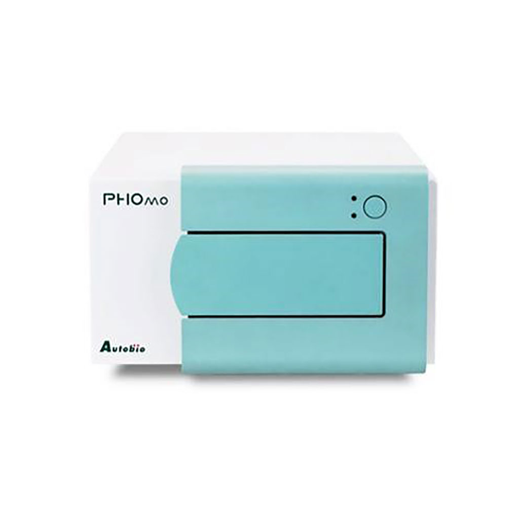
Image: The PHOMO Microplate Reader: the sophisticated optical design combined with the accurate plate transport system allows for high resolution scanning (29 points per well) to support agglutination assays (Photo courtesy of Autobio Diagnostics Co).
At present, the diagnosis of COVID-19 is mainly based on epidemiological history inquiry, laboratory testing, and chest radiology examination. Among these examinations, the detection of nucleic acid from SARS-CoV-2 is the direct evidence for COVID-19 diagnosis.
The detection of serum-specific IgM and IgG, especially the former, is routinely used in clinical laboratories to evaluate the acute phase infection of pathogens in the serum. In many infections, IgM can be detected as early as one week after infection. When the level of IgM reaches the detection limit of the assay kit, the detection of IgM can avoid false-negative results owing to sampling.
Medical Laboratory Scientists at the Affiliated Hospital of North Sichuan Medical College (Nanchong, P.R. China) used gold immunochromatography assay (GICA) and enzyme-linked immunosorbent assay (ELISA) to detect SARS-CoV-2 IgM in 86 serum samples, including five influenza A virus (Flu A) IgM-positive sera, five influenza B virus (Flu B) IgM-positive sera, five Mycoplasma pneumoniae IgM-positive sera, five Legionella pneumophila IgM-positive sera, six sera of HIV infection patients, 36 rheumatoid factor IgM (RF-IgM)-positive sera, five sera from hypertensive patients, five sera from diabetes mellitus patients, and 14 sera from novel coronavirus infection disease (COVID-19) patients.
The interference factors causing false-positive reactivity in the two methods were analyzed, and the urea dissociation test was employed to dissociate the SARS-CoV-2 IgM-positive serum using the best dissociation concentration. The IgM against Flu A and B, M. pneumoniae, and L. pneumophila were detected by indirect immunofluorescence assay (Respiratory tract 8 joint detection kit, EUROIMMUN, Inc., Lübeck, Germany). Rheumatoid Factor (RF-IgM) was detected by rate nephelometry assay (IMMAGE800, Beckman Coulter, Inc., Brea, CA, USA). HIV combi PT was detected by electrochemiluminescence assay (Cobas E602, Roche, Mannheim, Germany).
GICA and ELISA were used for SARS-CoV-2 IgM detection (kit provided by Beijing Hotgen Biotechnology Co., Beijing, China). Optical density in ELISA plates was measured using a PHOMO Microplate Reader (Autobio Diagnostics Co., Zhengzhou, China). Urea dissociation tests of GICA and ELISA were also performed.
The team reported that both GICA and ELISA detected positive SARS-CoV-2 IgM in 22 middle-high level RF-IgM-positive sera and in all the 14 sera from the patients with COVID-19 patients. The other 50 sera were negative. When urea dissociation concentration was 6 mol/L, SARS-CoV-2 IgM was positive in one middle-high level RF-IgM-positive sera and in the 14 COVID-19 samples detected using GICA. When urea dissociation concentration was 4 mol/L and the avidity index (AI) lower than 0.371 was set to negative, test results were positive for SARS-CoV-2 in 3 middle-high level RF-IgM-positive sera, as well as the 14 COVID-19 sera detected using ELISA.
The authors concluded that middle-high level of RF-IgM could lead to false-positive reactivity of SARS-CoV-2 IgM detected using GICA and ELISA, and urea dissociation tests would be helpful in reducing false-positive results of SARS-CoV-2 IgM. The study was published on April 10, 2020 in the Journal of Clinical Microbiology.
Latest Immunology News
- Blood Test Identifies Lung Cancer Patients Who Can Benefit from Immunotherapy Drug
- Whole-Genome Sequencing Approach Identifies Cancer Patients Benefitting From PARP-Inhibitor Treatment
- Ultrasensitive Liquid Biopsy Demonstrates Efficacy in Predicting Immunotherapy Response
- Blood Test Could Identify Colon Cancer Patients to Benefit from NSAIDs
- Blood Test Could Detect Adverse Immunotherapy Effects
- Routine Blood Test Can Predict Who Benefits Most from CAR T-Cell Therapy
- New Test Distinguishes Vaccine-Induced False Positives from Active HIV Infection
- Gene Signature Test Predicts Response to Key Breast Cancer Treatment
- Chip Captures Cancer Cells from Blood to Help Select Right Breast Cancer Treatment
- Blood-Based Liquid Biopsy Model Analyzes Immunotherapy Effectiveness
- Signature Genes Predict T-Cell Expansion in Cancer Immunotherapy
- Molecular Microscope Diagnostic System Assesses Lung Transplant Rejection
- Blood Test Tracks Treatment Resistance in High-Grade Serous Ovarian Cancer
- Luminescent Probe Measures Immune Cell Activity in Real Time
- Blood-Based Immune Cell Signatures Could Guide Treatment Decisions for Critically Ill Patients
- Novel Tool Predicts Most Effective Multiple Sclerosis Medication for Patients
Channels
Clinical Chemistry
view channel
New PSA-Based Prognostic Model Improves Prostate Cancer Risk Assessment
Prostate cancer is the second-leading cause of cancer death among American men, and about one in eight will be diagnosed in their lifetime. Screening relies on blood levels of prostate-specific antigen... Read more
Extracellular Vesicles Linked to Heart Failure Risk in CKD Patients
Chronic kidney disease (CKD) affects more than 1 in 7 Americans and is strongly associated with cardiovascular complications, which account for more than half of deaths among people with CKD.... Read moreMolecular Diagnostics
view channel
Diagnostic Device Predicts Treatment Response for Brain Tumors Via Blood Test
Glioblastoma is one of the deadliest forms of brain cancer, largely because doctors have no reliable way to determine whether treatments are working in real time. Assessing therapeutic response currently... Read more
Blood Test Detects Early-Stage Cancers by Measuring Epigenetic Instability
Early-stage cancers are notoriously difficult to detect because molecular changes are subtle and often missed by existing screening tools. Many liquid biopsies rely on measuring absolute DNA methylation... Read more
“Lab-On-A-Disc” Device Paves Way for More Automated Liquid Biopsies
Extracellular vesicles (EVs) are tiny particles released by cells into the bloodstream that carry molecular information about a cell’s condition, including whether it is cancerous. However, EVs are highly... Read more
Blood Test Identifies Inflammatory Breast Cancer Patients at Increased Risk of Brain Metastasis
Brain metastasis is a frequent and devastating complication in patients with inflammatory breast cancer, an aggressive subtype with limited treatment options. Despite its high incidence, the biological... Read moreHematology
view channel
New Guidelines Aim to Improve AL Amyloidosis Diagnosis
Light chain (AL) amyloidosis is a rare, life-threatening bone marrow disorder in which abnormal amyloid proteins accumulate in organs. Approximately 3,260 people in the United States are diagnosed... Read more
Fast and Easy Test Could Revolutionize Blood Transfusions
Blood transfusions are a cornerstone of modern medicine, yet red blood cells can deteriorate quietly while sitting in cold storage for weeks. Although blood units have a fixed expiration date, cells from... Read more
Automated Hemostasis System Helps Labs of All Sizes Optimize Workflow
High-volume hemostasis sections must sustain rapid turnaround while managing reruns and reflex testing. Manual tube handling and preanalytical checks can strain staff time and increase opportunities for error.... Read more
High-Sensitivity Blood Test Improves Assessment of Clotting Risk in Heart Disease Patients
Blood clotting is essential for preventing bleeding, but even small imbalances can lead to serious conditions such as thrombosis or dangerous hemorrhage. In cardiovascular disease, clinicians often struggle... Read moreMicrobiology
view channel
Comprehensive Review Identifies Gut Microbiome Signatures Associated With Alzheimer’s Disease
Alzheimer’s disease affects approximately 6.7 million people in the United States and nearly 50 million worldwide, yet early cognitive decline remains difficult to characterize. Increasing evidence suggests... Read moreAI-Powered Platform Enables Rapid Detection of Drug-Resistant C. Auris Pathogens
Infections caused by the pathogenic yeast Candida auris pose a significant threat to hospitalized patients, particularly those with weakened immune systems or those who have invasive medical devices.... Read morePathology
view channel
Engineered Yeast Cells Enable Rapid Testing of Cancer Immunotherapy
Developing new cancer immunotherapies is a slow, costly, and high-risk process, particularly for CAR T cell treatments that must precisely recognize cancer-specific antigens. Small differences in tumor... Read more
First-Of-Its-Kind Test Identifies Autism Risk at Birth
Autism spectrum disorder is treatable, and extensive research shows that early intervention can significantly improve cognitive, social, and behavioral outcomes. Yet in the United States, the average age... Read moreTechnology
view channel
Robotic Technology Unveiled for Automated Diagnostic Blood Draws
Routine diagnostic blood collection is a high‑volume task that can strain staffing and introduce human‑dependent variability, with downstream implications for sample quality and patient experience.... Read more
ADLM Launches First-of-Its-Kind Data Science Program for Laboratory Medicine Professionals
Clinical laboratories generate billions of test results each year, creating a treasure trove of data with the potential to support more personalized testing, improve operational efficiency, and enhance patient care.... Read moreAptamer Biosensor Technology to Transform Virus Detection
Rapid and reliable virus detection is essential for controlling outbreaks, from seasonal influenza to global pandemics such as COVID-19. Conventional diagnostic methods, including cell culture, antigen... Read more
AI Models Could Predict Pre-Eclampsia and Anemia Earlier Using Routine Blood Tests
Pre-eclampsia and anemia are major contributors to maternal and child mortality worldwide, together accounting for more than half a million deaths each year and leaving millions with long-term health complications.... Read moreIndustry
view channelNew Collaboration Brings Automated Mass Spectrometry to Routine Laboratory Testing
Mass spectrometry is a powerful analytical technique that identifies and quantifies molecules based on their mass and electrical charge. Its high selectivity, sensitivity, and accuracy make it indispensable... Read more
AI-Powered Cervical Cancer Test Set for Major Rollout in Latin America
Noul Co., a Korean company specializing in AI-based blood and cancer diagnostics, announced it will supply its intelligence (AI)-based miLab CER cervical cancer diagnostic solution to Mexico under a multi‑year... Read more
Diasorin and Fisher Scientific Enter into US Distribution Agreement for Molecular POC Platform
Diasorin (Saluggia, Italy) has entered into an exclusive distribution agreement with Fisher Scientific, part of Thermo Fisher Scientific (Waltham, MA, USA), for the LIAISON NES molecular point-of-care... Read more


