Melanoma Cells Secrete Factors to Promote Tumor Growth
|
By LabMedica International staff writers Posted on 12 Feb 2019 |


|
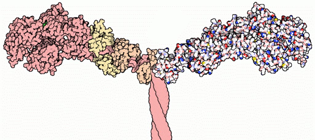
Image: Part of the Myosin II structure. Atoms in the heavy chain are colored pink (on the left-hand side); atoms in the light chains are colored faded-orange and faded-yellow (also on the left-hand side) (Photo courtesy of Wikimedia Commons).
A team of British researchers found that high Myosin II activity in invasive melanoma cells induced reprogramming of innate immune responses in the local microenvironment to support tumor growth.
Myosin II (also known as conventional myosin) is the myosin type responsible for producing contraction in muscle cells, and ROCK (Rho-associated protein kinase)-Myosin II was found to drive rounded-amoeboid migration in cancer cells during metastatic dissemination.
Following up this line of research, investigators at Queen Mary University of London (United Kingdom) reported in the January 31, 2019, online edition of the journal Cell that analysis of human melanoma biopsies revealed that amoeboid melanoma cells with high Myosin II activity were predominant in the invasive fronts of primary tumors in proximity to tumor-associated macrophages and vessels. Proteomic analysis showed that ROCK-Myosin II activity in amoeboid cancer cells controlled an immunomodulatory secretome – comprising all the factors secreted by the cell into the extracellular space - enabling the recruitment of monocytes and their differentiation into tumor-promoting macrophages. Both amoeboid cancer cells and their associated macrophages supported an abnormal system of blood vessels, which ultimately facilitated tumor progression.
Mechanistically, amoeboid cancer cells maintained their behavior via ROCK-Myosin II-driven interleukin 1 alpha (IL-1alpha) secretion and NF-kappaB (nuclear factor kappa-light-chain-enhancer of activated B cells) activation.
In addition, using an array of tumor models, the investigators demonstrated that high Myosin II activity in tumor cells reprogrammed the innate immune microenvironment to support tumor growth.
"This study highlights how cancer cells interact with and influence their surrounding environment to grow and spread. Developing treatments that target the chemicals that alter the immune system could help to prevent the spread of the disease," said senior author Dr. Victoria Sanz-Moreno, professor of cancer cell biology at Queen Mary University of London. "We are excited to find out whether inhibitor drugs could be used in combination with other targeted therapies. By identifying effective treatment combinations, we hope that in the future Myosin II and interleukin 1alpha inhibitors could be used to improve patient outcomes and reduce the risk of melanoma coming back."
Related Links:
Queen Mary University of London







Myosin II (also known as conventional myosin) is the myosin type responsible for producing contraction in muscle cells, and ROCK (Rho-associated protein kinase)-Myosin II was found to drive rounded-amoeboid migration in cancer cells during metastatic dissemination.
Following up this line of research, investigators at Queen Mary University of London (United Kingdom) reported in the January 31, 2019, online edition of the journal Cell that analysis of human melanoma biopsies revealed that amoeboid melanoma cells with high Myosin II activity were predominant in the invasive fronts of primary tumors in proximity to tumor-associated macrophages and vessels. Proteomic analysis showed that ROCK-Myosin II activity in amoeboid cancer cells controlled an immunomodulatory secretome – comprising all the factors secreted by the cell into the extracellular space - enabling the recruitment of monocytes and their differentiation into tumor-promoting macrophages. Both amoeboid cancer cells and their associated macrophages supported an abnormal system of blood vessels, which ultimately facilitated tumor progression.
Mechanistically, amoeboid cancer cells maintained their behavior via ROCK-Myosin II-driven interleukin 1 alpha (IL-1alpha) secretion and NF-kappaB (nuclear factor kappa-light-chain-enhancer of activated B cells) activation.
In addition, using an array of tumor models, the investigators demonstrated that high Myosin II activity in tumor cells reprogrammed the innate immune microenvironment to support tumor growth.
"This study highlights how cancer cells interact with and influence their surrounding environment to grow and spread. Developing treatments that target the chemicals that alter the immune system could help to prevent the spread of the disease," said senior author Dr. Victoria Sanz-Moreno, professor of cancer cell biology at Queen Mary University of London. "We are excited to find out whether inhibitor drugs could be used in combination with other targeted therapies. By identifying effective treatment combinations, we hope that in the future Myosin II and interleukin 1alpha inhibitors could be used to improve patient outcomes and reduce the risk of melanoma coming back."
Related Links:
Queen Mary University of London

Platinum Member
Automated Coagulation Analyzer
Hemolumi H6
LAIR2 Antibody Pair Set
LAIR2 Antibody Pair [Biotin]
Gold Member
Flocked Fiber Swabs
Puritan® Patented HydraFlock®
Latest BioResearch News
- Genomic Tool Supports Analysis of Repeat Expansions Linked to Inherited Disorders
- CSF Biomarker Meprin-Beta Increases in Alzheimer’s Disease
- Cancer DNA Trapping Mechanism Reveals Potential Target for Future Therapies
- Blood Antibody Signals May Offer Clues to the Origins of Childhood Cancer
- Genetic Study Links Inherited Blindness to Gene Tied to Rare Metabolic Disorder
- Population-Scale Proteomics Study Identifies New Genetic-Protein Links
- Circulating Tumor DNA Helps Identify Targets for Personalized Immunotherapy
- Researchers Classify 108 Lysosomal Disorders in Updated Diagnostic Framework
- New Autoantibody Target Identified in Neuromyelitis Optica Spectrum Disorder
- New Immune Target Could Support More Precise High Blood Pressure Treatment
- New Gene-Disease Link May Help Diagnose Rare Neurodevelopmental Disorders
- Researchers Identify Shared Molecular Networks Behind Fatigue-Related Illnesses
- Blood Metabolite Signature Predicts ALS Progression and Points to Treatment Strategy
- Whole-Blood Extracellular Vesicle Analysis Captures Molecular Signals Missed by Plasma Testing
- Age-Related Genomic Differences Could Refine Treatment Decisions in Lung Cancer
- Computational Tool Identifies Central Asthma Genes for Target Discovery
Channels
Clinical Chemistry
view channel
Blood Proteins Reveal Signals Linked to Prostate Cancer Risk Years Before Diagnosis
Prostate cancer is the most commonly diagnosed cancer in men worldwide. While many tumors grow slowly, aggressive forms can be life-threatening, underscoring the need for earlier risk identification.... Read more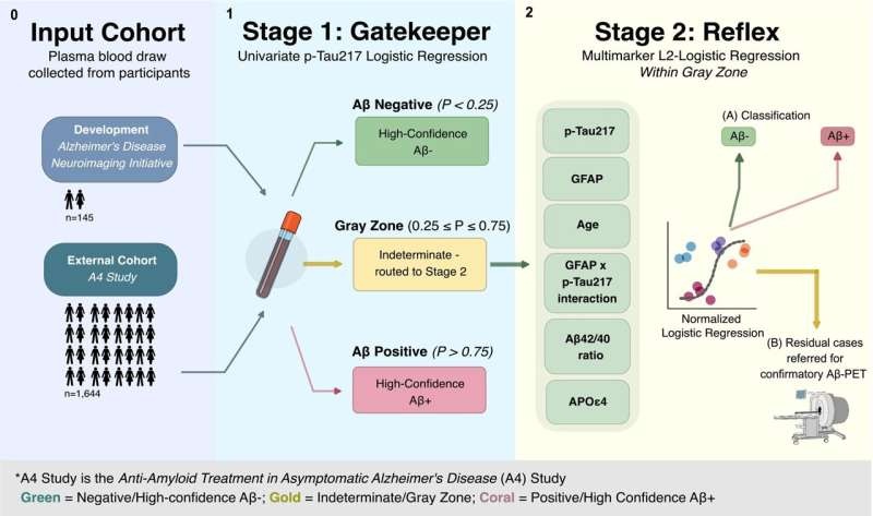
Two-Step Blood Testing Approach Reduces Uncertainty in Amyloid Assessment
Blood-based biomarkers offer a more accessible, less expensive way to detect Alzheimer’s-related brain changes than traditional methods such as amyloid-beta positron emission tomography (Aβ-PET).... Read more
Multimodal Platform to Combine Blood and Imaging Biomarkers for Lung Nodule Assessment
Lung nodules are commonly detected during chest imaging and require careful risk assessment to determine whether follow-up imaging, additional testing, or biopsy is needed. With millions of patients evaluated... Read more
Molecular Diagnostics
view channel
New Variant Tracking Option Expands Molecular Residual Disease Monitoring
Molecular residual disease (MRD) testing can detect traces of cancer that persist after treatment or emerge during recurrence. Because circulating tumor DNA (ctDNA) may be present at very low levels, residual... Read more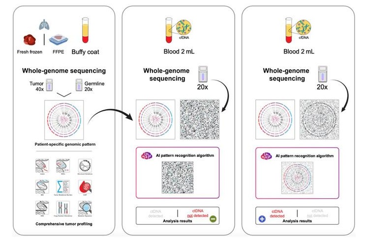
Liquid Biopsy Detects Lung Cancer Recurrence Signals Months Before Imaging
Non-small cell lung cancer (NSCLC) is the most common type of lung cancer and remains a major cause of cancer-related death. Early-stage tumors are often treated surgically, but recurrence can still occur... Read more

Hematology
view channel
New Myelofibrosis Guidelines Recommend Expanded Genetic Testing at Diagnosis
Myelofibrosis is a rare, life-threatening blood cancer affecting up to six per 100,000 people. The disease causes scarring in the bone marrow, limiting the production of healthy blood cells and potentially... Read more
Handheld Point-of-Care Test Aims to Enable Rapid Coagulation Assessment in Trauma
Severe blood loss and inadequate tissue oxygenation are leading causes of preventable death after traumatic injury. Blood products are essential in trauma care, but cold-chain storage, short shelf life,... Read more

Immunology
view channel
Antibody Profiling Predicts Inflammatory Bowel Disease Years Before Diagnosis
Inflammatory bowel disease (IBD), which includes Crohn’s disease and ulcerative colitis, involves abnormal immune responses that cause persistent inflammation and damage in the digestive system.... Read more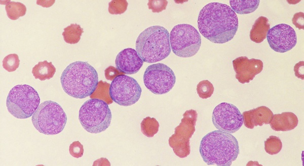
Immune Profiling Initiative Aims to Improve Bone Marrow Analysis in Leukemia
Acute myeloid leukemia (AML) is an aggressive blood cancer in which assessing disease burden after therapy can help guide clinical evaluation. Patients with measurable residual disease (MRD) after treatment... Read more
Immune Profiling Identifies Early Maternal Autoantibodies Linked to Preterm Birth Risk
A typical pregnancy lasts about 40 weeks, but labor begins prematurely in roughly one in 10 pregnancies. Preterm birth is the leading cause of newborn death worldwide, yet its underlying causes and which... Read more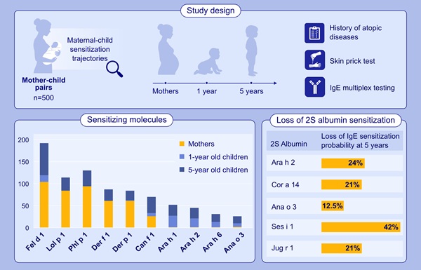
Longitudinal Blood Testing Reveals Changes in Allergen Sensitization During Childhood
Food allergy is a major childhood health concern, and distinguishing persistent risk from transient sensitization can guide clinical decisions. Specific IgE antibodies may be detectable even when a child... Read moreMicrobiology
view channel
Molecular Panel Gains WHO Emergency Use Listing to Support Ebola Outbreak Response
The ongoing Ebola Bundibugyo outbreak in the Democratic Republic of the Congo and neighboring countries has become one of the largest outbreaks of this Ebola virus species recorded to date.... Read more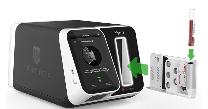
Rapid Host-Response Test Aims to Predict Corticosteroid Benefit in Sepsis
Sepsis and severe infections are difficult to treat because patients with similar clinical presentations may have very different underlying immune responses. Corticosteroids are commonly used in severe... Read more

Pathology
view channel
AI Tissue Analysis May Help Identify Missed Precancerous Colon Polyps
Colorectal cancer is diagnosed in an estimated 1.9 million people worldwide each year, often without symptoms in its early stages. Timely detection of precancerous lesions is therefore important, yet some... Read more
AI Pre-Screening Helps Identify Lung Cancer Cases for Additional Biomarker Testing
Tempus AI (Chicago, IL, USA) will present findings from a study evaluating an artificial intelligence (AI)-enabled pre-screening panel for advanced non-small cell lung cancer (NSCLC) at the ESMO Congress... Read more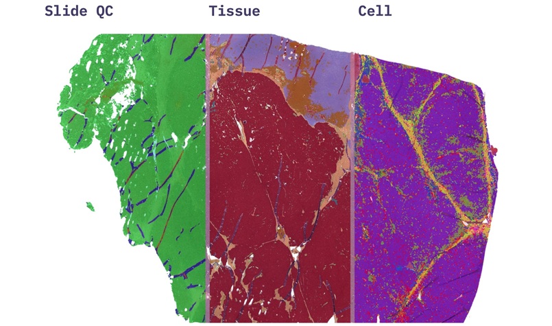
AI Pathology Platform Targets Workflow Delays and Molecular Test Failures
Pathology laboratories often rely on visual review of tissue slides to assess quality, identify tumors, count mitoses, and estimate tumor content. These tasks can be time-consuming and vary between readers,... Read more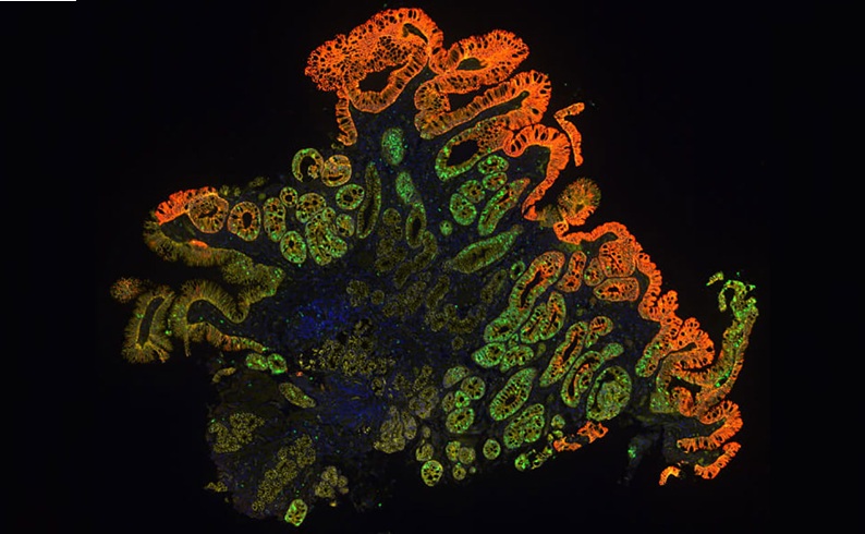
Molecular Testing Refines Barrett’s Esophagus Progression Risk Beyond Biopsy Findings
Barrett’s esophagus is a precancerous condition associated with increased risk of esophageal adenocarcinoma. Routine endoscopic biopsy findings guide surveillance and therapy, but histopathology may not... Read moreTechnology
view channel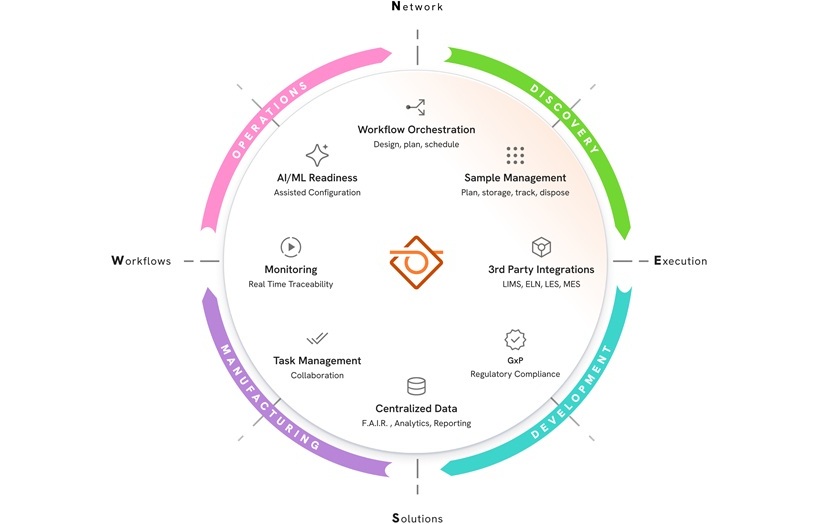
New AI-Powered System Aims to Streamline Laboratory and Manufacturing Workflows
Scientific laboratories supporting drug development, biotechnology, and materials research increasingly rely on coordinated digital workflows to move data, samples, and activities across teams.... Read more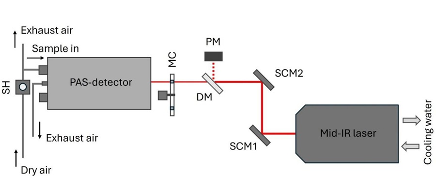
Laser-Based Swab Analysis Shows Promise for Detecting Disease-Linked Odor Patterns
Disease-related changes in volatile organic compounds can alter body odor, producing measurable patterns in exhaled breath and bodily fluids. Current analytical methods can be complex, time-consuming,... Read more

Industry
view channel
Hologic to Broaden Breast Cancer Testing Portfolio Through Reveal Genomics Acquisition
Hologic (Marlborough, MA, USA) has signed a definitive agreement to acquire Reveal Genomics (Barcelona, Spain), a precision oncology company developing genomic diagnostic tests to help personalize cancer treatment.... Read more