


Combined Treatments Cure Muscular Dystrophy in Model
|
By LabMedica International staff writers Posted on 03 Jan 2018 |
|
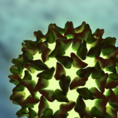
 restore human dystrophin (green) after transplantation significantly greater than previous methods (left) (Photo courtesy of the University of California, Los Angeles and Nature Cell Biology).")
Image: Skeletal muscle cells isolated using the ERBB3 and NGFR surface markers (right) restore human dystrophin (green) after transplantation significantly greater than previous methods (left) (Photo courtesy of the University of California, Los Angeles and Nature Cell Biology).
A new approach for generating functional muscle tissue from induced human pluripotent stem cells enabled replacement of muscle loss in a mouse model of Duchenne muscular dystrophy (DMD).
Duchenne muscular dystrophy (DMD) is caused by mutations in the gene that encodes dystrophin, a protein crucial for maintaining muscle cell integrity and function, and the subsequent disruption of the dystrophin-associated protein complex (DAPC). The mutation occurs on the X-chromosome, and the disease effects about one of every 3,500 boys whose muscle function is so degraded that they die usually before reaching the age of 30. The majority of DMD mutations are deletions that prematurely terminate the dystrophin protein. Deletions of exon 50 of the dystrophin gene are among the most common single exon deletions causing DMD. Such mutations can be corrected by skipping exon 51, thereby restoring the dystrophin reading frame.
Human pluripotent stem cells (hPSCs) can be directed to differentiate into skeletal muscle progenitor cells (SMPCs). However, the myogenicity of hPSC-SMPCs relative to human fetal or adult satellite cells has been unclear, and it has been observed that hPSC-SMPCs derived by directed differentiation were less functional in vitro and in vivo compared to human satellite cells.
To improve the efficiency of stem cell-derived muscle tissue, investigators at the University of California, Los Angeles (USA) first used RNA sequencing to identify cell surface receptors that marked fetal-derived muscle cell populations. They chose the proteins ERBB3 (receptor tyrosine-protein kinase erbB-3) and NGFR (low-affinity nerve growth factor receptor), and used them as markers in subsequent cell selection procedures.
The investigators found that stem cell-derived muscle cell populations were immature, but that inhibition of transforming growth factor-beta (TGF-beta) signaling during differentiation improved fusion efficiency, ultrastructural organization, and the expression of adult myosins. In the next step, cells from DMD patients were induced into becoming pluripotent stem cells. The investigators then corrected the DMD-causing genetic mutation using the gene editing technology CRISPR/Cas9.
CRISPR/Cas9 is regarded as the cutting edge of molecular biology technology. CRISPRs (clustered regularly interspaced short palindromic repeats) are segments of prokaryotic DNA containing short repetitions of base sequences. Each repetition is followed by short segments of "spacer DNA" from previous exposures to a bacterial virus or plasmid. Since 2013, the CRISPR/Cas9 system has been used in research for gene editing (adding, disrupting, or changing the sequence of specific genes) and gene regulation. By delivering the Cas9 enzyme and appropriate guide RNAs (sgRNAs) into a cell, the organism's genome can be cut at any desired location. The conventional CRISPR/Cas9 system is composed of two parts: the Cas9 enzyme, which cleaves the DNA molecule and specific RNA guides that shepherd the Cas9 protein to the target gene on a DNA strand. Efficient genome editing with Cas9-sgRNA in vivo has required the use of viral delivery systems, which have limitations for clinical applications.
The investigators used the ERBB3 and NGFR surface markers to isolate CRISPR-modified, stem cell-derived skeletal muscle cells, which were then injected into mice at the same time as a TGF-beta inhibitor. Results published in the December 18, 2017, online edition of the journal Nature Cell Biology revealed that this enrichment and maturation strategy restored dystrophin in hundreds of dystrophin-deficient myofibers after engraftment of CRISPR/Cas9-corrected DMD hPSC-SMPCs.
“We have found that just because a skeletal muscle cell produced in the lab expresses muscle markers, does not mean it is fully functional,” said senior author Dr. April Pyle, associate professor of microbiology, immunology, and molecular genetics at the University of California, Los Angeles. “For a stem cell therapy for Duchenne to move forward, we must have a better understanding of the cells we are generating from human pluripotent stem cells compared to the muscle stem cells found naturally in the human body and during the development process. The results were exactly what we had hoped for. This is the first study to demonstrate that functional muscle cells can be created in a laboratory and restore dystrophin in animal models of Duchenne using the human development process as a guide.”
Related Links:
University of California, Los Angeles
Gold Member
Respiratory Syncytial Virus Test
OSOM® RSV Test
HBV DNA Test
GENERIC HBV VIRAL LOAD VER 2.0
Homocysteine Quality Control
Liquichek Homocysteine Control
Latest BioResearch News
- Genome Analysis Predicts Likelihood of Neurodisability in Oxygen-Deprived Newborns
- Gene Panel Predicts Disease Progession for Patients with B-cell Lymphoma
- New Method Simplifies Preparation of Tumor Genomic DNA Libraries
- New Tool Developed for Diagnosis of Chronic HBV Infection
- Panel of Genetic Loci Accurately Predicts Risk of Developing Gout
- Disrupted TGFB Signaling Linked to Increased Cancer-Related Bacteria
- Gene Fusion Protein Proposed as Prostate Cancer Biomarker
- NIV Test to Diagnose and Monitor Vascular Complications in Diabetes
- Semen Exosome MicroRNA Proves Biomarker for Prostate Cancer
- Genetic Loci Link Plasma Lipid Levels to CVD Risk
- Newly Identified Gene Network Aids in Early Diagnosis of Autism Spectrum Disorder
- Link Confirmed between Living in Poverty and Developing Diseases
- Genomic Study Identifies Kidney Disease Loci in Type I Diabetes Patients
- Liquid Biopsy More Effective for Analyzing Tumor Drug Resistance Mutations
- New Liquid Biopsy Assay Reveals Host-Pathogen Interactions
- Method Developed for Enriching Trophoblast Population in Samples
Channels
Clinical Chemistry
view channel")
New PSA-Based Prognostic Model Improves Prostate Cancer Risk Assessment
Prostate cancer is the second-leading cause of cancer death among American men, and about one in eight will be diagnosed in their lifetime. Screening relies on blood levels of prostate-specific antigen... Read more
Extracellular Vesicles Linked to Heart Failure Risk in CKD Patients
Chronic kidney disease (CKD) affects more than 1 in 7 Americans and is strongly associated with cardiovascular complications, which account for more than half of deaths among people with CKD.... Read more")
")
Molecular Diagnostics
view channel")
Diagnostic Device Predicts Treatment Response for Brain Tumors Via Blood Test
Glioblastoma is one of the deadliest forms of brain cancer, largely because doctors have no reliable way to determine whether treatments are working in real time. Assessing therapeutic response currently... Read more")
Blood Test Detects Early-Stage Cancers by Measuring Epigenetic Instability
Early-stage cancers are notoriously difficult to detect because molecular changes are subtle and often missed by existing screening tools. Many liquid biopsies rely on measuring absolute DNA methylation... Read more")
“Lab-On-A-Disc” Device Paves Way for More Automated Liquid Biopsies
Extracellular vesicles (EVs) are tiny particles released by cells into the bloodstream that carry molecular information about a cell’s condition, including whether it is cancerous. However, EVs are highly... Read more in the area surrounding sEcad-high cancer cells (blue, center) (Photo courtesy of Debeb Laboratory)")
Blood Test Identifies Inflammatory Breast Cancer Patients at Increased Risk of Brain Metastasis
Brain metastasis is a frequent and devastating complication in patients with inflammatory breast cancer, an aggressive subtype with limited treatment options. Despite its high incidence, the biological... Read moreHematology
view channel")
New Guidelines Aim to Improve AL Amyloidosis Diagnosis
Light chain (AL) amyloidosis is a rare, life-threatening bone marrow disorder in which abnormal amyloid proteins accumulate in organs. Approximately 3,260 people in the United States are diagnosed... Read more")
Fast and Easy Test Could Revolutionize Blood Transfusions
Blood transfusions are a cornerstone of modern medicine, yet red blood cells can deteriorate quietly while sitting in cold storage for weeks. Although blood units have a fixed expiration date, cells from... Read more line (Photo courtesy of Sysmex America)")
Automated Hemostasis System Helps Labs of All Sizes Optimize Workflow
High-volume hemostasis sections must sustain rapid turnaround while managing reruns and reflex testing. Manual tube handling and preanalytical checks can strain staff time and increase opportunities for error.... Read more")
High-Sensitivity Blood Test Improves Assessment of Clotting Risk in Heart Disease Patients
Blood clotting is essential for preventing bleeding, but even small imbalances can lead to serious conditions such as thrombosis or dangerous hemorrhage. In cardiovascular disease, clinicians often struggle... Read moreImmunology
view channelBlood Test Identifies Lung Cancer Patients Who Can Benefit from Immunotherapy Drug
Small cell lung cancer (SCLC) is an aggressive disease with limited treatment options, and even newly approved immunotherapies do not benefit all patients. While immunotherapy can extend survival for some,... Read more")
Whole-Genome Sequencing Approach Identifies Cancer Patients Benefitting From PARP-Inhibitor Treatment
Targeted cancer therapies such as PARP inhibitors can be highly effective, but only for patients whose tumors carry specific DNA repair defects. Identifying these patients accurately remains challenging,... Read more")
Ultrasensitive Liquid Biopsy Demonstrates Efficacy in Predicting Immunotherapy Response
Immunotherapy has transformed cancer treatment, but only a small proportion of patients experience lasting benefit, with response rates often remaining between 10% and 20%. Clinicians currently lack reliable... Read more")
Microbiology
view channel")
Comprehensive Review Identifies Gut Microbiome Signatures Associated With Alzheimer’s Disease
Alzheimer’s disease affects approximately 6.7 million people in the United States and nearly 50 million worldwide, yet early cognitive decline remains difficult to characterize. Increasing evidence suggests... Read moreAI-Powered Platform Enables Rapid Detection of Drug-Resistant C. Auris Pathogens
Infections caused by the pathogenic yeast Candida auris pose a significant threat to hospitalized patients, particularly those with weakened immune systems or those who have invasive medical devices.... Read more")
")
Pathology
view channel system (Deichmann, M. et al., Nat Commun 16, 10306, 2025. DOI: 10.1038/s41467-025-65236-7)")
Engineered Yeast Cells Enable Rapid Testing of Cancer Immunotherapy
Developing new cancer immunotherapies is a slow, costly, and high-risk process, particularly for CAR T cell treatments that must precisely recognize cancer-specific antigens. Small differences in tumor... Read more")
First-Of-Its-Kind Test Identifies Autism Risk at Birth
Autism spectrum disorder is treatable, and extensive research shows that early intervention can significantly improve cognitive, social, and behavioral outcomes. Yet in the United States, the average age... Read more")
")
Technology
view channel")
Robotic Technology Unveiled for Automated Diagnostic Blood Draws
Routine diagnostic blood collection is a high‑volume task that can strain staffing and introduce human‑dependent variability, with downstream implications for sample quality and patient experience.... Read more")
ADLM Launches First-of-Its-Kind Data Science Program for Laboratory Medicine Professionals
Clinical laboratories generate billions of test results each year, creating a treasure trove of data with the potential to support more personalized testing, improve operational efficiency, and enhance patient care.... Read moreAptamer Biosensor Technology to Transform Virus Detection
Rapid and reliable virus detection is essential for controlling outbreaks, from seasonal influenza to global pandemics such as COVID-19. Conventional diagnostic methods, including cell culture, antigen... Read more")
AI Models Could Predict Pre-Eclampsia and Anemia Earlier Using Routine Blood Tests
Pre-eclampsia and anemia are major contributors to maternal and child mortality worldwide, together accounting for more than half a million deaths each year and leaving millions with long-term health complications.... Read moreIndustry
view channelNew Collaboration Brings Automated Mass Spectrometry to Routine Laboratory Testing
Mass spectrometry is a powerful analytical technique that identifies and quantifies molecules based on their mass and electrical charge. Its high selectivity, sensitivity, and accuracy make it indispensable... Read more")
AI-Powered Cervical Cancer Test Set for Major Rollout in Latin America
Noul Co., a Korean company specializing in AI-based blood and cancer diagnostics, announced it will supply its intelligence (AI)-based miLab CER cervical cancer diagnostic solution to Mexico under a multi‑year... Read more")
Diasorin and Fisher Scientific Enter into US Distribution Agreement for Molecular POC Platform
Diasorin (Saluggia, Italy) has entered into an exclusive distribution agreement with Fisher Scientific, part of Thermo Fisher Scientific (Waltham, MA, USA), for the LIAISON NES molecular point-of-care... Read more will be held at Dubai World Trade Centre from 10-13 February")