Glucocorticoid Receptor Suppresses Tumor Formation by Ensuring Accurate Chromosome Segregation During Mitosis
|
By LabMedica International staff writers Posted on 20 Apr 2015 |


|

Image: Electrophoretic data confirmed disruption of mitosis in cells lacking GR (Photo courtesy of the University of Manchester).
A team of British molecular biologists has defined the mechanism by which glucocorticoid receptor (GR) regulates the process of mitosis to prevent errors in cell division that could lead to tumor formation.
GR is a member of the nuclear receptor superfamily, which controls programs regulating cell proliferation, differentiation, and apoptosis. GR is expressed in almost every cell in the body and regulates genes controlling development, metabolism, and immune response. As the receptor gene is expressed in several forms, it has many different effects in various parts of the body. Unbound receptor resides in the cytosol of the cell. Binding of GR to glucocorticoids triggers its primary mechanism of action, which is the regulation of gene transcription. After the receptor is bound to glucocorticoid, the receptor-glucorticoid complex up-regulates the expression of anti-inflammatory proteins in the nucleus or represses the expression of pro-inflammatory proteins in the cytosol (by preventing the translocation of other transcription factors from the cytosol into the nucleus).
Investigators at the University of Manchester (United Kingdom) identified an additional role for GR related to regulation of cell division. They discovered that specifically modified GR species accumulated at the mitotic spindle during mitosis in a distribution that overlapped with enzymes of the Aurora kinase family. They found that Aurora A was required to mediate mitosis-driven GR phosphorylation, but not recruitment of GR to the spindle. GR was necessary for mitotic progression, with increased time to complete mitosis, frequency of mitotic aberrations, and death in mitosis observed following GR knockdown. Complementation studies revealed an essential role for the GR ligand-binding domain, but no clear requirement for ligand binding in regulating chromosome segregation.
GR haploinsufficient mice (animals with only a single functional copy of the GR gene that did not produce enough of the gene product to bring about the wild-type condition) had an increased incidence of tumor formation, and these tumors were further depleted for GR, implying additional GR loss as a consequence of cell transformation. Reduced GR expression was identified in a panel of human liver, lung, prostate, colon, and breast cancers.
The unexpected role for the GR in promoting accurate chromosome segregation during mitosis marked GR as an authentic tumor suppressor gene. "Cancer is caused by cell division going wrong, but no one has previously looked at the role GR has to play in this process," said senior author Dr. David Ray, professor of human development at the University of Manchester. "It is now clear that it is vital."
The study was published in the April 6, 2015, online edition of the journal Proceedings of the National Academy of Sciences of the United States of America (PNAS).
Related Links:
University of Manchester





GR is a member of the nuclear receptor superfamily, which controls programs regulating cell proliferation, differentiation, and apoptosis. GR is expressed in almost every cell in the body and regulates genes controlling development, metabolism, and immune response. As the receptor gene is expressed in several forms, it has many different effects in various parts of the body. Unbound receptor resides in the cytosol of the cell. Binding of GR to glucocorticoids triggers its primary mechanism of action, which is the regulation of gene transcription. After the receptor is bound to glucocorticoid, the receptor-glucorticoid complex up-regulates the expression of anti-inflammatory proteins in the nucleus or represses the expression of pro-inflammatory proteins in the cytosol (by preventing the translocation of other transcription factors from the cytosol into the nucleus).
Investigators at the University of Manchester (United Kingdom) identified an additional role for GR related to regulation of cell division. They discovered that specifically modified GR species accumulated at the mitotic spindle during mitosis in a distribution that overlapped with enzymes of the Aurora kinase family. They found that Aurora A was required to mediate mitosis-driven GR phosphorylation, but not recruitment of GR to the spindle. GR was necessary for mitotic progression, with increased time to complete mitosis, frequency of mitotic aberrations, and death in mitosis observed following GR knockdown. Complementation studies revealed an essential role for the GR ligand-binding domain, but no clear requirement for ligand binding in regulating chromosome segregation.
GR haploinsufficient mice (animals with only a single functional copy of the GR gene that did not produce enough of the gene product to bring about the wild-type condition) had an increased incidence of tumor formation, and these tumors were further depleted for GR, implying additional GR loss as a consequence of cell transformation. Reduced GR expression was identified in a panel of human liver, lung, prostate, colon, and breast cancers.
The unexpected role for the GR in promoting accurate chromosome segregation during mitosis marked GR as an authentic tumor suppressor gene. "Cancer is caused by cell division going wrong, but no one has previously looked at the role GR has to play in this process," said senior author Dr. David Ray, professor of human development at the University of Manchester. "It is now clear that it is vital."
The study was published in the April 6, 2015, online edition of the journal Proceedings of the National Academy of Sciences of the United States of America (PNAS).
Related Links:
University of Manchester

Platinum Member
Automated Coagulation Analyzer
Hemolumi H6
MR-proADM Test
B•R•A•H•M•S MR-proADM KRYPTOR test
Gold Member
Quantitative POC Immunoassay Analyzer
EASY READER+
Latest BioResearch News
- Cancer DNA Trapping Mechanism Reveals Potential Target for Future Therapies
- Blood Antibody Signals May Offer Clues to the Origins of Childhood Cancer
- Genetic Study Links Inherited Blindness to Gene Tied to Rare Metabolic Disorder
- Population-Scale Proteomics Study Identifies New Genetic-Protein Links
- Circulating Tumor DNA Helps Identify Targets for Personalized Immunotherapy
- Researchers Classify 108 Lysosomal Disorders in Updated Diagnostic Framework
- New Autoantibody Target Identified in Neuromyelitis Optica Spectrum Disorder
- New Immune Target Could Support More Precise High Blood Pressure Treatment
- New Gene-Disease Link May Help Diagnose Rare Neurodevelopmental Disorders
- Researchers Identify Shared Molecular Networks Behind Fatigue-Related Illnesses
- Blood Metabolite Signature Predicts ALS Progression and Points to Treatment Strategy
- Whole-Blood Extracellular Vesicle Analysis Captures Molecular Signals Missed by Plasma Testing
- Age-Related Genomic Differences Could Refine Treatment Decisions in Lung Cancer
- Computational Tool Identifies Central Asthma Genes for Target Discovery
- Molecular Pathway Reveals Driver of Triple-Negative Breast Cancer Spread
- D-Serine May Predict Immune Checkpoint Therapy Resistance in Gastric Cancer
Channels
Clinical Chemistry
view channel
New One-Step Syphilis Test Could Enable Earlier Detection and Faster Treatment
Syphilis can be difficult to diagnose because its symptoms may resemble other diseases and appear and disappear without treatment. Undetected infection can continue causing harm, spread to partners and... Read more
Multimodal Clinical Data Models Predict Cachexia Risk Across Cancers
Cancer cachexia is a severe wasting syndrome that affects roughly half of patients with cancer and cannot be reversed simply by increasing food intake. It can reduce treatment tolerance, impair quality... Read more

Molecular Diagnostics
view channel
RNA-Based Test Predicts Immunotherapy Response in Non-Small Cell Lung Cancer
Non-small cell lung cancer (NSCLC) represents the largest U.S. patient population eligible for immune checkpoint inhibitor therapy. Treatment selection often relies on PD-L1 expression, but a single protein... Read more
Modeling Study Shows Blood-Based CRC Screening Could Prevent More Deaths Than Stool Tests
Colorectal cancer (CRC) screening depends on patient participation and completion of follow-up colonoscopy after an abnormal noninvasive result. Access barriers can make both steps difficult, particularly... Read more
Hematology
view channel
New Myelofibrosis Guidelines Recommend Expanded Genetic Testing at Diagnosis
Myelofibrosis is a rare, life-threatening blood cancer affecting up to six per 100,000 people. The disease causes scarring in the bone marrow, limiting the production of healthy blood cells and potentially... Read more
Handheld Point-of-Care Test Aims to Enable Rapid Coagulation Assessment in Trauma
Severe blood loss and inadequate tissue oxygenation are leading causes of preventable death after traumatic injury. Blood products are essential in trauma care, but cold-chain storage, short shelf life,... Read more
Immunology
view channel
Immune Profiling Identifies Early Maternal Autoantibodies Linked to Preterm Birth Risk
A typical pregnancy lasts about 40 weeks, but labor begins prematurely in roughly one in 10 pregnancies. Preterm birth is the leading cause of newborn death worldwide, yet its underlying causes and which... Read more
Longitudinal Blood Testing Reveals Changes in Allergen Sensitization During Childhood
Food allergy is a major childhood health concern, and distinguishing persistent risk from transient sensitization can guide clinical decisions. Specific IgE antibodies may be detectable even when a child... Read more
Microbiology
view channel
Molecular Panel Gains WHO Emergency Use Listing to Support Ebola Outbreak Response
The ongoing Ebola Bundibugyo outbreak in the Democratic Republic of the Congo and neighboring countries has become one of the largest outbreaks of this Ebola virus species recorded to date.... Read more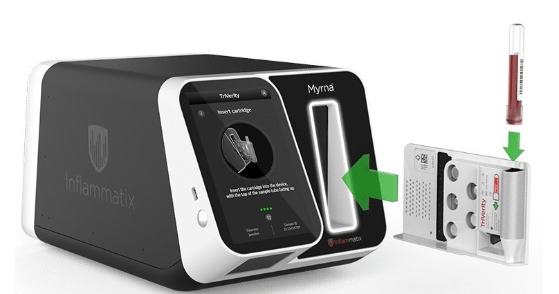
Rapid Host-Response Test Aims to Predict Corticosteroid Benefit in Sepsis
Sepsis and severe infections are difficult to treat because patients with similar clinical presentations may have very different underlying immune responses. Corticosteroids are commonly used in severe... Read more

Pathology
view channel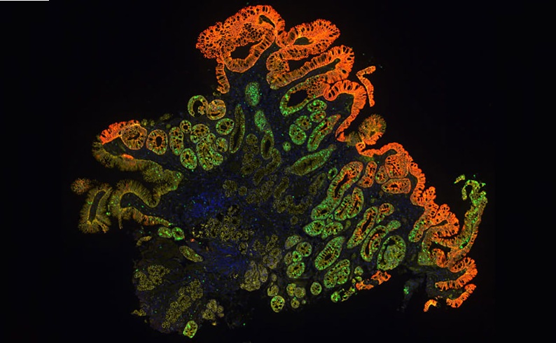
Molecular Testing Refines Barrett’s Esophagus Progression Risk Beyond Biopsy Findings
Barrett’s esophagus is a precancerous condition associated with increased risk of esophageal adenocarcinoma. Routine endoscopic biopsy findings guide surveillance and therapy, but histopathology may not... Read more
Updated Digital Pathology Platform Expands AI Integration for Cancer Diagnosis
Digital pathology is reshaping how laboratories manage case review, image analysis, and collaboration. As diagnostic workflows become more complex, pathology teams need platforms that can support digital... Read more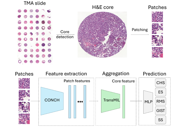
Technology
view channel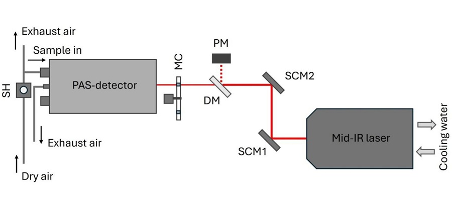
Laser-Based Swab Analysis Shows Promise for Detecting Disease-Linked Odor Patterns
Disease-related changes in volatile organic compounds can alter body odor, producing measurable patterns in exhaled breath and bodily fluids. Current analytical methods can be complex, time-consuming,... Read more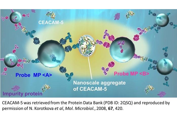
Laser-Enhanced Assay Boosts Sensitivity for Colorectal Cancer Biomarker Detection
Colorectal cancer is the third most commonly diagnosed cancer and the second leading cause of cancer-related death worldwide. Early detection remains critical, but cancer biomarkers can produce only faint... Read more

Industry
view channel
Expanding Screening and Biomarker Testing Drive Cancer Diagnostics Market Growth
Cancer diagnostics encompasses imaging, tissue pathology, laboratory testing, and molecular technologies used to detect, confirm, characterize, and monitor cancer. The global cancer diagnostics market... Read more