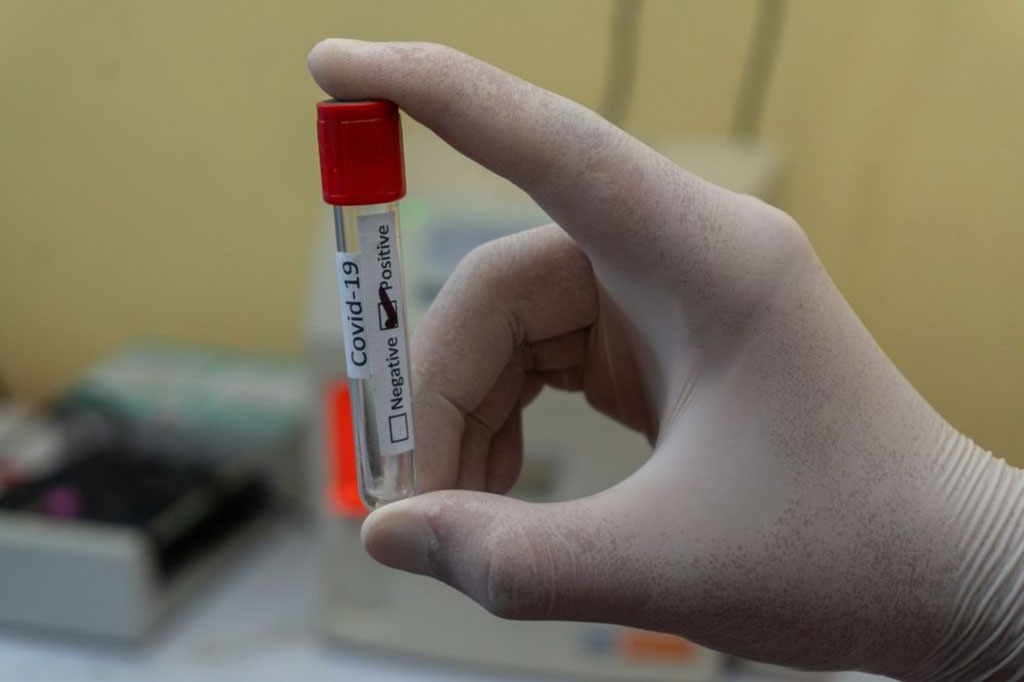
RNA Reference Materials Useful for Standardizing COVID-19 Tests, Finds Study
|
By LabMedica International staff writers Posted on 24 Jan 2022 |

A new study has shown that RNA reference materials are useful for standardizing COVID-19 tests.
The multiorganizational study led by researchers at the National Institute of Standards and Technology (NIST; Gaithersburg, MD, USA) looked at anchoring cycle threshold (Ct) values to a reference sample with known amounts of the virus in an effort to make the COVID-19 tests results more comparable between labs.
Scientists track and monitor the circulation of SARS-CoV-2, the virus that causes COVID-19, using methods based on a laboratory technique called polymerase chain reaction (PCR). Also used as the “gold standard” test to diagnose COVID-19 in individuals, PCR amplifies pieces of DNA by copying them numerous times through a series of chemical reactions. The number of cycles it takes to amplify DNA sequences of interest so that they are detectable by the PCR machine, known as Ct, is what researchers and medical professionals look at to detect the virus. However, not all labs get the same Ct values.
SARS-CoV-2 is an RNA virus: Its genetic material is single-stranded instead of double-stranded like DNA and contains some different molecular building blocks, namely uracil in place of thymine. But the PCR test only works with DNA, and labs first must convert the RNA to DNA to screen for COVID-19. For the test, RNA is isolated from a patient’s sample and combined with other ingredients, including short DNA sequences known as primers, to transform the RNA into DNA. When running a PCR test, labs often use the Ct value to make a positive or negative diagnosis of COVID-19. Labs often set a “cutoff” Ct value above which they interpret and can declare a patient “negative” if the virus is not detected after a certain number of cycles. But even though different labs can accurately detect the virus using their PCR tests, they use their own test methods and instruments, which could lead to different Ct values.
So, for this study, a multiorganizational research team set out to explore how much Ct values could vary among different labs when they ran PCR tests on the same reference samples containing known amounts of the SARS-CoV-2 virus. Two reference materials with carefully measured concentrations of the SARS-CoV-2 RNA were developed by a group of organizations and institutions. RM 1 had an estimated viral load of 10 million (107) copies per milliliter, and RM 2 had an estimated viral load of one million (106) copies per milliliter.
To ensure these values were accurate, metrology institutes measured and validated the reference materials using digital PCR. Digital PCR follows the same steps as traditional PCR but is a more advanced version. In digital PCR, the sample is partitioned into thousands of tiny droplets. A compound is also added so when the targeted DNA is detected it gives off a glow, allowing researchers to confirm if the sample is positive for the coronavirus with the presence of the fluorescent molecules.
The reference materials were sent out to a total of 305 laboratories in Germany, which yielded 1,109 data sets to be analyzed. The Ct values differed between labs depending on the test system or PCR equipment used and the targeted DNA sequences of the virus. For example, PCR assays aiming to detect a key gene (the “N” gene) in the COVID-19 virus had a range of Ct values between 17.6 and 26.9 for RM 1, while for RM 2 the range was between 20.7 and 30.1. The ranges between the two RMs overlap (20.7 to 26.9) even though they have different viral load concentrations, which shows it’s not possible to tell the precise concentration from Ct values alone.
The differences in Ct values among labs showed the usefulness of reference materials as a tool to help labs compare and standardize their results. Some have previously viewed the Ct value as a way to measure the viral load, the amount of virus in a person’s body. But the variation in Ct values across labs underscores that the two variables are merely correlated with one another. For example, if an individual has a higher viral load, then their Ct value would be low because it would take fewer cycles to amplify the virus’s genetic material. However, by using the appropriate reference materials, Ct values can potentially be converted to an actual virus concentration value in copies per microliter, which would be one way to quantify the viral load, though this wasn’t a focus of the study
Some have also suggested using Ct values to determine how infectious a patient is, another practice that the researchers caution against. Even though Ct values alone don’t determine how sick or infected an individual is, understanding these values could help inform decisions on setting criteria for monitoring people affected by the coronavirus. And RNA reference materials can help make these results more reliable and comparable.
“A lower Ct value means more DNA with the coronavirus starting out in the sample. It correlates to the viral load. But Ct can vary depending on the extraction material or the method used. All of these things can affect at which point the DNA is detected,” said NIST researcher Megan Cleveland, a co-author of the study. “People should use well-characterized reference materials along with their test methods instead of just relying on the Ct values. On its own, the Ct values are not easily comparable between testing laboratories, but researchers having access to reference materials and understanding their importance is beneficial.”
Related Links:
NIST
Latest COVID-19 News
- New Immunosensor Paves Way to Rapid POC Testing for COVID-19 and Emerging Infectious Diseases
- Long COVID Etiologies Found in Acute Infection Blood Samples
- Novel Device Detects COVID-19 Antibodies in Five Minutes
- CRISPR-Powered COVID-19 Test Detects SARS-CoV-2 in 30 Minutes Using Gene Scissors
- Gut Microbiome Dysbiosis Linked to COVID-19
- Novel SARS CoV-2 Rapid Antigen Test Validated for Diagnostic Accuracy
- New COVID + Flu + R.S.V. Test to Help Prepare for `Tripledemic`
- AI Takes Guesswork Out Of Lateral Flow Testing
- Fastest Ever SARS-CoV-2 Antigen Test Designed for Non-Invasive COVID-19 Testing in Any Setting
- Rapid Antigen Tests Detect Omicron, Delta SARS-CoV-2 Variants
- Health Care Professionals Showed Increased Interest in POC Technologies During Pandemic, Finds Study
- Set Up Reserve Lab Capacity Now for Faster Response to Next Pandemic, Say Researchers
- Blood Test Performed During Initial Infection Predicts Long COVID Risk
- Low-Cost COVID-19 Testing Platform Combines Sensitivity of PCR and Speed of Antigen Tests
- Finger-Prick Blood Test Identifies Immunity to COVID-19
- Quick Test Kit Determines Immunity Against COVID-19 and Its Variants
Channels
Clinical Chemistry
view channel
3D Printed Point-Of-Care Mass Spectrometer Outperforms State-Of-The-Art Models
Mass spectrometry is a precise technique for identifying the chemical components of a sample and has significant potential for monitoring chronic illness health states, such as measuring hormone levels... Read more.jpg)
POC Biomedical Test Spins Water Droplet Using Sound Waves for Cancer Detection
Exosomes, tiny cellular bioparticles carrying a specific set of proteins, lipids, and genetic materials, play a crucial role in cell communication and hold promise for non-invasive diagnostics.... Read more
Highly Reliable Cell-Based Assay Enables Accurate Diagnosis of Endocrine Diseases
The conventional methods for measuring free cortisol, the body's stress hormone, from blood or saliva are quite demanding and require sample processing. The most common method, therefore, involves collecting... Read moreMolecular Diagnostics
view channel
Blood Test Accurately Predicts Lung Cancer Risk and Reduces Need for Scans
Lung cancer is extremely hard to detect early due to the limitations of current screening technologies, which are costly, sometimes inaccurate, and less commonly endorsed by healthcare professionals compared... Read more
Unique Autoantibody Signature to Help Diagnose Multiple Sclerosis Years before Symptom Onset
Autoimmune diseases such as multiple sclerosis (MS) are thought to occur partly due to unusual immune responses to common infections. Early MS symptoms, including dizziness, spasms, and fatigue, often... Read more
Blood Test Could Detect HPV-Associated Cancers 10 Years before Clinical Diagnosis
Human papilloma virus (HPV) is known to cause various cancers, including those of the genitals, anus, mouth, throat, and cervix. HPV-associated oropharyngeal cancer (HPV+OPSCC) is the most common HPV-associated... Read moreHematology
view channel
Next Generation Instrument Screens for Hemoglobin Disorders in Newborns
Hemoglobinopathies, the most widespread inherited conditions globally, affect about 7% of the population as carriers, with 2.7% of newborns being born with these conditions. The spectrum of clinical manifestations... Read more
First 4-in-1 Nucleic Acid Test for Arbovirus Screening to Reduce Risk of Transfusion-Transmitted Infections
Arboviruses represent an emerging global health threat, exacerbated by climate change and increased international travel that is facilitating their spread across new regions. Chikungunya, dengue, West... Read more
POC Finger-Prick Blood Test Determines Risk of Neutropenic Sepsis in Patients Undergoing Chemotherapy
Neutropenia, a decrease in neutrophils (a type of white blood cell crucial for fighting infections), is a frequent side effect of certain cancer treatments. This condition elevates the risk of infections,... Read more
First Affordable and Rapid Test for Beta Thalassemia Demonstrates 99% Diagnostic Accuracy
Hemoglobin disorders rank as some of the most prevalent monogenic diseases globally. Among various hemoglobin disorders, beta thalassemia, a hereditary blood disorder, affects about 1.5% of the world's... Read moreImmunology
view channel
Diagnostic Blood Test for Cellular Rejection after Organ Transplant Could Replace Surgical Biopsies
Transplanted organs constantly face the risk of being rejected by the recipient's immune system which differentiates self from non-self using T cells and B cells. T cells are commonly associated with acute... Read more
AI Tool Precisely Matches Cancer Drugs to Patients Using Information from Each Tumor Cell
Current strategies for matching cancer patients with specific treatments often depend on bulk sequencing of tumor DNA and RNA, which provides an average profile from all cells within a tumor sample.... Read more
Genetic Testing Combined With Personalized Drug Screening On Tumor Samples to Revolutionize Cancer Treatment
Cancer treatment typically adheres to a standard of care—established, statistically validated regimens that are effective for the majority of patients. However, the disease’s inherent variability means... Read moreMicrobiology
view channel
New CE-Marked Hepatitis Assays to Help Diagnose Infections Earlier
According to the World Health Organization (WHO), an estimated 354 million individuals globally are afflicted with chronic hepatitis B or C. These viruses are the leading causes of liver cirrhosis, liver... Read more
1 Hour, Direct-From-Blood Multiplex PCR Test Identifies 95% of Sepsis-Causing Pathogens
Sepsis contributes to one in every three hospital deaths in the US, and globally, septic shock carries a mortality rate of 30-40%. Diagnosing sepsis early is challenging due to its non-specific symptoms... Read morePathology
view channelAI-Powered Digital Imaging System to Revolutionize Cancer Diagnosis
The process of biopsy is important for confirming the presence of cancer. In the conventional histopathology technique, tissue is excised, sliced, stained, mounted on slides, and examined under a microscope... Read more
New Mycobacterium Tuberculosis Panel to Support Real-Time Surveillance and Combat Antimicrobial Resistance
Tuberculosis (TB), the leading cause of death from an infectious disease globally, is a contagious bacterial infection that primarily spreads through the coughing of patients with active pulmonary TB.... Read moreTechnology
view channel
New Diagnostic System Achieves PCR Testing Accuracy
While PCR tests are the gold standard of accuracy for virology testing, they come with limitations such as complexity, the need for skilled lab operators, and longer result times. They also require complex... Read more
DNA Biosensor Enables Early Diagnosis of Cervical Cancer
Molybdenum disulfide (MoS2), recognized for its potential to form two-dimensional nanosheets like graphene, is a material that's increasingly catching the eye of the scientific community.... Read more
Self-Heating Microfluidic Devices Can Detect Diseases in Tiny Blood or Fluid Samples
Microfluidics, which are miniature devices that control the flow of liquids and facilitate chemical reactions, play a key role in disease detection from small samples of blood or other fluids.... Read more
Breakthrough in Diagnostic Technology Could Make On-The-Spot Testing Widely Accessible
Home testing gained significant importance during the COVID-19 pandemic, yet the availability of rapid tests is limited, and most of them can only drive one liquid across the strip, leading to continued... Read moreIndustry
view channel
ECCMID Congress Name Changes to ESCMID Global
Over the last few years, the European Society of Clinical Microbiology and Infectious Diseases (ESCMID, Basel, Switzerland) has evolved remarkably. The society is now stronger and broader than ever before... Read more
Bosch and Randox Partner to Make Strategic Investment in Vivalytic Analysis Platform
Given the presence of so many diseases, determining whether a patient is presenting the symptoms of a simple cold, the flu, or something as severe as life-threatening meningitis is usually only possible... Read more
Siemens to Close Fast Track Diagnostics Business
Siemens Healthineers (Erlangen, Germany) has announced its intention to close its Fast Track Diagnostics unit, a small collection of polymerase chain reaction (PCR) testing products that is part of the... Read more


.jpg)

