


Pathology Lab on Wheels Can Revolutionize Brain Surgery
|
By LabMedica International staff writers Posted on 27 May 2022 |
|
")
Guesswork defines a central challenge of brain surgery. Cut too little and the residual tumor cells will reboot and kill the patient. Cut too much and critical brain functions could be irreversibly damaged. Studies show that in up to three-quarters of patients with brain cancer, portions of the tumor that could be safely removed are left behind, simply because the surgeon cannot see them. Without visual certainty, the risk of removing precious healthy tissue that could be involved in speech, memory, movement, or virtually any other important function of the brain is simply too high to cut beyond the known boundaries. Now, a radically new kind of imaging system helps brain surgeons cut with more confidence.
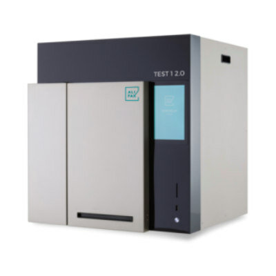
The new imaging system, now employed at the Brain and Spine Tumor Center at NYU Langone’s Perlmutter Cancer Center NYU Langone (New York, NY, USA), is called stimulated Raman histology, or SRH, a method that distinguishes tumor regions, rich in protein and DNA, from normal lipid-rich brain tissue, creating contrasted images akin to conventional histology slides. The technology is based on an old technique, Raman spectroscopy, used in chemistry since the 1920s, that involves shining a laser beam at a sample. The unique vibrational properties of different molecules change the optical properties of the laser, helping to create an image of the sample’s structure.
Pathologists can just as readily distinguish between cancerous and healthy tissue using the SRH images made in the operating room compared to the conventional ones created in the lab. The system works in concert with a powerful new diagnostic technique that leverages AI to distinguish among tumor types in less than two minutes, compared to the 20 to 30 minutes it typically takes human pathologists. The speed of diagnosis is a game changer, eliminating the time that a patient remains on the operating table while surgeons await lab results - a dangerous gap that increases the odds of infection or complications. Housed in a metal box about the size of a mini-fridge and mounted on wheels, the technology, which is now available to all NYU Langone Health patients with brain tumors, can be rolled into any operating room to provide a surgeon with near real-time analysis of a tissue sample.
“If we have a patient with a tumor of unknown etiology, for example, we might not know whether it’s glioblastoma or lymphoma - two tumors with very different treatments. Making the distinction in the operating room is extremely important,” said Daniel A. Orringer, MD. “Surgical decision-making is like operating brake and gas pedals. We are taking the guesswork out of the picture by allowing the surgeon to interrogate the tissue on a microscopic level and use imaging data to inform surgical strategy.”
Related Links:
NYU Langone


Latest Pathology News
- Engineered Yeast Cells Enable Rapid Testing of Cancer Immunotherapy
- First-Of-Its-Kind Test Identifies Autism Risk at Birth
- AI Algorithms Improve Genetic Mutation Detection in Cancer Diagnostics
- Skin Biopsy Offers New Diagnostic Method for Neurodegenerative Diseases
- Fast Label-Free Method Identifies Aggressive Cancer Cells
- New X-Ray Method Promises Advances in Histology
- Single-Cell Profiling Technique Could Guide Early Cancer Detection
- Intraoperative Tumor Histology to Improve Cancer Surgeries
- Rapid Stool Test Could Help Pinpoint IBD Diagnosis
- AI-Powered Label-Free Optical Imaging Accurately Identifies Thyroid Cancer During Surgery
- Deep Learning–Based Method Improves Cancer Diagnosis
- ADLM Updates Expert Guidance on Urine Drug Testing for Patients in Emergency Departments
- New Age-Based Blood Test Thresholds to Catch Ovarian Cancer Earlier
- Genetics and AI Improve Diagnosis of Aortic Stenosis
- AI Tool Simultaneously Identifies Genetic Mutations and Disease Type
- Rapid Low-Cost Tests Can Prevent Child Deaths from Contaminated Medicinal Syrups
Channels
Clinical Chemistry
view channel")
New PSA-Based Prognostic Model Improves Prostate Cancer Risk Assessment
Prostate cancer is the second-leading cause of cancer death among American men, and about one in eight will be diagnosed in their lifetime. Screening relies on blood levels of prostate-specific antigen... Read more
Extracellular Vesicles Linked to Heart Failure Risk in CKD Patients
Chronic kidney disease (CKD) affects more than 1 in 7 Americans and is strongly associated with cardiovascular complications, which account for more than half of deaths among people with CKD.... Read more")
")
Molecular Diagnostics
view channel")
Diagnostic Device Predicts Treatment Response for Brain Tumors Via Blood Test
Glioblastoma is one of the deadliest forms of brain cancer, largely because doctors have no reliable way to determine whether treatments are working in real time. Assessing therapeutic response currently... Read more")
Blood Test Detects Early-Stage Cancers by Measuring Epigenetic Instability
Early-stage cancers are notoriously difficult to detect because molecular changes are subtle and often missed by existing screening tools. Many liquid biopsies rely on measuring absolute DNA methylation... Read more")
“Lab-On-A-Disc” Device Paves Way for More Automated Liquid Biopsies
Extracellular vesicles (EVs) are tiny particles released by cells into the bloodstream that carry molecular information about a cell’s condition, including whether it is cancerous. However, EVs are highly... Read more in the area surrounding sEcad-high cancer cells (blue, center) (Photo courtesy of Debeb Laboratory)")
Blood Test Identifies Inflammatory Breast Cancer Patients at Increased Risk of Brain Metastasis
Brain metastasis is a frequent and devastating complication in patients with inflammatory breast cancer, an aggressive subtype with limited treatment options. Despite its high incidence, the biological... Read moreHematology
view channel")
New Guidelines Aim to Improve AL Amyloidosis Diagnosis
Light chain (AL) amyloidosis is a rare, life-threatening bone marrow disorder in which abnormal amyloid proteins accumulate in organs. Approximately 3,260 people in the United States are diagnosed... Read more")
Fast and Easy Test Could Revolutionize Blood Transfusions
Blood transfusions are a cornerstone of modern medicine, yet red blood cells can deteriorate quietly while sitting in cold storage for weeks. Although blood units have a fixed expiration date, cells from... Read more line (Photo courtesy of Sysmex America)")
Automated Hemostasis System Helps Labs of All Sizes Optimize Workflow
High-volume hemostasis sections must sustain rapid turnaround while managing reruns and reflex testing. Manual tube handling and preanalytical checks can strain staff time and increase opportunities for error.... Read more")
High-Sensitivity Blood Test Improves Assessment of Clotting Risk in Heart Disease Patients
Blood clotting is essential for preventing bleeding, but even small imbalances can lead to serious conditions such as thrombosis or dangerous hemorrhage. In cardiovascular disease, clinicians often struggle... Read moreImmunology
view channelBlood Test Identifies Lung Cancer Patients Who Can Benefit from Immunotherapy Drug
Small cell lung cancer (SCLC) is an aggressive disease with limited treatment options, and even newly approved immunotherapies do not benefit all patients. While immunotherapy can extend survival for some,... Read more")
Whole-Genome Sequencing Approach Identifies Cancer Patients Benefitting From PARP-Inhibitor Treatment
Targeted cancer therapies such as PARP inhibitors can be highly effective, but only for patients whose tumors carry specific DNA repair defects. Identifying these patients accurately remains challenging,... Read more")
Ultrasensitive Liquid Biopsy Demonstrates Efficacy in Predicting Immunotherapy Response
Immunotherapy has transformed cancer treatment, but only a small proportion of patients experience lasting benefit, with response rates often remaining between 10% and 20%. Clinicians currently lack reliable... Read more")
Microbiology
view channel")
Comprehensive Review Identifies Gut Microbiome Signatures Associated With Alzheimer’s Disease
Alzheimer’s disease affects approximately 6.7 million people in the United States and nearly 50 million worldwide, yet early cognitive decline remains difficult to characterize. Increasing evidence suggests... Read moreAI-Powered Platform Enables Rapid Detection of Drug-Resistant C. Auris Pathogens
Infections caused by the pathogenic yeast Candida auris pose a significant threat to hospitalized patients, particularly those with weakened immune systems or those who have invasive medical devices.... Read more")
")
Pathology
view channel system (Deichmann, M. et al., Nat Commun 16, 10306, 2025. DOI: 10.1038/s41467-025-65236-7)")
Engineered Yeast Cells Enable Rapid Testing of Cancer Immunotherapy
Developing new cancer immunotherapies is a slow, costly, and high-risk process, particularly for CAR T cell treatments that must precisely recognize cancer-specific antigens. Small differences in tumor... Read more")
First-Of-Its-Kind Test Identifies Autism Risk at Birth
Autism spectrum disorder is treatable, and extensive research shows that early intervention can significantly improve cognitive, social, and behavioral outcomes. Yet in the United States, the average age... Read more")
")
Technology
view channel")
Robotic Technology Unveiled for Automated Diagnostic Blood Draws
Routine diagnostic blood collection is a high‑volume task that can strain staffing and introduce human‑dependent variability, with downstream implications for sample quality and patient experience.... Read more")
ADLM Launches First-of-Its-Kind Data Science Program for Laboratory Medicine Professionals
Clinical laboratories generate billions of test results each year, creating a treasure trove of data with the potential to support more personalized testing, improve operational efficiency, and enhance patient care.... Read moreAptamer Biosensor Technology to Transform Virus Detection
Rapid and reliable virus detection is essential for controlling outbreaks, from seasonal influenza to global pandemics such as COVID-19. Conventional diagnostic methods, including cell culture, antigen... Read more")
AI Models Could Predict Pre-Eclampsia and Anemia Earlier Using Routine Blood Tests
Pre-eclampsia and anemia are major contributors to maternal and child mortality worldwide, together accounting for more than half a million deaths each year and leaving millions with long-term health complications.... Read moreIndustry
view channelNew Collaboration Brings Automated Mass Spectrometry to Routine Laboratory Testing
Mass spectrometry is a powerful analytical technique that identifies and quantifies molecules based on their mass and electrical charge. Its high selectivity, sensitivity, and accuracy make it indispensable... Read more")
AI-Powered Cervical Cancer Test Set for Major Rollout in Latin America
Noul Co., a Korean company specializing in AI-based blood and cancer diagnostics, announced it will supply its intelligence (AI)-based miLab CER cervical cancer diagnostic solution to Mexico under a multi‑year... Read more")
Diasorin and Fisher Scientific Enter into US Distribution Agreement for Molecular POC Platform
Diasorin (Saluggia, Italy) has entered into an exclusive distribution agreement with Fisher Scientific, part of Thermo Fisher Scientific (Waltham, MA, USA), for the LIAISON NES molecular point-of-care... Read more will be held at Dubai World Trade Centre from 10-13 February")