Novel Technique Uses ‘Sugar’ Signatures to Identify and Classify Pancreatic Cancer Cell Subtypes
Posted on 14 Apr 2025
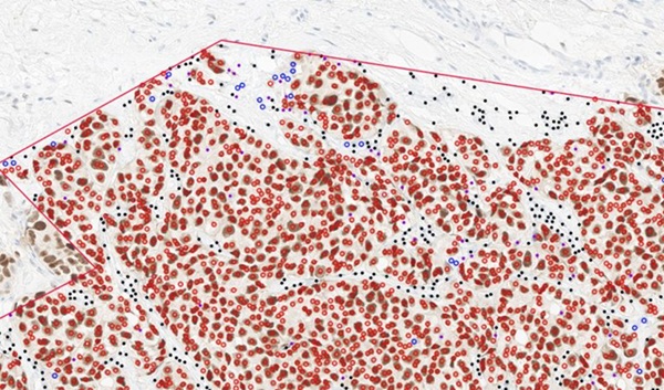
Pancreatic cancer is often asymptomatic in its early stages, making it difficult to detect until it has progressed. Consequently, only 15% of pancreatic cancers are diagnosed early enough to allow for surgical intervention. Complicating the situation, pancreatic tumors are made up of various subtypes of malignant cells, each of which responds differently to treatment. However, a newly developed technique can now identify and classify these cancer cell subtypes based on the sugars found on the surface of the cancer cells. These sugars, known as glycans, are involved in cell communication and recognition and serve as a cellular “signature,” with each subtype of pancreatic cancer cells having a unique glycan composition.
This innovative technique, known as multiplexed glycan immunofluorescence, was developed by researchers at the Van Andel Institute (Grand Rapids, MI, USA) in collaboration with other scientists, and has been detailed in the journal Science Advances. The method utilizes advanced software and imaging technology to precisely identify the mix of pancreatic cancer cell subtypes within a tumor. In the future, this approach may help in achieving earlier and more accurate diagnoses. The team initially identified glycan signatures by analyzing tumor tissue and then refined their technique to detect these glycans secreted into the blood by cancer cells.
")
This development is crucial, as blood tests are less invasive for patients, offering a quicker, more affordable, and non-invasive alternative to surgery. While the method is not yet available for use in clinical settings, the researchers are optimistic that it could become a standard diagnostic tool for pancreatic cancer in the coming years. In the meantime, the team continues to validate their method and explore the possibility of detecting rarer cell types. Additionally, they are investigating whether this technique can be applied to better detect and characterize other gastrointestinal cancers.
“Our new method allows us to go one step beyond cancer diagnosis by revealing which subtypes of pancreatic cancer cells make up a tumor. The more we know about which cells are present, the better physicians can tailor treatments for each patient,” said Brian Haab, Ph.D., a VAI professor and corresponding author of the study.