Computer-Designed Proteins Programmed to Deactivate Flu Viruses
|
By LabMedica International staff writers Posted on 20 Jun 2012 |


|
Computer-designed proteins are now being constructed to fight the flu. Researchers are demonstrating that proteins found in nature, but that do not normally bind the flu, can be engineered to act as broad-spectrum antiviral agents against a range of flu virus strains, including H1N1 pandemic influenza.
“One of these engineered proteins has a flu-fighting potency that rivals that of several human monoclonal antibodies,” said Dr. David Baker, a professor of biochemistry at the University of Washington (Seattle, USA), in a report June 7, 2012, published in the journal Nature Biotechnology.
Dr. Baker’s research team is making major inroads in optimizing the function of computer-designed influenza inhibitors. These proteins are constructed via computer modeling to fit exquisitely into a specific nano-sized target on flu viruses. By binding the target areas similar to a key into a lock, they keep the virus from changing shape, a tactic that the virus uses to infect living cells. The research efforts, analogous to docking a space station but on a molecular level, are made possible by computers that can describe the panoramas of forces involved on the submicroscopic scale.
Dr. Baker is head of the new Institute for Protein Design Center at the University of Washington. Biochemists, engineers, computer scientists, and medical specialists at the center are engineering innovative proteins with new functions for specific purposes in medicine, environmental protection, and other areas. Proteins underlie all typical activities and structures of living cells, and also control disease actions of pathogens such as viruses. Abnormal protein formation and interactions are also implicated in many inherited and later-life chronic disorders.
Because influenza is a serious worldwide public health problem due to its genetic shifts and drifts that sporadically become more virulent, the flu is one of the key interests of the Institutes for Protein Design and its collaborators in the United States and worldwide. Researchers are trying to meet the vital need for better therapeutic agents to protect against this very adaptable and extremely infective virus. Vaccines for new strains of influenza take months to develop, evaluate, and manufacture, and are not helpful for those already sick. The long response time for vaccine creation and distribution is unsettling when a more lethal strain abruptly emerges and spreads rapidly. The speed of transmission is accelerated by the lack of widespread immunity in the general population to the latest form of the virus.
Flu trackers refer to strains by their H and N subtypes. H stands for hemagglutinins, which are the molecules on the flu virus that enable it to invade the cells of respiratory passages. The virus’s hemagglutinin molecules attach to the surface of cells lining the respiratory tract. When the cell tries to engulf the virus, it makes the error of pulling it into a more acidic location. The drop in pH changes the shape of the viral hemagglutinin, thereby allowing the virus to fuse to the cell and open an entry for the virus’ RNA to come in and start making fresh viruses. It is hypothesized that the Baker Lab protein inhibits this shape change by binding the hemagglutinin in a very specific orientation and thus keeps the virus from invading cells.
Dr. Baker and his team wanted to create antivirals that could react against a wide variety of H subtypes, as this versatility could lead to a comprehensive therapy for influenza. Specifically, viruses that have hemagglutinins of the H2 subtype are responsible for the deadly pandemic of 1957 and continued to circulate until 1968. People born after that date have not been exposed to H2 viruses. The recent avian flu has a new version of H1 hemagglutinin. Data suggest that Dr. Baker’s proteins bind to all types of the group I hemagglutinin, a group that includes not only H1 but also the pandemic H2 and avian H5 strains.
The methods developed for the influenza inhibitor protein design, according to Dr. Baker, could be “a powerful route to inhibitors or binders for any surface patch on any desired target of interest.” For example, if a new disease pathogen arises, scientists could figure out how it interacts with human cells or other hosts on a molecular level. Scientists could then employ protein interface design to create a diversity of small proteins that they predict would block the pathogen’s interaction surface.
Genes for large numbers of the most promising, computer-designed proteins could be tested using yeast cells. After additional molecular chemistry research to search for the best binding among those proteins, those could be reprogrammed in the laboratory to undergo mutations, and all the mutated forms could be stored in a “library” for an in-depth examination of their amino acids, molecular architecture, and energy bonds.
Sophisticated technologies would allow the scientists to rapidly browse through the library to find those tiny proteins that clung to the pathogen surface target with pinpoint accuracy. The finalists would be selected from this pool for excelling at blocking the pathogen from attaching to, entering, and infecting human or animal cells.
The utilization of deep sequencing, the same technology now used to sequence human genomes cheaply, was particularly central in creating detailed maps relating sequencing to function. These maps were used to reprogram the design to achieve a more exact interaction between the inhibitor protein and the virus molecule. It also enabled the scientists, they said, “to leapfrog over bottlenecks” to improve the activity of the binder. They were able to see how small contributions from many small alterations in the protein, too difficult to see individually, could together create a binder with better attachment strength.
“We anticipate that our approach combining computational design followed by comprehensive energy landscape mapping,” Dr. Baker said, “will be widely useful in generating high-affinity and high-specificity binders to a broad range of targets for use in therapeutics and diagnostics.”
Related Links:
University of Washington


.jpg)





“One of these engineered proteins has a flu-fighting potency that rivals that of several human monoclonal antibodies,” said Dr. David Baker, a professor of biochemistry at the University of Washington (Seattle, USA), in a report June 7, 2012, published in the journal Nature Biotechnology.
Dr. Baker’s research team is making major inroads in optimizing the function of computer-designed influenza inhibitors. These proteins are constructed via computer modeling to fit exquisitely into a specific nano-sized target on flu viruses. By binding the target areas similar to a key into a lock, they keep the virus from changing shape, a tactic that the virus uses to infect living cells. The research efforts, analogous to docking a space station but on a molecular level, are made possible by computers that can describe the panoramas of forces involved on the submicroscopic scale.
Dr. Baker is head of the new Institute for Protein Design Center at the University of Washington. Biochemists, engineers, computer scientists, and medical specialists at the center are engineering innovative proteins with new functions for specific purposes in medicine, environmental protection, and other areas. Proteins underlie all typical activities and structures of living cells, and also control disease actions of pathogens such as viruses. Abnormal protein formation and interactions are also implicated in many inherited and later-life chronic disorders.
Because influenza is a serious worldwide public health problem due to its genetic shifts and drifts that sporadically become more virulent, the flu is one of the key interests of the Institutes for Protein Design and its collaborators in the United States and worldwide. Researchers are trying to meet the vital need for better therapeutic agents to protect against this very adaptable and extremely infective virus. Vaccines for new strains of influenza take months to develop, evaluate, and manufacture, and are not helpful for those already sick. The long response time for vaccine creation and distribution is unsettling when a more lethal strain abruptly emerges and spreads rapidly. The speed of transmission is accelerated by the lack of widespread immunity in the general population to the latest form of the virus.
Flu trackers refer to strains by their H and N subtypes. H stands for hemagglutinins, which are the molecules on the flu virus that enable it to invade the cells of respiratory passages. The virus’s hemagglutinin molecules attach to the surface of cells lining the respiratory tract. When the cell tries to engulf the virus, it makes the error of pulling it into a more acidic location. The drop in pH changes the shape of the viral hemagglutinin, thereby allowing the virus to fuse to the cell and open an entry for the virus’ RNA to come in and start making fresh viruses. It is hypothesized that the Baker Lab protein inhibits this shape change by binding the hemagglutinin in a very specific orientation and thus keeps the virus from invading cells.
Dr. Baker and his team wanted to create antivirals that could react against a wide variety of H subtypes, as this versatility could lead to a comprehensive therapy for influenza. Specifically, viruses that have hemagglutinins of the H2 subtype are responsible for the deadly pandemic of 1957 and continued to circulate until 1968. People born after that date have not been exposed to H2 viruses. The recent avian flu has a new version of H1 hemagglutinin. Data suggest that Dr. Baker’s proteins bind to all types of the group I hemagglutinin, a group that includes not only H1 but also the pandemic H2 and avian H5 strains.
The methods developed for the influenza inhibitor protein design, according to Dr. Baker, could be “a powerful route to inhibitors or binders for any surface patch on any desired target of interest.” For example, if a new disease pathogen arises, scientists could figure out how it interacts with human cells or other hosts on a molecular level. Scientists could then employ protein interface design to create a diversity of small proteins that they predict would block the pathogen’s interaction surface.
Genes for large numbers of the most promising, computer-designed proteins could be tested using yeast cells. After additional molecular chemistry research to search for the best binding among those proteins, those could be reprogrammed in the laboratory to undergo mutations, and all the mutated forms could be stored in a “library” for an in-depth examination of their amino acids, molecular architecture, and energy bonds.
Sophisticated technologies would allow the scientists to rapidly browse through the library to find those tiny proteins that clung to the pathogen surface target with pinpoint accuracy. The finalists would be selected from this pool for excelling at blocking the pathogen from attaching to, entering, and infecting human or animal cells.
The utilization of deep sequencing, the same technology now used to sequence human genomes cheaply, was particularly central in creating detailed maps relating sequencing to function. These maps were used to reprogram the design to achieve a more exact interaction between the inhibitor protein and the virus molecule. It also enabled the scientists, they said, “to leapfrog over bottlenecks” to improve the activity of the binder. They were able to see how small contributions from many small alterations in the protein, too difficult to see individually, could together create a binder with better attachment strength.
“We anticipate that our approach combining computational design followed by comprehensive energy landscape mapping,” Dr. Baker said, “will be widely useful in generating high-affinity and high-specificity binders to a broad range of targets for use in therapeutics and diagnostics.”
Related Links:
University of Washington

Platinum Member
Automated Coagulation Analyzer
Hemolumi H6
Gold Member
Clinical Chemistry Assay
Sorbitol Dehydrogenase (SDH)
HPV Test
Allplex HPV28 Detection
New
Gold Member
Serum Indices Control
Acusera Serum Indices Control
Latest BioResearch News
- Cancer DNA Trapping Mechanism Reveals Potential Target for Future Therapies
- Blood Antibody Signals May Offer Clues to the Origins of Childhood Cancer
- Genetic Study Links Inherited Blindness to Gene Tied to Rare Metabolic Disorder
- Population-Scale Proteomics Study Identifies New Genetic-Protein Links
- Circulating Tumor DNA Helps Identify Targets for Personalized Immunotherapy
- Researchers Classify 108 Lysosomal Disorders in Updated Diagnostic Framework
- New Autoantibody Target Identified in Neuromyelitis Optica Spectrum Disorder
- New Immune Target Could Support More Precise High Blood Pressure Treatment
- New Gene-Disease Link May Help Diagnose Rare Neurodevelopmental Disorders
- Researchers Identify Shared Molecular Networks Behind Fatigue-Related Illnesses
- Blood Metabolite Signature Predicts ALS Progression and Points to Treatment Strategy
- Whole-Blood Extracellular Vesicle Analysis Captures Molecular Signals Missed by Plasma Testing
- Age-Related Genomic Differences Could Refine Treatment Decisions in Lung Cancer
- Computational Tool Identifies Central Asthma Genes for Target Discovery
- Molecular Pathway Reveals Driver of Triple-Negative Breast Cancer Spread
- D-Serine May Predict Immune Checkpoint Therapy Resistance in Gastric Cancer
Channels
Clinical Chemistry
view channel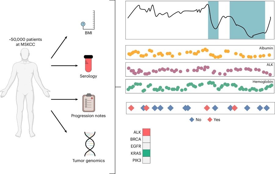
Multimodal Clinical Data Models Predict Cachexia Risk Across Cancers
Cancer cachexia is a severe wasting syndrome that affects roughly half of patients with cancer and cannot be reversed simply by increasing food intake. It can reduce treatment tolerance, impair quality... Read more
Study Evaluates Alzheimer’s Blood Biomarker Across Racial and Ethnic Groups
Alzheimer’s disease trials increasingly depend on identifying cognitively unimpaired people who already have elevated brain amyloid. Blood biomarkers may help reduce reliance on positron emission tomography... Read more
Molecular Diagnostics
view channel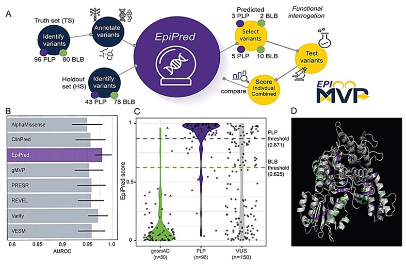
AI Tool Classifies Pathogenic Variants in Severe Childhood Epilepsy
Developmental and epileptic encephalopathies are rare neurodevelopmental disorders characterized by early-life seizures and significant developmental delays. Genetic testing can identify variants associated... Read more
Noninvasive Cervical Test Detects Endometrial Cancer for Triage
Endometrial cancer is rising in incidence and mortality in the United States. The disease has no population screening program or FDA-cleared noninvasive test for early identification, and evaluation can... Read more

Hematology
view channel
New Myelofibrosis Guidelines Recommend Expanded Genetic Testing at Diagnosis
Myelofibrosis is a rare, life-threatening blood cancer affecting up to six per 100,000 people. The disease causes scarring in the bone marrow, limiting the production of healthy blood cells and potentially... Read more
Handheld Point-of-Care Test Aims to Enable Rapid Coagulation Assessment in Trauma
Severe blood loss and inadequate tissue oxygenation are leading causes of preventable death after traumatic injury. Blood products are essential in trauma care, but cold-chain storage, short shelf life,... Read more

Immunology
view channel
FDA Approval Adds Automated Testing Option for Parvovirus B19 Serology
Human Parvovirus B19 is a highly contagious erythrovirus transmitted mainly through respiratory droplets and close contact. Diagnosis relies on IgM antibodies to detect active infection and IgG antibodies... Read more
Blood-Based Immune Aging Map Could Help Spot Disease Risk Earlier
Chronological age does not always reflect immune health or future disease risk. Two adults of the same age can have immune systems that age very differently, with some remaining resilient while others... Read more
Microbiology
view channel
Rapid Host-Response Test Aims to Predict Corticosteroid Benefit in Sepsis
Sepsis and severe infections are difficult to treat because patients with similar clinical presentations may have very different underlying immune responses. Corticosteroids are commonly used in severe... Read more
Oral and Lung Microbiome Changes Linked to Severe Long Covid Symptoms
Long Covid can involve persistent respiratory, cognitive, and gastrointestinal symptoms after acute infection, creating a difficult diagnostic and management challenge. Because COVID-19 initially affects... Read more

Pathology
view channel
Molecular Testing Refines Barrett’s Esophagus Progression Risk Beyond Biopsy Findings
Barrett’s esophagus is a precancerous condition associated with increased risk of esophageal adenocarcinoma. Routine endoscopic biopsy findings guide surveillance and therapy, but histopathology may not... Read more
Updated Digital Pathology Platform Expands AI Integration for Cancer Diagnosis
Digital pathology is reshaping how laboratories manage case review, image analysis, and collaboration. As diagnostic workflows become more complex, pathology teams need platforms that can support digital... Read more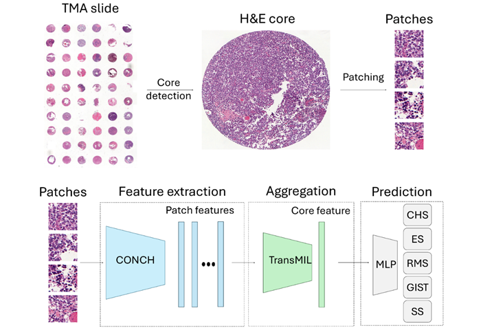
Technology
view channel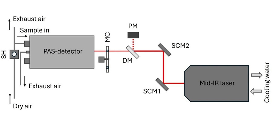
Laser-Based Swab Analysis Shows Promise for Detecting Disease-Linked Odor Patterns
Disease-related changes in volatile organic compounds can alter body odor, producing measurable patterns in exhaled breath and bodily fluids. Current analytical methods can be complex, time-consuming,... Read more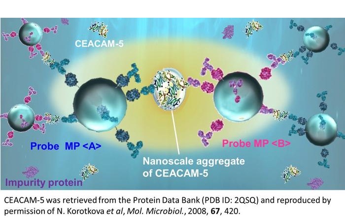
Laser-Enhanced Assay Boosts Sensitivity for Colorectal Cancer Biomarker Detection
Colorectal cancer is the third most commonly diagnosed cancer and the second leading cause of cancer-related death worldwide. Early detection remains critical, but cancer biomarkers can produce only faint... Read more

Industry
view channel
Global TB Summit Connects Diagnostic Testing With Prevention and Clinical Care
Tuberculosis remains a major global infectious disease challenge despite declining deaths. In 2024, an estimated 10.7 million people worldwide developed the disease and nearly 1.2 million died from it.... Read more