Aerobic Bacteria Rapidly Identified by MALDI-TOF Mass Spectrometry
|
By LabMedica International staff writers Posted on 27 Jan 2016 |

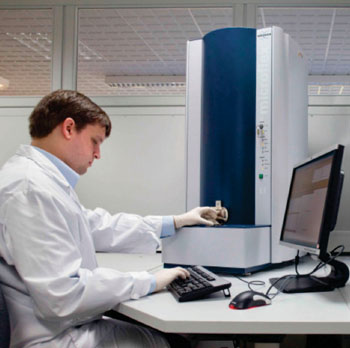
Image: The Microflex LT bench-top MALDI-TOF mass spectrometer and BioTyper (Photo courtesy of Bruker Daltonics).

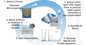
Image: Schematics of the Bruker MALDI BioTyper Workflow (Photo courtesy of Professor Melissa B. Miller).
Matrix-assisted laser desorption ionization time-of-flight mass spectrometry (MALDI-TOF MS) is a diagnostic tool for the identification of organisms routinely found in the microbiology laboratory.
MALDI-TOF MS has been proven to be a rapid and cost-effective diagnostic method for routine use in the clinical microbiology laboratory and MALDI-TOF MS technology uses ionizing laser for structural elements of the isolate to generate isolate-derived spectra, which is then compared to a reference database.
Scientists at the Children's Hospital Los Angeles (CA, USA) and their colleague at the Keck School of Medicine (Los Angeles, CA, USA) used isolates previously recovered by routine culture and workup from clinical specimens were cultured to appropriate media, identified directly by MALDI-TOF MS, and compared to results from various biochemical identification methods. A total of 996 aerobic Gram-positive and Gram-negative organism isolates were included in the study.
All MALDI-TOF MS testing was performed in duplicate with the same swab spotted onto two spots on the target plate, one spot representing a “heavy” inoculum and the second spot representing a “light” inoculum. Acquisition and analysis of mass spectra was performed using the Microflex LT mass spectrometer (Bruker Daltonics; Fremont, CA, USA). For media studies, 84 Gram-negative bacteria were cultured on blood agar, chocolate agar, and MacConkey agar; 74 Gram-positive bacteria were cultured on blood agar, chocolate agar, and colistin nalidixic acid (CNA) agar. For temperature and stability studies, four Gram-negative bacteria and four Gram-positive bacteria were cultured onto blood agar. Results of MALDI-TOF MS were compared to routine methods performed in the microbiology laboratory. All discordant identifications were confirmed by additional biochemical or 16S rRNA sequencing.
In MALDI-TOF MS data interpretation, a score of equal to or greater than 2.0 is equivocal to reliable species-level identification, a score of 1.7–1.99 is considered reliable to the genus level and a score of less than 1.7 is considered unreliable for bacteria identification. Using the direct-smear method, 99.5% and 98.0% of aerobic Gram-negative and Gram-positive bacteria, respectively, were identified to the genus level. At a score of equal to or greater than 1.9, 97.6% Gram-negative organisms and 94.6% Gram-positive organisms were correctly identified to the species level by direct-smear method. Only 1.1% of isolates required further reflex to direct-plate extraction. The direct-smear method proved to be robust, as various growth temperatures, media, culture age, and different operators had no notable impact on the bacterial identification rate.
The authors concluded that the direct-smear method was accurate and effective in streamlining the workflow of Gram-positive and Gram-negative bacterial identification in our clinical laboratory. For simplicity, they recommend using the direct-smear method for the majority of specimens commonly found in the clinical laboratory and an optimized spectral score cutoff of equal to or greater than 1.9 for enhanced species-level identification rates. The study was first published on December 14, 2015, in the Journal of Clinical Laboratory Analysis.
Related Links:
Children's Hospital Los Angeles
Keck School of Medicine
Bruker Daltonics
Latest Microbiology News
- Comprehensive Review Identifies Gut Microbiome Signatures Associated With Alzheimer’s Disease
- AI-Powered Platform Enables Rapid Detection of Drug-Resistant C. Auris Pathogens
- New Test Measures How Effectively Antibiotics Kill Bacteria
- New Antimicrobial Stewardship Standards for TB Care to Optimize Diagnostics
- New UTI Diagnosis Method Delivers Antibiotic Resistance Results 24 Hours Earlier
- Breakthroughs in Microbial Analysis to Enhance Disease Prediction
- Blood-Based Diagnostic Method Could Identify Pediatric LRTIs
- Rapid Diagnostic Test Matches Gold Standard for Sepsis Detection
- Rapid POC Tuberculosis Test Provides Results Within 15 Minutes
- Rapid Assay Identifies Bloodstream Infection Pathogens Directly from Patient Samples
- Blood-Based Molecular Signatures to Enable Rapid EPTB Diagnosis
- 15-Minute Blood Test Diagnoses Life-Threatening Infections in Children
- High-Throughput Enteric Panels Detect Multiple GI Bacterial Infections from Single Stool Swab Sample
- Fast Noninvasive Bedside Test Uses Sugar Fingerprint to Detect Fungal Infections
- Rapid Sepsis Diagnostic Device to Enable Personalized Critical Care for ICU Patients
- Microfluidic Platform Assesses Neutrophil Function in Sepsis Patients
Channels
Clinical Chemistry
view channel
New PSA-Based Prognostic Model Improves Prostate Cancer Risk Assessment
Prostate cancer is the second-leading cause of cancer death among American men, and about one in eight will be diagnosed in their lifetime. Screening relies on blood levels of prostate-specific antigen... Read more
Extracellular Vesicles Linked to Heart Failure Risk in CKD Patients
Chronic kidney disease (CKD) affects more than 1 in 7 Americans and is strongly associated with cardiovascular complications, which account for more than half of deaths among people with CKD.... Read moreMolecular Diagnostics
view channel
Diagnostic Device Predicts Treatment Response for Brain Tumors Via Blood Test
Glioblastoma is one of the deadliest forms of brain cancer, largely because doctors have no reliable way to determine whether treatments are working in real time. Assessing therapeutic response currently... Read more
Blood Test Detects Early-Stage Cancers by Measuring Epigenetic Instability
Early-stage cancers are notoriously difficult to detect because molecular changes are subtle and often missed by existing screening tools. Many liquid biopsies rely on measuring absolute DNA methylation... Read more
“Lab-On-A-Disc” Device Paves Way for More Automated Liquid Biopsies
Extracellular vesicles (EVs) are tiny particles released by cells into the bloodstream that carry molecular information about a cell’s condition, including whether it is cancerous. However, EVs are highly... Read more
Blood Test Identifies Inflammatory Breast Cancer Patients at Increased Risk of Brain Metastasis
Brain metastasis is a frequent and devastating complication in patients with inflammatory breast cancer, an aggressive subtype with limited treatment options. Despite its high incidence, the biological... Read moreHematology
view channel
New Guidelines Aim to Improve AL Amyloidosis Diagnosis
Light chain (AL) amyloidosis is a rare, life-threatening bone marrow disorder in which abnormal amyloid proteins accumulate in organs. Approximately 3,260 people in the United States are diagnosed... Read more
Fast and Easy Test Could Revolutionize Blood Transfusions
Blood transfusions are a cornerstone of modern medicine, yet red blood cells can deteriorate quietly while sitting in cold storage for weeks. Although blood units have a fixed expiration date, cells from... Read more
Automated Hemostasis System Helps Labs of All Sizes Optimize Workflow
High-volume hemostasis sections must sustain rapid turnaround while managing reruns and reflex testing. Manual tube handling and preanalytical checks can strain staff time and increase opportunities for error.... Read more
High-Sensitivity Blood Test Improves Assessment of Clotting Risk in Heart Disease Patients
Blood clotting is essential for preventing bleeding, but even small imbalances can lead to serious conditions such as thrombosis or dangerous hemorrhage. In cardiovascular disease, clinicians often struggle... Read moreImmunology
view channelBlood Test Identifies Lung Cancer Patients Who Can Benefit from Immunotherapy Drug
Small cell lung cancer (SCLC) is an aggressive disease with limited treatment options, and even newly approved immunotherapies do not benefit all patients. While immunotherapy can extend survival for some,... Read more
Whole-Genome Sequencing Approach Identifies Cancer Patients Benefitting From PARP-Inhibitor Treatment
Targeted cancer therapies such as PARP inhibitors can be highly effective, but only for patients whose tumors carry specific DNA repair defects. Identifying these patients accurately remains challenging,... Read more
Ultrasensitive Liquid Biopsy Demonstrates Efficacy in Predicting Immunotherapy Response
Immunotherapy has transformed cancer treatment, but only a small proportion of patients experience lasting benefit, with response rates often remaining between 10% and 20%. Clinicians currently lack reliable... Read morePathology
view channel
Engineered Yeast Cells Enable Rapid Testing of Cancer Immunotherapy
Developing new cancer immunotherapies is a slow, costly, and high-risk process, particularly for CAR T cell treatments that must precisely recognize cancer-specific antigens. Small differences in tumor... Read more
First-Of-Its-Kind Test Identifies Autism Risk at Birth
Autism spectrum disorder is treatable, and extensive research shows that early intervention can significantly improve cognitive, social, and behavioral outcomes. Yet in the United States, the average age... Read moreTechnology
view channel
Robotic Technology Unveiled for Automated Diagnostic Blood Draws
Routine diagnostic blood collection is a high‑volume task that can strain staffing and introduce human‑dependent variability, with downstream implications for sample quality and patient experience.... Read more
ADLM Launches First-of-Its-Kind Data Science Program for Laboratory Medicine Professionals
Clinical laboratories generate billions of test results each year, creating a treasure trove of data with the potential to support more personalized testing, improve operational efficiency, and enhance patient care.... Read moreAptamer Biosensor Technology to Transform Virus Detection
Rapid and reliable virus detection is essential for controlling outbreaks, from seasonal influenza to global pandemics such as COVID-19. Conventional diagnostic methods, including cell culture, antigen... Read more
AI Models Could Predict Pre-Eclampsia and Anemia Earlier Using Routine Blood Tests
Pre-eclampsia and anemia are major contributors to maternal and child mortality worldwide, together accounting for more than half a million deaths each year and leaving millions with long-term health complications.... Read moreIndustry
view channelNew Collaboration Brings Automated Mass Spectrometry to Routine Laboratory Testing
Mass spectrometry is a powerful analytical technique that identifies and quantifies molecules based on their mass and electrical charge. Its high selectivity, sensitivity, and accuracy make it indispensable... Read more
AI-Powered Cervical Cancer Test Set for Major Rollout in Latin America
Noul Co., a Korean company specializing in AI-based blood and cancer diagnostics, announced it will supply its intelligence (AI)-based miLab CER cervical cancer diagnostic solution to Mexico under a multi‑year... Read more
Diasorin and Fisher Scientific Enter into US Distribution Agreement for Molecular POC Platform
Diasorin (Saluggia, Italy) has entered into an exclusive distribution agreement with Fisher Scientific, part of Thermo Fisher Scientific (Waltham, MA, USA), for the LIAISON NES molecular point-of-care... Read more


