A One-step Rapid Point-of-Care Assay for Diagnosis of Celiac Disease
|
By LabMedica International staff writers Posted on 12 Dec 2019 |

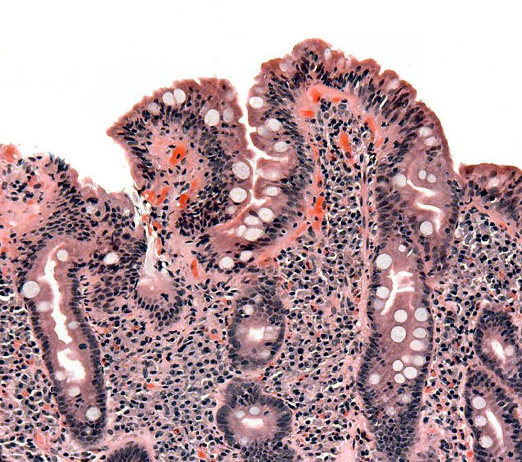
Image: Biopsy of small bowel showing celiac disease manifested by blunting of villi, crypt hypertrophy, and lymphocyte infiltration of crypts (Photo courtesy of Wikimedia Commons)
A spectrophotometric assay has been developed that shows strong potential for use as a point-of-care (POC) assay for the rapid detection of celiac disease antibodies.
Celiac disease (CD) is an autoimmune disorder usually presenting with non-specific gastrointestinal symptoms. It affects approximately 1% of world population, with more than 90% of cases being undiagnosed or misdiagnosed. The diagnosis of CD is currently based on serology and intestinal biopsy, with detection of anti-tissue transglutaminase (tTG) IgA antibodies recommended as the first-line test. Enzyme immunoassays (EIAs) are the established approach for anti-tTG antibody detection, while the existing point-of-care (POC) tests lack sensitivity and/or specificity. Improved POC methods could help reduce the under diagnosis and diagnostic delay of CD.
In this regard, investigators at the University of Helsinki (Finland) formulated an assay for the detection of anti-tTG antibodies by modifying a rapid homogeneous immunoassay based on time-resolved Förster resonance energy transfer (TR-FRET) that they had developed for serodiagnostics of hanta- and Zika virus infections.
TR-FRET is a phenomenon occurring when two fluorophores, donor and acceptor, are in close proximity. Excitation of the donor leads to energy transfer to the acceptor, which then emits the energy at a characteristic wavelength. The TR-FRET efficiency depends inversely on the distance between the two fluorophores. Background autofluorescence is minimized by time-resolved measurement, enabled by chelated lanthanide fluorophores with a long fluorescence half-life. TR-FRET has been employed widely in research and diagnosis to investigate protein-protein interactions and disease markers.
The investigators modified a rapid wash-free TR-FRET-based method for antibody detection by coupling it to the antibody-binding protein “protein L” (LFRET). LFRET employs a donor-labeled antigen, and an acceptor-labeled protein L, which binds the kappa light chains of all immunoglobulin classes. If the clinical sample contains antibodies against the antigen, they will bring the fluorophores to close proximity. Thus, the TR-FRET signal shows that the sample contains the antibodies of interest. The LFRET signal can be measured without additional steps shortly after combining the sample with the reagent mix, allowing for rapid point-of-care diagnosis.
For the anti-tTG antibodies study, the investigators evaluated 74 patients with biopsy-confirmed CD and 70 healthy controls, with 1) the new tTG-LFRET assay, and for reference 2) a well-established EIA, and 3) an existing commercial POC test. IgG depletion was employed to differentiate between anti-tTG IgA and IgG positivity.
Results revealed that the sensitivity and specificity of the first-generation tTG-LFRET POC assay in detection of CD were 87.8% and 94.3%, respectively, in line with those of the reference POC test. For comparison, the sensitivity and specificity of EIA were 95.9% and 91.9%, respectively.
"The performance of the test was comparable to that of current methods. The prevailing method involves transporting the sample to a central laboratory and a multi-step procedure taking hours. With the new method, results can be achieved in less than half an hour by simply combining the sample and a reagent mix, waiting for a while and reading the result," said first author Dr. Juuso Rusanen, a researcher at the University of Helsinki. "We hope our rapid method could lower the threshold for screening of celiac disease and thus help overcome the vast under diagnosis of this relatively common condition. Additionally, this is the first time the new method has been used for diagnostics of autoimmune disease. This is a promising result, and prompts the development of similar tests for diagnostics of other autoimmune disorders."
The study was published in the November 26, 2019, online edition of the journal PLOS One.
Related Links:
University of Helsinki
Latest Molecular Diagnostics News
- Diagnostic Device Predicts Treatment Response for Brain Tumors Via Blood Test
- Blood Test Detects Early-Stage Cancers by Measuring Epigenetic Instability
- Two-in-One DNA Analysis Improves Diagnostic Accuracy While Saving Time and Costs
- “Lab-On-A-Disc” Device Paves Way for More Automated Liquid Biopsies
- New Tool Maps Chromosome Shifts in Cancer Cells to Predict Tumor Evolution
- Blood Test Identifies Inflammatory Breast Cancer Patients at Increased Risk of Brain Metastasis
- Newly-Identified Parkinson’s Biomarkers to Enable Early Diagnosis Via Blood Tests
- New Blood Test Could Detect Pancreatic Cancer at More Treatable Stage
- Liquid Biopsy Could Replace Surgical Biopsy for Diagnosing Primary Central Nervous Lymphoma
- New Tool Reveals Hidden Metabolic Weakness in Blood Cancers
- World's First Blood Test Distinguishes Between Benign and Cancerous Lung Nodules
- Rapid Test Uses Mobile Phone to Identify Severe Imported Malaria Within Minutes
- Gut Microbiome Signatures Predict Long-Term Outcomes in Acute Pancreatitis
- Blood Test Promises Faster Answers for Deadly Fungal Infections
- Blood Test Could Detect Infection Exposure History
- Urine-Based MRD Test Tracks Response to Bladder Cancer Surgery
Channels
Clinical Chemistry
view channel
New PSA-Based Prognostic Model Improves Prostate Cancer Risk Assessment
Prostate cancer is the second-leading cause of cancer death among American men, and about one in eight will be diagnosed in their lifetime. Screening relies on blood levels of prostate-specific antigen... Read more
Extracellular Vesicles Linked to Heart Failure Risk in CKD Patients
Chronic kidney disease (CKD) affects more than 1 in 7 Americans and is strongly associated with cardiovascular complications, which account for more than half of deaths among people with CKD.... Read moreHematology
view channel
New Guidelines Aim to Improve AL Amyloidosis Diagnosis
Light chain (AL) amyloidosis is a rare, life-threatening bone marrow disorder in which abnormal amyloid proteins accumulate in organs. Approximately 3,260 people in the United States are diagnosed... Read more
Fast and Easy Test Could Revolutionize Blood Transfusions
Blood transfusions are a cornerstone of modern medicine, yet red blood cells can deteriorate quietly while sitting in cold storage for weeks. Although blood units have a fixed expiration date, cells from... Read more
Automated Hemostasis System Helps Labs of All Sizes Optimize Workflow
High-volume hemostasis sections must sustain rapid turnaround while managing reruns and reflex testing. Manual tube handling and preanalytical checks can strain staff time and increase opportunities for error.... Read more
High-Sensitivity Blood Test Improves Assessment of Clotting Risk in Heart Disease Patients
Blood clotting is essential for preventing bleeding, but even small imbalances can lead to serious conditions such as thrombosis or dangerous hemorrhage. In cardiovascular disease, clinicians often struggle... Read moreImmunology
view channelBlood Test Identifies Lung Cancer Patients Who Can Benefit from Immunotherapy Drug
Small cell lung cancer (SCLC) is an aggressive disease with limited treatment options, and even newly approved immunotherapies do not benefit all patients. While immunotherapy can extend survival for some,... Read more
Whole-Genome Sequencing Approach Identifies Cancer Patients Benefitting From PARP-Inhibitor Treatment
Targeted cancer therapies such as PARP inhibitors can be highly effective, but only for patients whose tumors carry specific DNA repair defects. Identifying these patients accurately remains challenging,... Read more
Ultrasensitive Liquid Biopsy Demonstrates Efficacy in Predicting Immunotherapy Response
Immunotherapy has transformed cancer treatment, but only a small proportion of patients experience lasting benefit, with response rates often remaining between 10% and 20%. Clinicians currently lack reliable... Read moreMicrobiology
view channel
Comprehensive Review Identifies Gut Microbiome Signatures Associated With Alzheimer’s Disease
Alzheimer’s disease affects approximately 6.7 million people in the United States and nearly 50 million worldwide, yet early cognitive decline remains difficult to characterize. Increasing evidence suggests... Read moreAI-Powered Platform Enables Rapid Detection of Drug-Resistant C. Auris Pathogens
Infections caused by the pathogenic yeast Candida auris pose a significant threat to hospitalized patients, particularly those with weakened immune systems or those who have invasive medical devices.... Read morePathology
view channel
Engineered Yeast Cells Enable Rapid Testing of Cancer Immunotherapy
Developing new cancer immunotherapies is a slow, costly, and high-risk process, particularly for CAR T cell treatments that must precisely recognize cancer-specific antigens. Small differences in tumor... Read more
First-Of-Its-Kind Test Identifies Autism Risk at Birth
Autism spectrum disorder is treatable, and extensive research shows that early intervention can significantly improve cognitive, social, and behavioral outcomes. Yet in the United States, the average age... Read moreTechnology
view channel
Robotic Technology Unveiled for Automated Diagnostic Blood Draws
Routine diagnostic blood collection is a high‑volume task that can strain staffing and introduce human‑dependent variability, with downstream implications for sample quality and patient experience.... Read more
ADLM Launches First-of-Its-Kind Data Science Program for Laboratory Medicine Professionals
Clinical laboratories generate billions of test results each year, creating a treasure trove of data with the potential to support more personalized testing, improve operational efficiency, and enhance patient care.... Read moreAptamer Biosensor Technology to Transform Virus Detection
Rapid and reliable virus detection is essential for controlling outbreaks, from seasonal influenza to global pandemics such as COVID-19. Conventional diagnostic methods, including cell culture, antigen... Read more
AI Models Could Predict Pre-Eclampsia and Anemia Earlier Using Routine Blood Tests
Pre-eclampsia and anemia are major contributors to maternal and child mortality worldwide, together accounting for more than half a million deaths each year and leaving millions with long-term health complications.... Read moreIndustry
view channelNew Collaboration Brings Automated Mass Spectrometry to Routine Laboratory Testing
Mass spectrometry is a powerful analytical technique that identifies and quantifies molecules based on their mass and electrical charge. Its high selectivity, sensitivity, and accuracy make it indispensable... Read more
AI-Powered Cervical Cancer Test Set for Major Rollout in Latin America
Noul Co., a Korean company specializing in AI-based blood and cancer diagnostics, announced it will supply its intelligence (AI)-based miLab CER cervical cancer diagnostic solution to Mexico under a multi‑year... Read more
Diasorin and Fisher Scientific Enter into US Distribution Agreement for Molecular POC Platform
Diasorin (Saluggia, Italy) has entered into an exclusive distribution agreement with Fisher Scientific, part of Thermo Fisher Scientific (Waltham, MA, USA), for the LIAISON NES molecular point-of-care... Read more


