Flow Cytometry Improved Updated Spectral Analyzer
|
By LabMedica International staff writers Posted on 31 Oct 2019 |
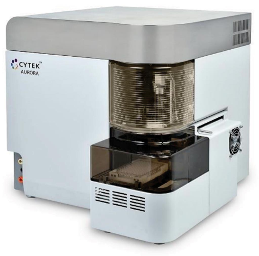
Image: The Aurora advanced flow cytometry system is now available with five lasers to enable seeing more than 30 colors from a single sample (Photo courtesy of Cytek Biosciences).
Flow cytometry aims to count the number, size, granularity, and other properties of cells in a heterogeneous population. Standard flow cytometry lasers excite certain fluorescent markers (fluorochromes, antibodies, or stains) on a cell as it passes through the beam.
Detectors in the instrument record and quantify the relative amount of light emitted by fluorescent markers in the cell, which the tool presents to scientists through a histogram. However, flow cytometer users can run into a myriad of technical issues, such as dealing with limited sample volumes and lacking enough lasers to excite a target amount of fluorochromes. Each laser also can only excite a certain number of fluorochromes on a cell before inducing spectral overlap.
An updated model of its Aurora flow cytometry system has been released by Cytek Biosciences (Fremont, CA, USA), which offers scientists the ability to multiplex 40 fluorescent biomarkers on a cell in a blood sample for scientific and clinical purposes. The updated Aurora platform uses five optical lasers (ultraviolet, violet, blue, yellow-green, and red) to excite 40 fluorochromes on cellular antibodies, which are then recorded by 64 detectors.
With standard flow cytometry panels, a patient's blood sample must be separated into multiple tubes to identify biomarkers linked to different types of leukemia; however the Aurora platform only needs a single tube of blood to identify the fluorescent antibodies. While scientists still need to run controls prior to running a multicolor tube to measure the different emission spectra recorded by Aurora, they can save the controls in the software and reuse them with the same panel in future tests.
Wenbin Jiang, PhD, CEO of Cytek Biosciences, said, “After chemotherapy, no one really has that many bone marrow samples available for testing and splitting into several different tubes. But because you don't need to split blood samples into several tubes with Aurora, you can have more cells per tube, which leads to more specific results." Dr. Jiang also argued that the updated Aurora system can analyze up to 30,000 to 40,000 cells per second while maintaining a "competitive sensitivity." Flow cytometry platforms on the market currently offer anywhere from 20,000 to 100,000 cells per second, but not always at the sample level of multiplexing.
Steven A. Porcelli, MD, scientific director at the Albert Einstein College of Medicine Flow Cytometry Core (Bronx, NY, USA), said, “Cytek's tool collected light coming out over a wide range of wavelengths for each cell and for each laser that we've used to excite the cell. Instead of giving you a high or low value for a tracer, it allows you to distinguish many different tracers from each other because you create a kind of fingerprint of the wavelengths being emitted.”
Related Links:
Cytek Biosciences
Albert Einstein College of Medicine Flow Cytometry Core
Latest Technology News
- AI-Powered Biomarker Predicts Liver Cancer Risk
- Robotic Technology Unveiled for Automated Diagnostic Blood Draws
- ADLM Launches First-of-Its-Kind Data Science Program for Laboratory Medicine Professionals
- Aptamer Biosensor Technology to Transform Virus Detection
- AI Models Could Predict Pre-Eclampsia and Anemia Earlier Using Routine Blood Tests
- AI-Generated Sensors Open New Paths for Early Cancer Detection
- Pioneering Blood Test Detects Lung Cancer Using Infrared Imaging
- AI Predicts Colorectal Cancer Survival Using Clinical and Molecular Features
Channels
Clinical Chemistry
view channel
Rapid Blood Testing Method Aids Safer Decision-Making in Drug-Related Emergencies
Acute recreational drug toxicity is a frequent reason for emergency department visits, yet clinicians rarely have access to confirmatory toxicology results in real time. Instead, treatment decisions are... Read more
New PSA-Based Prognostic Model Improves Prostate Cancer Risk Assessment
Prostate cancer is the second-leading cause of cancer death among American men, and about one in eight will be diagnosed in their lifetime. Screening relies on blood levels of prostate-specific antigen... Read moreMolecular Diagnostics
view channel
AI-Powered Liquid Biopsy Classifies Pediatric Brain Tumors with High Accuracy
Liquid biopsies offer a noninvasive way to study cancer by analyzing circulating tumor DNA in body fluids. However, in pediatric brain tumors, the small amount of ctDNA in cerebrospinal fluid has limited... Read more
New CSF Liquid Biopsy Assay Reveals Genomic Insights for CNS Tumors
Central nervous system (CNS) malignancies pose distinctive diagnostic challenges because tissue-based testing is often infeasible and the blood–brain barrier limits the usefulness of plasma liquid biopsy.... Read more
Group A Strep Molecular Test Delivers Definitive Results at POC in 15 Minutes
Strep throat is a bacterial infection caused by Group A Streptococcus (GAS). It is a leading bacterial cause of acute pharyngitis, particularly in children and adolescents, and one of the most common reasons... Read more
Rapid Molecular Test Identifies Sepsis Patients Most Likely to Have Positive Blood Cultures
Sepsis is caused by a patient’s overwhelming immune response to an infection. If undetected or left untreated, sepsis leads to tissue damage, organ failure, permanent disability, and often death.... Read moreHematology
view channel
Rapid Cartridge-Based Test Aims to Expand Access to Hemoglobin Disorder Diagnosis
Sickle cell disease and beta thalassemia are hemoglobin disorders that often require referral to specialized laboratories for definitive diagnosis, delaying results for patients and clinicians.... Read more
New Guidelines Aim to Improve AL Amyloidosis Diagnosis
Light chain (AL) amyloidosis is a rare, life-threatening bone marrow disorder in which abnormal amyloid proteins accumulate in organs. Approximately 3,260 people in the United States are diagnosed... Read moreImmunology
view channel
New Biomarker Predicts Chemotherapy Response in Triple-Negative Breast Cancer
Triple-negative breast cancer is an aggressive form of breast cancer in which patients often show widely varying responses to chemotherapy. Predicting who will benefit from treatment remains challenging,... Read moreBlood Test Identifies Lung Cancer Patients Who Can Benefit from Immunotherapy Drug
Small cell lung cancer (SCLC) is an aggressive disease with limited treatment options, and even newly approved immunotherapies do not benefit all patients. While immunotherapy can extend survival for some,... Read more
Whole-Genome Sequencing Approach Identifies Cancer Patients Benefitting From PARP-Inhibitor Treatment
Targeted cancer therapies such as PARP inhibitors can be highly effective, but only for patients whose tumors carry specific DNA repair defects. Identifying these patients accurately remains challenging,... Read more
Ultrasensitive Liquid Biopsy Demonstrates Efficacy in Predicting Immunotherapy Response
Immunotherapy has transformed cancer treatment, but only a small proportion of patients experience lasting benefit, with response rates often remaining between 10% and 20%. Clinicians currently lack reliable... Read moreMicrobiology
view channel
Rapid Test Promises Faster Answers for Drug-Resistant Infections
Drug-resistant pathogens continue to pose a growing threat in healthcare facilities, where delayed detection can impede outbreak control and increase mortality. Candida auris is notoriously difficult to... Read more
CRISPR-Based Technology Neutralizes Antibiotic-Resistant Bacteria
Antibiotic resistance has accelerated into a global health crisis, with projections estimating more than 10 million deaths per year by 2050 as drug-resistant “superbugs” continue to spread.... Read more
Comprehensive Review Identifies Gut Microbiome Signatures Associated With Alzheimer’s Disease
Alzheimer’s disease affects approximately 6.7 million people in the United States and nearly 50 million worldwide, yet early cognitive decline remains difficult to characterize. Increasing evidence suggests... Read morePathology
view channel
AI Tool Uses Blood Biomarkers to Predict Transplant Complications Before Symptoms Appear
Stem cell and bone marrow transplants can be lifesaving, but serious complications may arise months after patients leave the hospital. One of the most dangerous is chronic graft-versus-host disease, in... Read more
Research Consortium Harnesses AI and Spatial Biology to Advance Cancer Discovery
AI has the potential to transform cancer care, yet progress remains constrained by fragmented, inaccessible data that hinder advances in early diagnosis and precision therapy. Unlocking patterns missed... Read moreIndustry
view channel
QuidelOrtho Collaborates with Lifotronic to Expand Global Immunoassay Portfolio
QuidelOrtho (San Diego, CA, USA) has entered a long-term strategic supply agreement with Lifotronic Technology (Shenzhen, China) to expand its global immunoassay portfolio and accelerate customer access... Read more


