



Microfluidic Device Brings Single-Cell Technology to Bedside
|
By LabMedica International staff writers Posted on 06 Mar 2018 |


|
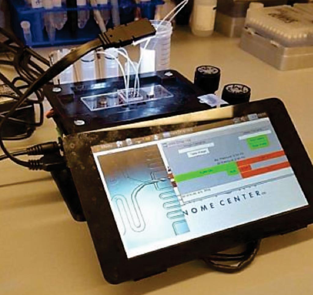
Image: The microfluidic control instrument performing a Drop-seq run (Photo courtesy of the New York Genome Center).
The complex architecture and associated higher-order function of human tissues relies on functionally and molecularly diverse cell populations. Defining the cellular subsets found in pathologic tissues provides insights into disease etiology and treatment options.
Traditional methods such as flow cytometry, which require a priori knowledge of cell type-specific markers, have begun to define this landscape, but fall short in comprehensively identifying cellular states in a tissue, with particular difficulty detecting extremely rare subpopulations.
Scientists at the New York Genome Center (New York, NY, USA) and their colleagues have facilitated broad access to single-cell sequencing by developing a 3D-printed, portable and low-cost microfluidic controller. They adapted this device to perform massively parallel single-cell RNA-seq (Drop-seq), observing metrics and performance that are indistinguishable from a study level Drop-seq setup.
The group used the instrument to profile joint synovial tissue from rheumatoid arthritis (RA) patients. RA is an autoimmune disease that affects 1% of the population and is associated with painful swelling in the joints. The precise cause of RA is undetermined and muddled by the diversity of cells found in the swollen joints of patients. The portability of the controller permitted patient samples to be processed on-site and immediately after surgery, minimizing handling and transport to optimize sample quality. The team collected samples from five RA patients totaling 20,387 cells and looked at the individual gene expression patterns for each cell.
By analyzing the complete dataset and searching for clusters of similar cells, the scientists identified 13 groups, representing both infiltrating immune and inflamed stromal populations. Of particular interest were distinct groups of fibroblasts with strikingly different gene expression patterns. They were able to validate the presence of these multiple groups using BD FACSAria II flow cytometry sorter, and discovered that they exhibited distinct localization patterns with the joint tissue as well. The instrument processes 1 mL of cells at a concentration of 150–200 cells/µL in about 30 minutes, generating over one million droplets at a generation rate of approximately 700 Hz.
Instructions and assembly manuals for the instrument can be found online at the popular microfluidics repository Metafluidics. The 3D-printed custom device, which, along with its electronic and pneumatic components, can be easily obtained and assembled for a total cost of about USD 600, a fraction of the cost of comparable commercial systems. The device occupies a small footprint as well, not much larger than a tissue box.
William Stephenson PhD, a Senior Research Engineer, and lead author of the study, said, “Most commercial microfluidic instruments are very costly; as a result, not every lab has access to exciting technology for single-cell analysis. We designed the instrument to perform droplet microfluidics and in particular Drop-seq, a massively parallel technology for single cell RNA-sequencing.” The study was published on February 23, 2018, in the journal Nature Communications.
Related Links:
New York Genome Center




Traditional methods such as flow cytometry, which require a priori knowledge of cell type-specific markers, have begun to define this landscape, but fall short in comprehensively identifying cellular states in a tissue, with particular difficulty detecting extremely rare subpopulations.
Scientists at the New York Genome Center (New York, NY, USA) and their colleagues have facilitated broad access to single-cell sequencing by developing a 3D-printed, portable and low-cost microfluidic controller. They adapted this device to perform massively parallel single-cell RNA-seq (Drop-seq), observing metrics and performance that are indistinguishable from a study level Drop-seq setup.
The group used the instrument to profile joint synovial tissue from rheumatoid arthritis (RA) patients. RA is an autoimmune disease that affects 1% of the population and is associated with painful swelling in the joints. The precise cause of RA is undetermined and muddled by the diversity of cells found in the swollen joints of patients. The portability of the controller permitted patient samples to be processed on-site and immediately after surgery, minimizing handling and transport to optimize sample quality. The team collected samples from five RA patients totaling 20,387 cells and looked at the individual gene expression patterns for each cell.
By analyzing the complete dataset and searching for clusters of similar cells, the scientists identified 13 groups, representing both infiltrating immune and inflamed stromal populations. Of particular interest were distinct groups of fibroblasts with strikingly different gene expression patterns. They were able to validate the presence of these multiple groups using BD FACSAria II flow cytometry sorter, and discovered that they exhibited distinct localization patterns with the joint tissue as well. The instrument processes 1 mL of cells at a concentration of 150–200 cells/µL in about 30 minutes, generating over one million droplets at a generation rate of approximately 700 Hz.
Instructions and assembly manuals for the instrument can be found online at the popular microfluidics repository Metafluidics. The 3D-printed custom device, which, along with its electronic and pneumatic components, can be easily obtained and assembled for a total cost of about USD 600, a fraction of the cost of comparable commercial systems. The device occupies a small footprint as well, not much larger than a tissue box.
William Stephenson PhD, a Senior Research Engineer, and lead author of the study, said, “Most commercial microfluidic instruments are very costly; as a result, not every lab has access to exciting technology for single-cell analysis. We designed the instrument to perform droplet microfluidics and in particular Drop-seq, a massively parallel technology for single cell RNA-sequencing.” The study was published on February 23, 2018, in the journal Nature Communications.
Related Links:
New York Genome Center

Platinum Member
Automated Coagulation Analyzer
Hemolumi H6
New
Slide Scanner System
NanoZoomer S540MD
Gold Member
Quantitative POC Immunoassay Analyzer
EASY READER+
Latest Technology News
- New AI-Powered System Aims to Streamline Laboratory and Manufacturing Workflows
- Laser-Based Swab Analysis Shows Promise for Detecting Disease-Linked Odor Patterns
- Laser-Enhanced Assay Boosts Sensitivity for Colorectal Cancer Biomarker Detection
- New Automation Workflow Streamlines NGS Library Preparation for High-Volume Labs
- ADLM Calls for CLIA Updates to Support Safe AI Use in Laboratory Medicine
- Multimodal AI Framework Aims to Guide Cancer Immunotherapy Decisions
- New Multipurpose Centrifuge Combines High Capacity with Sustainable Cooling
- Bacterial Vesicle Expression System Streamlines Production of Cancer Diagnostic Proteins
- Autonomous Robotic System Receives FDA Authorization for Blood Collection
- Interoperable Data Platform Standardizes Multi-Cancer Blood Test Results
- Training Device Improves Accuracy of Pooled Molecular Diagnostics
- New CE-Certified Software Advances Whole-Genome Cancer Testing
- National Rare Disease Registry Standardizes Genetic and Clinical Data for Coordinated Care
- AI Platform Links Biomarker Results to Cancer Clinical Trials and Guidelines
- Agentic AI Platform Supports Genomic Decision-Making in Oncology
- Algorithm Panel Aids Liver Fibrosis Assessment and Liver Cancer Surveillance
Channels
Clinical Chemistry
view channel
Point-of-Care Blood Biomarker Test Detects Brain Injury Missed by CT
Traumatic brain injury remains difficult to assess in emergency settings because some patients continue to have symptoms despite normal computed tomography findings. Approximately 2.8 million people in the U.... Read more
Daily Blood Testing May Help Guide Earlier Antibiotic Stopping in Sepsis
Sepsis is a life-threatening condition caused by the body’s overwhelming response to infection and accounts for thousands of deaths each year. In intensive care, concern about stopping treatment too early... Read more

Hematology
view channel
New Myelofibrosis Guidelines Recommend Expanded Genetic Testing at Diagnosis
Myelofibrosis is a rare, life-threatening blood cancer affecting up to six per 100,000 people. The disease causes scarring in the bone marrow, limiting the production of healthy blood cells and potentially... Read more
Handheld Point-of-Care Test Aims to Enable Rapid Coagulation Assessment in Trauma
Severe blood loss and inadequate tissue oxygenation are leading causes of preventable death after traumatic injury. Blood products are essential in trauma care, but cold-chain storage, short shelf life,... Read more

Immunology
view channel
Antibody Profiling Predicts Inflammatory Bowel Disease Years Before Diagnosis
Inflammatory bowel disease (IBD), which includes Crohn’s disease and ulcerative colitis, involves abnormal immune responses that cause persistent inflammation and damage in the digestive system.... Read more
Immune Profiling Initiative Aims to Improve Bone Marrow Analysis in Leukemia
Acute myeloid leukemia (AML) is an aggressive blood cancer in which assessing disease burden after therapy can help guide clinical evaluation. Patients with measurable residual disease (MRD) after treatment... Read more
Immune Profiling Identifies Early Maternal Autoantibodies Linked to Preterm Birth Risk
A typical pregnancy lasts about 40 weeks, but labor begins prematurely in roughly one in 10 pregnancies. Preterm birth is the leading cause of newborn death worldwide, yet its underlying causes and which... Read more
Longitudinal Blood Testing Reveals Changes in Allergen Sensitization During Childhood
Food allergy is a major childhood health concern, and distinguishing persistent risk from transient sensitization can guide clinical decisions. Specific IgE antibodies may be detectable even when a child... Read moreMicrobiology
view channel
Molecular Panel Gains WHO Emergency Use Listing to Support Ebola Outbreak Response
The ongoing Ebola Bundibugyo outbreak in the Democratic Republic of the Congo and neighboring countries has become one of the largest outbreaks of this Ebola virus species recorded to date.... Read more
Rapid Host-Response Test Aims to Predict Corticosteroid Benefit in Sepsis
Sepsis and severe infections are difficult to treat because patients with similar clinical presentations may have very different underlying immune responses. Corticosteroids are commonly used in severe... Read more

Pathology
view channel
Digital Pathology Test Stratifies Recurrence Risk in Early Breast Cancer
In hormone receptor-positive, human epidermal growth factor receptor 2 (HER2)-negative early breast cancer, accurate recurrence-risk assessment helps guide treatment decisions. However, individual risks... Read more
AI Pathology Tool Predicts DNA Repair Deficiency in High-Grade Serous Ovarian Cancer
High-grade serous tubo-ovarian carcinoma is an aggressive cancer arising in the ovaries or fallopian tubes. Some tumors exhibit homologous recombination deficiency (HRD), a feature reflecting impaired DNA repair.... Read more

Technology
view channel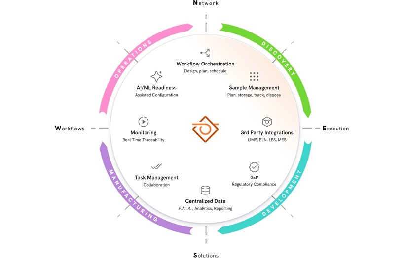
New AI-Powered System Aims to Streamline Laboratory and Manufacturing Workflows
Scientific laboratories supporting drug development, biotechnology, and materials research increasingly rely on coordinated digital workflows to move data, samples, and activities across teams.... Read more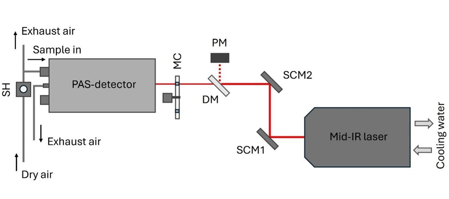
Laser-Based Swab Analysis Shows Promise for Detecting Disease-Linked Odor Patterns
Disease-related changes in volatile organic compounds can alter body odor, producing measurable patterns in exhaled breath and bodily fluids. Current analytical methods can be complex, time-consuming,... Read more
Industry
view channel
Qiagen Agrees to Go Private Through EQT-Led Acquisition
QIAGEN N.V. (Venlo, the Netherlands) and EQT Private Equity (Stockholm, Sweden) have agreed to an all-cash public takeover offer for all outstanding QIAGEN shares at EUR 46.50 per share.... Read more
Hologic and Inpeco Partner to Advance Molecular Laboratory Automation in Europe
Hologic, Inc. (Marlborough, MA, USA) and Inpeco S.A. (Novazzano, Switzerland) have signed a co-marketing partnership in Europe for Inpeco’s recently launched FlexLab X automation solution and Hologic’s... Read more