Flow Cytometry Detects Lymphoproliferative Disorders in Fluid Specimens
|
By LabMedica International staff writers Posted on 26 Aug 2014 |
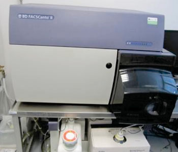
Image: The fluorescence-activated cell sorting FACSCanto II flow cytometer (Photo courtesy of BD Biosystems).
Immunophenotypic analysis of hematopoietic cell populations by flow cytometry has emerged as a useful ancillary study in the diagnostic evaluation of serous effusions and cerebrospinal fluids (CSFs).
The evaluation of lymphoid proliferations in these specimens can be particularly problematic, given the frequent presence of coexisting inflammatory conditions and the manner in which these specimens are processed.
Laboratory scientists at the Hospital of the University of Pennsylvania (Philadelphia, PA, USA) searched for all serous effusions specimens such as pleural fluid, peritoneal fluid, and pericardial fluid, and CSF specimens in which flow cytometry was performed, during the time period June 2008 to July 2012. Six-color flow cytometry was performed according to the manufacturer's protocol on a FACSCanto II flow cytometer (BD Biosystems; San Jose, CA, USA). A panel using different antibodies from Becton Dickinson Immunocytometry Systems, (San Jose, CA, USA) was utilized.
Flow cytometry was performed in 184 of 6,925 total cases (2.7% of all fluids). Flow cytometry was performed in 4.8% of pleural fluids (positive findings in 38%, negative in 40%, and atypical in 18%), 1.1% of peritoneal fluids (positive in 40%, negative in 50%, and atypical in 10%), 1.9% of pericardial fluids (positive in 67%, negative in 33%), and 1.9% of CSFs (positive in 23%, negative in 55%, atypical in 3%).
The authors concluded that atypical flow cytometry findings and atypical morphologic findings in the context of negative flow cytometry results led to the definitive diagnosis of a lymphoproliferative disorder in a significant number of cases when repeat procedures and ancillary studies were performed. Flow cytometry is especially suited for cells in fluid specimens, where aggregates of cells and extracellular matrix elements that may interfere with obtaining single cells for analysis are less prominent than in solid tissue specimens. In addition, it is well known to increase the sensitivity of detecting neoplastic populations of cells over morphology alone. The study was published in the August 2014 issue of the journal Diagnostic Cytopathology.
Related Links:
Hospital of the University of Pennsylvania
BD Biosystems
Becton Dickinson
Latest Immunology News
- New Biomarker Predicts Chemotherapy Response in Triple-Negative Breast Cancer
- Blood Test Identifies Lung Cancer Patients Who Can Benefit from Immunotherapy Drug
- Whole-Genome Sequencing Approach Identifies Cancer Patients Benefitting From PARP-Inhibitor Treatment
- Ultrasensitive Liquid Biopsy Demonstrates Efficacy in Predicting Immunotherapy Response
- Blood Test Could Identify Colon Cancer Patients to Benefit from NSAIDs
- Blood Test Could Detect Adverse Immunotherapy Effects
- Routine Blood Test Can Predict Who Benefits Most from CAR T-Cell Therapy
- New Test Distinguishes Vaccine-Induced False Positives from Active HIV Infection
- Gene Signature Test Predicts Response to Key Breast Cancer Treatment
- Chip Captures Cancer Cells from Blood to Help Select Right Breast Cancer Treatment
- Blood-Based Liquid Biopsy Model Analyzes Immunotherapy Effectiveness
- Signature Genes Predict T-Cell Expansion in Cancer Immunotherapy
- Molecular Microscope Diagnostic System Assesses Lung Transplant Rejection
- Blood Test Tracks Treatment Resistance in High-Grade Serous Ovarian Cancer
- Luminescent Probe Measures Immune Cell Activity in Real Time
- Blood-Based Immune Cell Signatures Could Guide Treatment Decisions for Critically Ill Patients
Channels
Clinical Chemistry
view channel
Electronic Nose Smells Early Signs of Ovarian Cancer in Blood
Ovarian cancer is often diagnosed at a late stage because its symptoms are vague and resemble those of more common conditions. Unlike breast cancer, there is currently no reliable screening method, and... Read more
Simple Blood Test Offers New Path to Alzheimer’s Assessment in Primary Care
Timely evaluation of cognitive symptoms in primary care is often limited by restricted access to specialized diagnostics and invasive confirmatory procedures. Clinicians need accessible tools to determine... Read moreMolecular Diagnostics
view channel
New Blood Test Score Detects Hidden Alcohol-Related Liver Disease
Fatty liver disease affects nearly one in three adults worldwide and can be driven by metabolic conditions such as obesity and diabetes or by excessive alcohol use. In routine care, it is often difficult... Read more
New Blood Test Predicts Who Will Most Likely Live Longer
As people age, it becomes increasingly difficult to determine who is likely to maintain stable health and who may face serious decline. Traditional indicators such as age, cholesterol, and physical activity... Read moreHematology
view channel
Rapid Cartridge-Based Test Aims to Expand Access to Hemoglobin Disorder Diagnosis
Sickle cell disease and beta thalassemia are hemoglobin disorders that often require referral to specialized laboratories for definitive diagnosis, delaying results for patients and clinicians.... Read more
New Guidelines Aim to Improve AL Amyloidosis Diagnosis
Light chain (AL) amyloidosis is a rare, life-threatening bone marrow disorder in which abnormal amyloid proteins accumulate in organs. Approximately 3,260 people in the United States are diagnosed... Read moreMicrobiology
view channel
Hidden Gut Viruses Linked to Colorectal Cancer Risk
Colorectal cancer (CRC) remains a leading cause of cancer mortality in many Western countries, and existing risk-stratification approaches leave substantial room for improvement. Although age, diet, and... Read more
Three-Test Panel Launched for Detection of Liver Fluke Infections
Parasitic liver fluke infections remain endemic in parts of Asia, where transmission commonly occurs through consumption of raw freshwater fish or aquatic plants. Chronic infection is a well-established... Read morePathology
view channel
Urine Specimen Collection System Improves Diagnostic Accuracy and Efficiency
Urine testing is a critical, non-invasive diagnostic tool used to detect conditions such as pregnancy, urinary tract infections, metabolic disorders, cancer, and kidney disease. However, contaminated or... Read more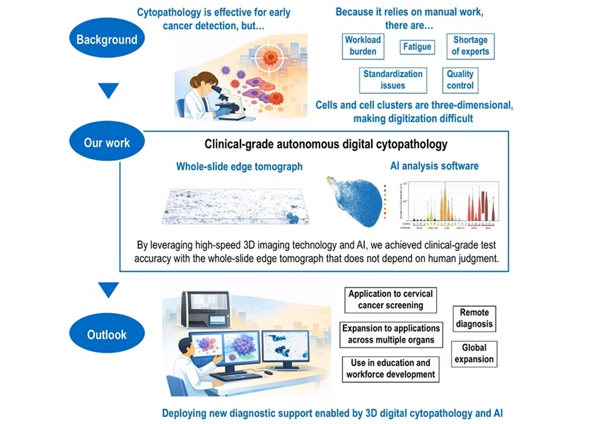
AI-Powered 3D Scanning System Speeds Cancer Screening
Cytology remains a cornerstone of cancer detection, requiring specialists to examine bodily fluids and cells under a microscope. This labor-intensive process involves inspecting up to one million cells... Read moreTechnology
view channel
Blood Test “Clocks” Predict Start of Alzheimer’s Symptoms
More than 7 million Americans live with Alzheimer’s disease, and related health and long-term care costs are projected to reach nearly USD 400 billion in 2025. The disease has no cure, and symptoms often... Read more
AI-Powered Biomarker Predicts Liver Cancer Risk
Liver cancer, or hepatocellular carcinoma, causes more than 800,000 deaths worldwide each year and often goes undetected until late stages. Even after treatment, recurrence rates reach 70% to 80%, contributing... Read more
Robotic Technology Unveiled for Automated Diagnostic Blood Draws
Routine diagnostic blood collection is a high‑volume task that can strain staffing and introduce human‑dependent variability, with downstream implications for sample quality and patient experience.... Read more
ADLM Launches First-of-Its-Kind Data Science Program for Laboratory Medicine Professionals
Clinical laboratories generate billions of test results each year, creating a treasure trove of data with the potential to support more personalized testing, improve operational efficiency, and enhance patient care.... Read moreIndustry
view channel
Cepheid Joins CDC Initiative to Strengthen U.S. Pandemic Testing Preparednesss
Cepheid (Sunnyvale, CA, USA) has been selected by the U.S. Centers for Disease Control and Prevention (CDC) as one of four national collaborators in a federal initiative to speed rapid diagnostic technologies... Read more
QuidelOrtho Collaborates with Lifotronic to Expand Global Immunoassay Portfolio
QuidelOrtho (San Diego, CA, USA) has entered a long-term strategic supply agreement with Lifotronic Technology (Shenzhen, China) to expand its global immunoassay portfolio and accelerate customer access... Read more


 Analyzer.jpg)


