Donor Platelet Activation Varies by Time of Day
|
By LabMedica International staff writers Posted on 13 Jun 2022 |
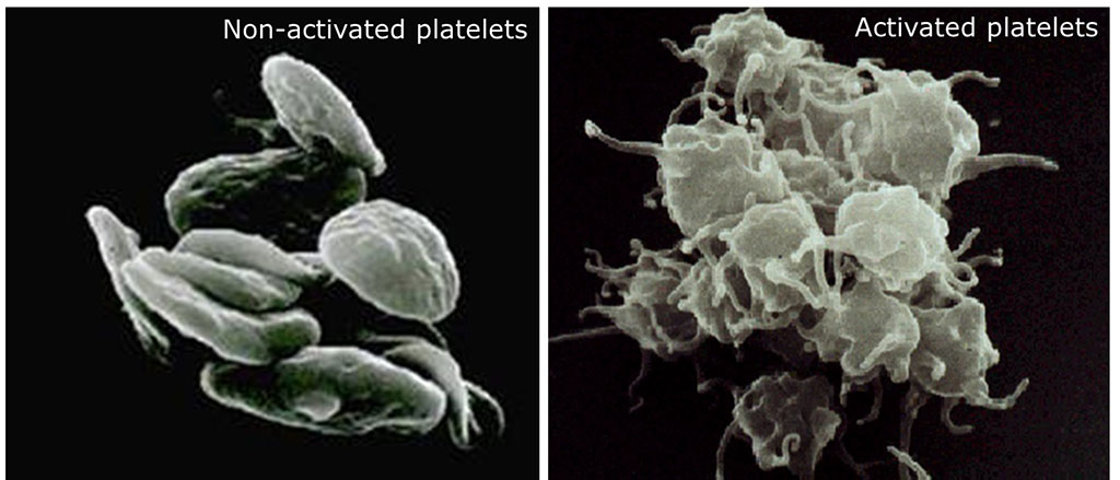
Platelets, are anucleate cells produced by megakaryocytes in the bone marrow that circulate in the bloodstream for about 10 days. Circulating platelets play a central role in the hemostatic response to vascular injury. They are also involved in maintaining vascular integrity and contributed to immune and inflammatory responses.
Platelet donors, like whole blood and plasma donors, are considered to be one of the healthiest groups of the general population. A number of factors may influence the in vivo platelet activation state in healthy individuals. In particular, platelet activation has been shown to vary according to the time of day, with the highest measured levels during the morning hours.
Hematologist at the Akershus University Hospital (Lørenskog, Norway) collected whole blood samples from 86 platelet donors presenting at their Blood Bank between April 2019 and February 2020. The mean age was 50.8 ± 9.7 years with 56% of donors were female. Samples were collected from the attached sampling bags (containing 42 mL whole blood collected immediately before plateletpheresis) using standard vacutainers. A blood sample was collected into a citrated tube for flow cytometric determination of platelet surface P-selectin expression.
For flow cytometry, 100 µL of citrated blood (diluted 1:20 with phosphate-buffered saline [PBS] containing 0.1% bovine serum albumin [BSA], pH 7.4) was incubated at room temperature with FITC-conjugated anti-CD61 (5 µL) and PE-conjugated anti-CD62P (10 µL) for 15 minutes. Background fluorescence was assessed by labeling diluted samples with 10 µL PE-conjugated isotype control antibody. After incubating, the samples were diluted to 4 mL with PBS/BSA and analyzed immediately on a FACS Canto II flow cytometer (BD Bioscience, San Jose, CA, USA). The percentage of activated platelets (i.e., CD61 and CD62P double-positive platelets) was obtained from a total of 10,000 recorded events with the CD61+ gate.
The investigators reported that the percentage of activated (i.e., P-selectin-expressing) platelets in pre-donation blood samples ranged from 0.2% to 7.5% (1.85 ± 1.57%; median, 1.3%; IQR, 0.8–2.3%). Twenty-seven donors (32%) had less than 1.0% activated platelets, and five donors (6%) had more than 5.0% activated platelets, indicating considerable donor variability in the levels of circulating activated platelets.
The percentage of activated platelets was significantly and inversely correlated with the collection time (i.e., the time of day blood samples were collected) and positively correlated to mean platelet volume (MPV). A weaker positive correlation was also observed with ABO blood group. Analysis of the collection time as a categorical variable showed a greater degree of activated platelets in samples collected between 08:00 and 10:00 than in samples collected during the hours after 10:00 to 14:00 (2.5 ± 1.8 versus 1.1 ± 0.74).
The authors concluded that their results showed that platelet activation was significantly and inversely associated with the collection time by univariate analysis and after adjustment for covariates. The study also showed that blood group A was associated with higher platelet activation. This work may have implications for optimizing the timing of platelet donation, if, for example, the temporal variation in platelet activation at the time of donation can impact the final heterogeneity of the stored platelet concentrates and, potentially, transfusion outcomes. The study was published on June 3 2022 in the Journal of Blood Medicine.
Related Links:
Akershus University Hospital
BD Bioscience
Latest Hematology News
- Next Generation Instrument Screens for Hemoglobin Disorders in Newborns
- First 4-in-1 Nucleic Acid Test for Arbovirus Screening to Reduce Risk of Transfusion-Transmitted Infections
- POC Finger-Prick Blood Test Determines Risk of Neutropenic Sepsis in Patients Undergoing Chemotherapy
- First Affordable and Rapid Test for Beta Thalassemia Demonstrates 99% Diagnostic Accuracy
- Handheld White Blood Cell Tracker to Enable Rapid Testing For Infections
- Smart Palm-size Optofluidic Hematology Analyzer Enables POCT of Patients’ Blood Cells
- Automated Hematology Platform Offers High Throughput Analytical Performance
- New Tool Analyzes Blood Platelets Faster, Easily and Accurately
- First Rapid-Result Hematology Analyzer Reports Measures of Infection and Severity at POC
- Bleeding Risk Diagnostic Test to Reduce Preventable Complications in Hospitals
- True POC Hematology Analyzer with Direct Capillary Sampling Enhances Ease-of-Use and Testing Throughput
- Point of Care CBC Analyzer with Direct Capillary Sampling Enhances Ease-of-Use and Testing Throughput
- Blood Test Could Predict Outcomes in Emergency Department and Hospital Admissions
- Novel Technology Diagnoses Immunothrombosis Using Breath Gas Analysis
- Advanced Hematology System Allows Labs to Process Up To 119 Complete Blood Count Results per Hour
- Unique AI-Based Approach Automates Clinical Analysis of Blood Data
Channels
Clinical Chemistry
view channel
3D Printed Point-Of-Care Mass Spectrometer Outperforms State-Of-The-Art Models
Mass spectrometry is a precise technique for identifying the chemical components of a sample and has significant potential for monitoring chronic illness health states, such as measuring hormone levels... Read more.jpg)
POC Biomedical Test Spins Water Droplet Using Sound Waves for Cancer Detection
Exosomes, tiny cellular bioparticles carrying a specific set of proteins, lipids, and genetic materials, play a crucial role in cell communication and hold promise for non-invasive diagnostics.... Read more
Highly Reliable Cell-Based Assay Enables Accurate Diagnosis of Endocrine Diseases
The conventional methods for measuring free cortisol, the body's stress hormone, from blood or saliva are quite demanding and require sample processing. The most common method, therefore, involves collecting... Read moreMolecular Diagnostics
view channelBlood Proteins Could Warn of Cancer Seven Years before Diagnosis
Two studies have identified proteins in the blood that could potentially alert individuals to the presence of cancer more than seven years before the disease is clinically diagnosed. Researchers found... Read moreUltrasound-Aided Blood Testing Detects Cancer Biomarkers from Cells
Ultrasound imaging serves as a noninvasive method to locate and monitor cancerous tumors effectively. However, crucial details about the cancer, such as the specific types of cells and genetic mutations... Read moreImmunology
view channel.jpg)
AI Predicts Tumor-Killing Cells with High Accuracy
Cellular immunotherapy involves extracting immune cells from a patient's tumor, potentially enhancing their cancer-fighting capabilities through engineering, and then expanding and reintroducing them into the body.... Read more
Diagnostic Blood Test for Cellular Rejection after Organ Transplant Could Replace Surgical Biopsies
Transplanted organs constantly face the risk of being rejected by the recipient's immune system which differentiates self from non-self using T cells and B cells. T cells are commonly associated with acute... Read more
AI Tool Precisely Matches Cancer Drugs to Patients Using Information from Each Tumor Cell
Current strategies for matching cancer patients with specific treatments often depend on bulk sequencing of tumor DNA and RNA, which provides an average profile from all cells within a tumor sample.... Read more
Genetic Testing Combined With Personalized Drug Screening On Tumor Samples to Revolutionize Cancer Treatment
Cancer treatment typically adheres to a standard of care—established, statistically validated regimens that are effective for the majority of patients. However, the disease’s inherent variability means... Read moreMicrobiology
view channel
Integrated Solution Ushers New Era of Automated Tuberculosis Testing
Tuberculosis (TB) is responsible for 1.3 million deaths every year, positioning it as one of the top killers globally due to a single infectious agent. In 2022, around 10.6 million people were diagnosed... Read more
Automated Sepsis Test System Enables Rapid Diagnosis for Patients with Severe Bloodstream Infections
Sepsis affects up to 50 million people globally each year, with bacteraemia, formerly known as blood poisoning, being a major cause. In the United States alone, approximately two million individuals are... Read moreEnhanced Rapid Syndromic Molecular Diagnostic Solution Detects Broad Range of Infectious Diseases
GenMark Diagnostics (Carlsbad, CA, USA), a member of the Roche Group (Basel, Switzerland), has rebranded its ePlex® system as the cobas eplex system. This rebranding under the globally renowned cobas name... Read more
Clinical Decision Support Software a Game-Changer in Antimicrobial Resistance Battle
Antimicrobial resistance (AMR) is a serious global public health concern that claims millions of lives every year. It primarily results from the inappropriate and excessive use of antibiotics, which reduces... Read morePathology
view channel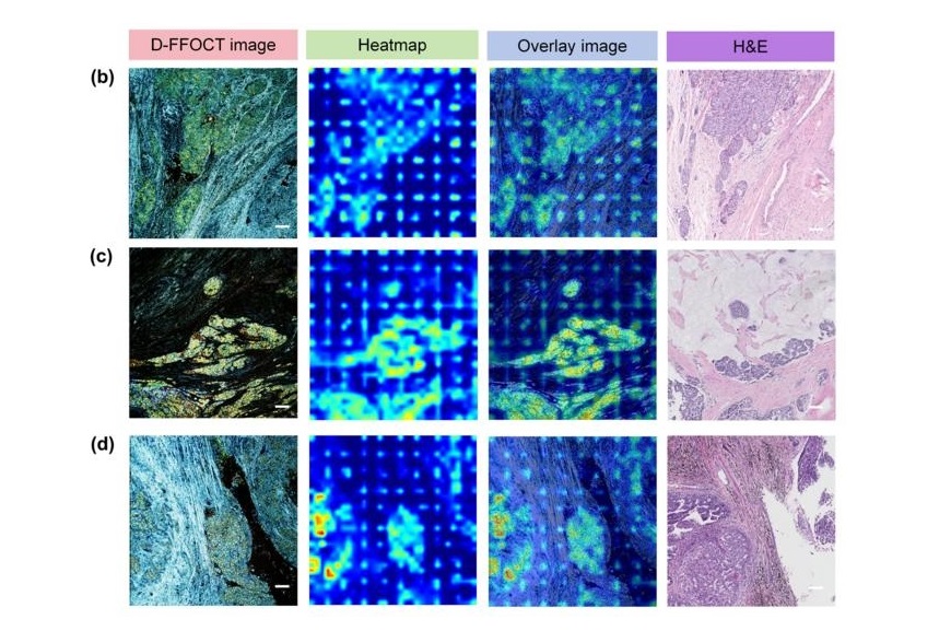
AI Integrated With Optical Imaging Technology Enables Rapid Intraoperative Diagnosis
Rapid and accurate intraoperative diagnosis is essential for tumor surgery as it guides surgical decisions with precision. Traditional intraoperative assessments, such as frozen sections based on H&E... Read more
HPV Self-Collection Solution Improves Access to Cervical Cancer Testing
Annually, over 604,000 women across the world are diagnosed with cervical cancer, and about 342,000 die from this disease, which is preventable and primarily caused by the Human Papillomavirus (HPV).... Read moreHyperspectral Dark-Field Microscopy Enables Rapid and Accurate Identification of Cancerous Tissues
Breast cancer remains a major cause of cancer-related mortality among women. Breast-conserving surgery (BCS), also known as lumpectomy, is the removal of the cancerous lump and a small margin of surrounding tissue.... Read moreTechnology
view channel
New Diagnostic System Achieves PCR Testing Accuracy
While PCR tests are the gold standard of accuracy for virology testing, they come with limitations such as complexity, the need for skilled lab operators, and longer result times. They also require complex... Read more
DNA Biosensor Enables Early Diagnosis of Cervical Cancer
Molybdenum disulfide (MoS2), recognized for its potential to form two-dimensional nanosheets like graphene, is a material that's increasingly catching the eye of the scientific community.... Read more
Self-Heating Microfluidic Devices Can Detect Diseases in Tiny Blood or Fluid Samples
Microfluidics, which are miniature devices that control the flow of liquids and facilitate chemical reactions, play a key role in disease detection from small samples of blood or other fluids.... Read more
Breakthrough in Diagnostic Technology Could Make On-The-Spot Testing Widely Accessible
Home testing gained significant importance during the COVID-19 pandemic, yet the availability of rapid tests is limited, and most of them can only drive one liquid across the strip, leading to continued... Read moreIndustry
view channel
Danaher and Johns Hopkins University Collaborate to Improve Neurological Diagnosis
Unlike severe traumatic brain injury (TBI), mild TBI often does not show clear correlations with abnormalities detected through head computed tomography (CT) scans. Consequently, there is a pressing need... Read more
Beckman Coulter and MeMed Expand Host Immune Response Diagnostics Partnership
Beckman Coulter Diagnostics (Brea, CA, USA) and MeMed BV (Haifa, Israel) have expanded their host immune response diagnostics partnership. Beckman Coulter is now an authorized distributor of the MeMed... Read more_1.jpg)


_1.jpg)
.jpg)
