Lack of Autoantibodies Against Related Proteins in Collagenous Colitis
|
By LabMedica International staff writers Posted on 21 Jun 2022 |


|

Collagenous colitis (CC) is an inflammatory disorder in the colonic mucosa that predominantly affects elderly women and causes chronic, non-bloody diarrhea with normal or close to normal endoscopic findings.
The histological criteria for CC are a thickened sub-epithelial collagen layer (> 10 µm) in the extracellular matrix (ECM) of the mucosa, epithelial damage and presence of an inflammatory infiltrate in the lamina propria. CC shares many of the characteristics found in autoimmune diseases, but no autoantibodies have been identified. In CC, an imbalance in collagen turnover is evident.
Gastroenterologists at the Skåne University Hospital (Malmö, Sweden) included in a study, 66 women with CC. The mean age at inclusion was 60 years (range 31–74 years) and mean age at diagnosis was 55 years (range 28–69 years). The size of the control groups differed, but they were selected from the same cohort consisting of 100 healthy female blood donors. The mean age of the total control group was 41.7 years (range 19–69).
In-house enzyme-linked immunosorbent assays (ELISAs) were set up for analysis of anti-IgM and IgG antibodies against collagen type III (Col III and IV). Analyses of antibodies against matrix metalloproteinase-9 (MMP-9 and Tenascin-C (TNC) were conducted by in-house ELISAs and another in-house ELISA was set up for analysis of IgM and IgG antibodies against tissue inhibitors of metalloproteinase (TIMP-1). Anti-Saccharomyces cerevisiae antibodies (ASCA) IgG were analyzed by a fluorescent enzyme immunoassay method (FEIA) for which the reference value is set > 10 U/mL (Orgentec Diagnostika, Mainz, Germany). Thyroid peroxidase (TPO) antibodies were analyzed by a chemiluminescence enzyme immunological method (Siemens Healthcare GmbH, Erlangen, Germany).
The investigators reported that there was no difference in prevalence of these collage-associated autoantibodies between CC patients and controls. Sixteen patients expressed one type of autoantibody, whereas two patients expressed two different types. The mean disease duration was significantly lower in the subjects who expressed collagen-associated autoantibodies (3.7 years; range 1–14), compared to those who did not (6.4 years; range 1–22). The presence of autoantibodies against TPO and ASCA was slightly increased compared to controls, but not statistically significant. Increased levels of total IgE were equally present in the CC patients and the control group, and there was no correlation between total IgE-positivity and presence of any autoantibodies.
The authors concluded that no increased presence of the investigated antibodies could be found in the present study of CC. Neither could antibodies against ASCA or TPO, or elevated levels of IgE, be found. Consequently, no association was found between CC and these proteins, even though this may not be generalizable to other compounds in the collagen layer. The study was published on June 6, 2022 in the journal BMC Immunology.
Related Links:
Skåne University Hospital
Orgentec Diagnostika
Siemens Healthcare GmbH






Latest Immunology News
- FDA Approval Adds Automated Testing Option for Parvovirus B19 Serology
- Blood-Based Immune Aging Map Could Help Spot Disease Risk Earlier
- Blood-Based Immune Signal Could Reveal Lung Cancer Risk Before Tumors Develop
- Prototype Point-of-Care Test Aims to Improve Neonatal Sepsis Diagnosis
- Single-Tier Lyme Disease Test Shows High Sensitivity in Early Infection
- New Web-Based Platform Helps Autoimmune Laboratories Manage Testing Complexity
- Routine Newborn Blood Spots Can Measure GBS Antibody Levels
- New Autoantibody Test Targets Pre-Symptomatic Type 1 Diabetes Screening
- Blood Test Differentiates Bacterial and Viral Infections in Febrile Infants
- HIV Monitoring Strategy Uses Drug Levels to Guide Resistance Testing
- Werfen Expands Automated APS Testing with FDA-Cleared and CE-Marked IgA Reagent
- Blood Biomarker May Predict Colitis Risk in Checkpoint Inhibitor Therapy
- Blood EBV Activity Biomarkers May Predict Multiple Sclerosis Relapse Months Ahead
- New Cellular Map May Help Predict Crohn’s Disease Course in Children
- Temporal Immune Profiling Reveals How Sepsis States Change Over Time
- Study Identifies Immune Cells That Drive Harmful Autoantibody Responses in COVID-19
Channels
Clinical Chemistry
view channel
Multimodal Clinical Data Models Predict Cachexia Risk Across Cancers
Cancer cachexia is a severe wasting syndrome that affects roughly half of patients with cancer and cannot be reversed simply by increasing food intake. It can reduce treatment tolerance, impair quality... Read more
Study Evaluates Alzheimer’s Blood Biomarker Across Racial and Ethnic Groups
Alzheimer’s disease trials increasingly depend on identifying cognitively unimpaired people who already have elevated brain amyloid. Blood biomarkers may help reduce reliance on positron emission tomography... Read more
Molecular Diagnostics
view channel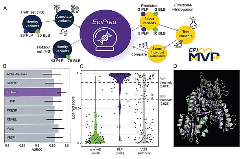
AI Tool Classifies Pathogenic Variants in Severe Childhood Epilepsy
Developmental and epileptic encephalopathies are rare neurodevelopmental disorders characterized by early-life seizures and significant developmental delays. Genetic testing can identify variants associated... Read more
Noninvasive Cervical Test Detects Endometrial Cancer for Triage
Endometrial cancer is rising in incidence and mortality in the United States. The disease has no population screening program or FDA-cleared noninvasive test for early identification, and evaluation can... Read more

Hematology
view channel
New Myelofibrosis Guidelines Recommend Expanded Genetic Testing at Diagnosis
Myelofibrosis is a rare, life-threatening blood cancer affecting up to six per 100,000 people. The disease causes scarring in the bone marrow, limiting the production of healthy blood cells and potentially... Read more
Handheld Point-of-Care Test Aims to Enable Rapid Coagulation Assessment in Trauma
Severe blood loss and inadequate tissue oxygenation are leading causes of preventable death after traumatic injury. Blood products are essential in trauma care, but cold-chain storage, short shelf life,... Read more

Microbiology
view channel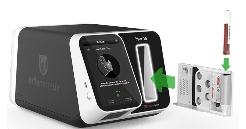
Rapid Host-Response Test Aims to Predict Corticosteroid Benefit in Sepsis
Sepsis and severe infections are difficult to treat because patients with similar clinical presentations may have very different underlying immune responses. Corticosteroids are commonly used in severe... Read more
Oral and Lung Microbiome Changes Linked to Severe Long Covid Symptoms
Long Covid can involve persistent respiratory, cognitive, and gastrointestinal symptoms after acute infection, creating a difficult diagnostic and management challenge. Because COVID-19 initially affects... Read more

Pathology
view channel
Molecular Testing Refines Barrett’s Esophagus Progression Risk Beyond Biopsy Findings
Barrett’s esophagus is a precancerous condition associated with increased risk of esophageal adenocarcinoma. Routine endoscopic biopsy findings guide surveillance and therapy, but histopathology may not... Read more
Updated Digital Pathology Platform Expands AI Integration for Cancer Diagnosis
Digital pathology is reshaping how laboratories manage case review, image analysis, and collaboration. As diagnostic workflows become more complex, pathology teams need platforms that can support digital... Read more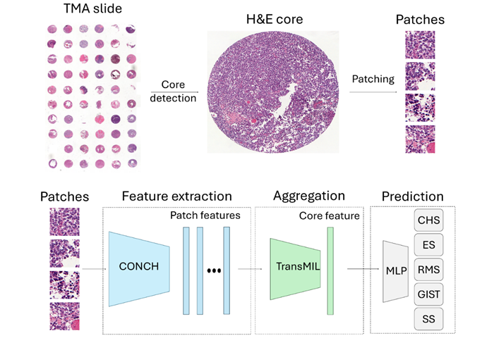
Technology
view channel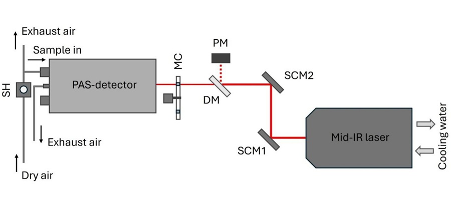
Laser-Based Swab Analysis Shows Promise for Detecting Disease-Linked Odor Patterns
Disease-related changes in volatile organic compounds can alter body odor, producing measurable patterns in exhaled breath and bodily fluids. Current analytical methods can be complex, time-consuming,... Read more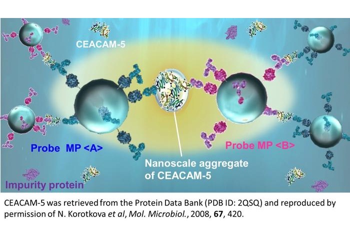
Laser-Enhanced Assay Boosts Sensitivity for Colorectal Cancer Biomarker Detection
Colorectal cancer is the third most commonly diagnosed cancer and the second leading cause of cancer-related death worldwide. Early detection remains critical, but cancer biomarkers can produce only faint... Read more

Industry
view channel
Global TB Summit Connects Diagnostic Testing With Prevention and Clinical Care
Tuberculosis remains a major global infectious disease challenge despite declining deaths. In 2024, an estimated 10.7 million people worldwide developed the disease and nearly 1.2 million died from it.... Read more