Immunohistochemistry Evaluated for Tissue-Invasive Cytomegalovirus in Gastrointestinal Mucosal Biopsies
|
By LabMedica International staff writers Posted on 04 Mar 2022 |


|
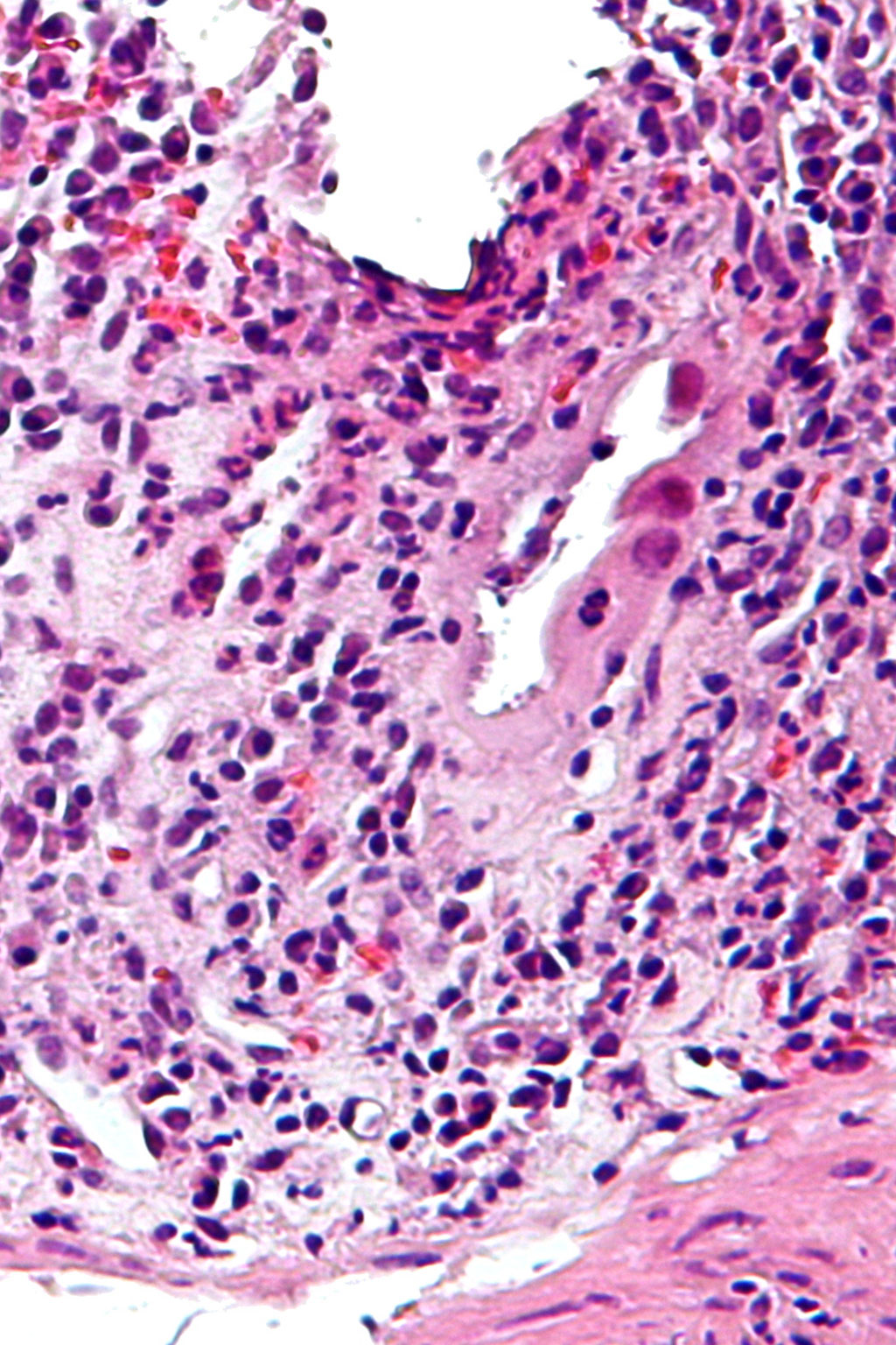
Cytomegalovirus (CMV) immunohistochemistry (IHC) is the most widely used method to diagnose CMV infection/reactivation in tissues in a pathology laboratory.
Cytomegalovirus (CMV), an encapsulated double-stranded DNA β-herpesvirus, often causes asymptomatic or mild mononucleosis-like primary infection followed by latent infection or chronic infection. About 40% to 100% of the world's adult population is seropositive for CMV.
Gastrointestinal (GI) CMV disease can manifest during primary infection or reactivation of latent virus. Reactivation of CMV is frequently seen in adults, especially in patients with immunocompromised conditions such as human immunodeficiency virus (HIV) infection, recipients of solid organ and stem cell transplant, and those with disease treated by immunosuppressive medications.
A team of pathologists at the New York University Langone Health (New York, NY, USA) included in a study total of 1,479 mucosal biopsies from the GI system. Their aim was to improve the efficiency of CMV immunohistochemistry (IHC) testing by evaluating immunopositive staining trends of tissue-invasive CMV in the gastrointestinal system. Immunohistochemistry was performed with specific antibodies against CMV (DAKO, Santa Clara, CA, USA).
The investigators reported that the overall positivity rate of CMV IHC was 4.73% (70/1,479). The positivity rate from physician-requested and pathologist-initiated tests was significantly different (7.54% versus 3.83%). Cases with severe inflammation showed a higher positive CMV rate than those with mild inflammation (5.37% versus 2.6%). Cytomegalovirus positivity in biopsies from post-transplant patients, inflammatory bowel disease, human immunodeficiency virus (HIV)/common variable immunodeficiency (CVID), cancer, and others was 19.69%, 3.84%, 23.33%, 9.0%, and 2.84%, respectively.
The positivity rate among post-transplant, HIV/CVID, or cancer patients was significantly higher than in other populations. Cases tested with multiple tissue blocks generated a higher positivity rate than those with a single block (7.77% versus 3.23%). Testing three to four blocks per case almost tripled the positive CMV detection rate (9.04%). Interestingly, using five or more blocks did not further ameliorate the positive CMV detection rate.
The authors concluded that physician request, immunosuppression, multiple blocks, and severe inflammation were strongly related to positive CMV IHC detection rate. These findings might provide value in helping pathologists manage CMV IHC testing more efficiently. The study published in the March 2022 issue of the journal Archives of Pathology and Laboratory Medicine.
Related Links:
New York University Langone Health
DAKO







Latest Pathology News
- Digital Pathology Test Stratifies Recurrence Risk in Early Breast Cancer
- AI Pathology Tool Predicts DNA Repair Deficiency in High-Grade Serous Ovarian Cancer
- AI Tissue Analysis May Help Identify Missed Precancerous Colon Polyps
- AI Pre-Screening Helps Identify Lung Cancer Cases for Additional Biomarker Testing
- AI Pathology Platform Targets Workflow Delays and Molecular Test Failures
- Updated Digital Pathology Platform Expands AI Integration for Cancer Diagnosis
- Molecular Testing Refines Barrett’s Esophagus Progression Risk Beyond Biopsy Findings
- AI Histopathology Tool Distinguishes Ewing Sarcoma for Diagnosis
- Rapid Mass Spectrometry Test May Aid Glioma Margin Decisions
- Genomic Classifier Predicts Benefit From Adding Hormone Therapy to Salvage Prostate Radiation
- Multimodal AI Supports Prostate Cancer Risk Assessment Across Diverse Cohorts
- New AI Model Maps Where Glioblastoma Could Return After Surgery
- Simple Test Could Reduce the Need for Invasive Uterine Cancer Checks
- Tumor Blood Vessel Features May Help Predict Colorectal Cancer Survival
- Multimodal AI Improves Breast Cancer Recurrence Risk Prediction Beyond Standard Genomic Testing
- Machine Learning Cytology Tool Improves Cancer Cell Identification
Channels
Clinical Chemistry
view channel
Common Kidney Function Tests May Miss CKD in Cancer Patients
Chronic kidney disease (CKD) can complicate cancer care, as reduced kidney filtration may affect treatment eligibility and dosing. In oncology, kidney function assessment often relies on estimated glomerular... Read more
Point-of-Care Blood Biomarker Test Detects Brain Injury Missed by CT
Traumatic brain injury remains difficult to assess in emergency settings because some patients continue to have symptoms despite normal computed tomography findings. Approximately 2.8 million people in the U.... Read more

Molecular Diagnostics
view channel
Molecular Vaginitis Panel Gains FDA Clearance and CLIA Waiver for Point-of-Care Use
Vaginitis affects most women at least once in their lifetime and can cause painful, disruptive symptoms. Common causes include bacterial vaginosis, vulvovaginal candidiasis, and trichomoniasis, reflecting... Read more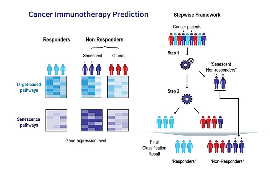
AI Tool Predicts Immunotherapy Response Using Tumor Senescence Signals
Patients receiving the same cancer immunotherapy can have markedly different outcomes, with some showing little or no response. Identifying these patients earlier could reduce the costs of ineffective... Read more
Hematology
view channel
New Risk-Stratification Tool Predicts Relapse After Blinatumomab in Childhood ALL
Acute lymphoblastic leukemia in children and young people requires careful assessment of relapse risk, particularly as treatment approaches move beyond standard chemotherapy. Traditional relapse predictors... Read more
New Myelofibrosis Guidelines Recommend Expanded Genetic Testing at Diagnosis
Myelofibrosis is a rare, life-threatening blood cancer affecting up to six per 100,000 people. The disease causes scarring in the bone marrow, limiting the production of healthy blood cells and potentially... Read more

Immunology
view channel
Antibody Profiling Predicts Inflammatory Bowel Disease Years Before Diagnosis
Inflammatory bowel disease (IBD), which includes Crohn’s disease and ulcerative colitis, involves abnormal immune responses that cause persistent inflammation and damage in the digestive system.... Read more
Immune Profiling Initiative Aims to Improve Bone Marrow Analysis in Leukemia
Acute myeloid leukemia (AML) is an aggressive blood cancer in which assessing disease burden after therapy can help guide clinical evaluation. Patients with measurable residual disease (MRD) after treatment... Read more
Immune Profiling Identifies Early Maternal Autoantibodies Linked to Preterm Birth Risk
A typical pregnancy lasts about 40 weeks, but labor begins prematurely in roughly one in 10 pregnancies. Preterm birth is the leading cause of newborn death worldwide, yet its underlying causes and which... Read more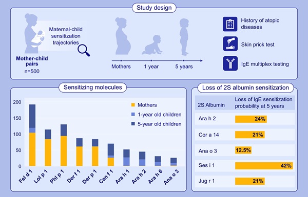
Longitudinal Blood Testing Reveals Changes in Allergen Sensitization During Childhood
Food allergy is a major childhood health concern, and distinguishing persistent risk from transient sensitization can guide clinical decisions. Specific IgE antibodies may be detectable even when a child... Read moreMicrobiology
view channel
Molecular Panel Gains WHO Emergency Use Listing to Support Ebola Outbreak Response
The ongoing Ebola Bundibugyo outbreak in the Democratic Republic of the Congo and neighboring countries has become one of the largest outbreaks of this Ebola virus species recorded to date.... Read more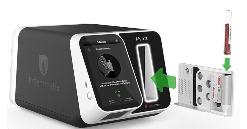
Rapid Host-Response Test Aims to Predict Corticosteroid Benefit in Sepsis
Sepsis and severe infections are difficult to treat because patients with similar clinical presentations may have very different underlying immune responses. Corticosteroids are commonly used in severe... Read more
Technology
view channel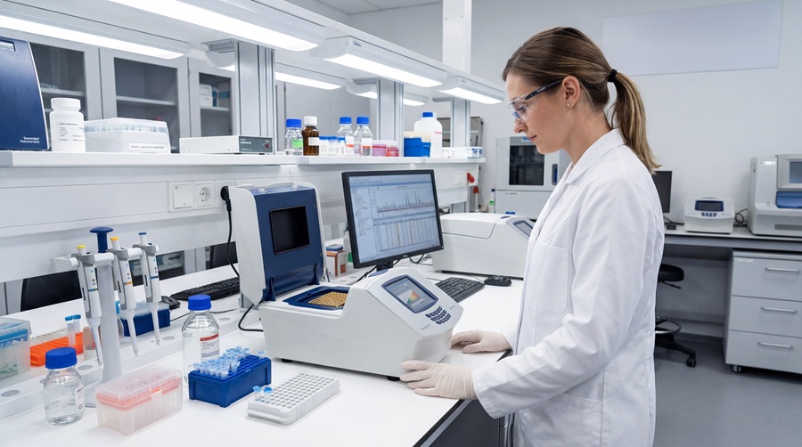
Compact PCR Thermal Cycler Launches for Routine Molecular Laboratory Workflows
Molecular laboratories rely on nucleic acid analysis to detect molecular information accurately and reproducibly across a wide range of applications. Supporting these routine workflows, a newly launched... Read more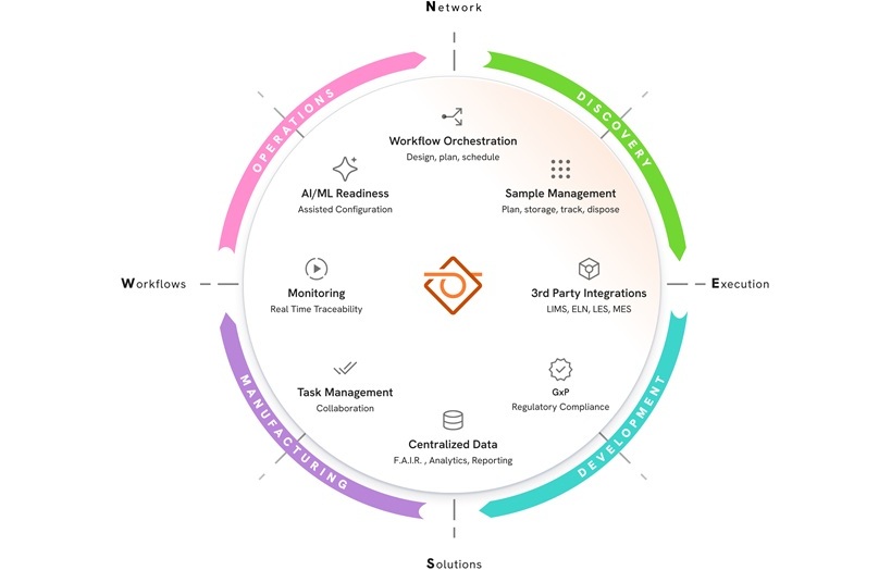
New AI-Powered System Aims to Streamline Laboratory and Manufacturing Workflows
Scientific laboratories supporting drug development, biotechnology, and materials research increasingly rely on coordinated digital workflows to move data, samples, and activities across teams.... Read more

Industry
view channel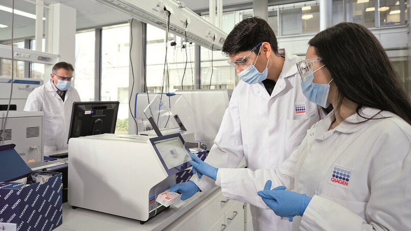
Qiagen Agrees to Go Private Through EQT-Led Acquisition
QIAGEN N.V. (Venlo, the Netherlands) and EQT Private Equity (Stockholm, Sweden) have agreed to an all-cash public takeover offer for all outstanding QIAGEN shares at EUR 46.50 per share.... Read more
Hologic and Inpeco Partner to Advance Molecular Laboratory Automation in Europe
Hologic, Inc. (Marlborough, MA, USA) and Inpeco S.A. (Novazzano, Switzerland) have signed a co-marketing partnership in Europe for Inpeco’s recently launched FlexLab X automation solution and Hologic’s... Read more