Intratumor Heterogeneity Analyzed in Neuroblastoma
|
By LabMedica International staff writers Posted on 22 Dec 2021 |


|
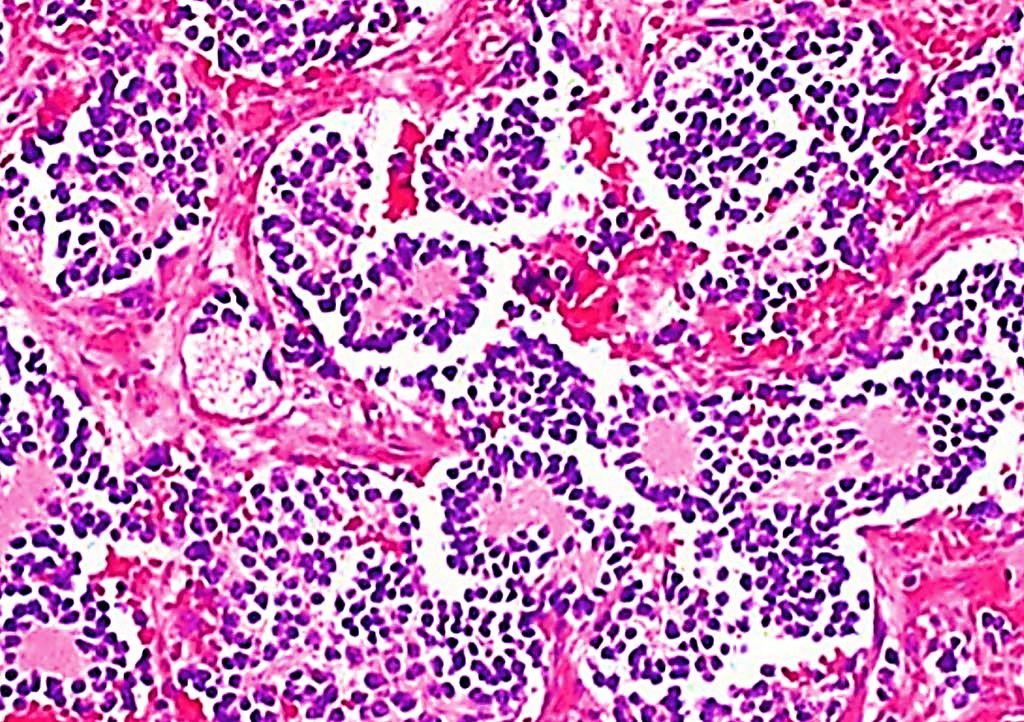
Image: Histopathology of a typical neuroblastoma with rosette formation (Photo courtesy of Dr. Mark Applebaum, MD)
Neuroblastoma is the most common solid pediatric tumour, accounting for 15% of cancer-related deaths in early childhood and especially for patients directly diagnosed with high-risk neuroblastoma have a poor prognosis.
Tumors are heterogeneous, which means that different parts of the same tumor can be genetically distinct. This phenomenon, known as intratumor heterogeneity, is steadily gaining in significance within the field of oncology. Cellular and molecular differences within the same tumor play an important role in many different cancers due to their implications for diagnosis and the use of targeted therapies.
A large team of pediatric oncologists at the Charité-Universitätsmedizin Berlin (Berlin, Germany) and their colleagues collected tumor samples from 10 patients who were enrolled in a study between 2014 and 2018. Samples were collected by open surgical biopsy either at diagnosis, at tumour resection after 4–6 cycles of chemotherapy or at diagnosis of relapse. Fresh samples were immediately snap-frozen in liquid nitrogen and stored at −80 °C. Portions of tumour material were formalin-fixed and paraffin-embedded (FFPE) in parallel for diagnostics and preservation in the pathology unit. The team analyzed a total of 140 neuroblastoma samples.
Peripheral blood collected from each patient was used as a matched germline control for tumor samples. DNA was prepared using the Qiagen DNA Mini kit (Qiagen, Venlo, the Netherlands). The SureSelect Human All Exon V6 kit (Agilent Technologies, Santa Clara, CA, USA) was used to prepare libraries enriched with exonic sequences. The libraries were further prepared for sequencing using the Illumina TruSeq Exome Kit and sequenced on Illumina HiSeq 2500 and Illumina NextSeq sequencers (Illumina, San Diego, CA, USA). A list of genes potentially involved in neuroblastoma development was compiled by combining lists of potential cancer driver genes from several sources.
The investigators focused their analysis on the neuroblastoma-associated genes ALK, MYCN and FGFR1, which play an important role in both clinical course and treatment. According to their results, changes in the ALK and MYCN genes were not present continuously throughout the course of the disease, nor were they found in all tumor cells. Changes in the ALK and FGFR1 genes can offer useful treatment targets, particularly in relapsed patients. They found that, in some patients, ALK mutations which were present at the time of diagnosis had disappeared by the time the tumor was surgically removed. Changes in the FGFR1 gene were only found in distinct tumor regions. The scientists were also able to identify an instability in the number of gene copies present in neuroblastoma cells.
Angelika Eggert, MD, a Professor of Pediatric Oncology and a senior author of the study, said, “We are now in a better position to understand how neuroblastoma cells behave. This knowledge is essential in relation to patients who suffer a recurrence of their disease because their treatment often requires the use of personalized and targeted therapies. When a tumor presents as genetically heterogeneous, targeted molecular therapy may well capture a majority of the abnormal tissue but, crucially, will not capture all of the affected cells. The cancer will then be able to regrow from those remaining cells.”
The authors conclude that their in-depth analysis of neuroblastoma intratumor heterogeneity reveals that extensive genetic heterogeneity and subclonal diversification evolves under therapy and may have important implications for the clinical interpretation of molecular diagnostic results and for the selection of appropriate second-line treatment approaches. The study was published on November 23, 2021 in the journal Nature Communications.
Related Links:
Charité-Universitätsmedizin Berlin
Qiagen
Agilent Technologies
Illumina





Tumors are heterogeneous, which means that different parts of the same tumor can be genetically distinct. This phenomenon, known as intratumor heterogeneity, is steadily gaining in significance within the field of oncology. Cellular and molecular differences within the same tumor play an important role in many different cancers due to their implications for diagnosis and the use of targeted therapies.
A large team of pediatric oncologists at the Charité-Universitätsmedizin Berlin (Berlin, Germany) and their colleagues collected tumor samples from 10 patients who were enrolled in a study between 2014 and 2018. Samples were collected by open surgical biopsy either at diagnosis, at tumour resection after 4–6 cycles of chemotherapy or at diagnosis of relapse. Fresh samples were immediately snap-frozen in liquid nitrogen and stored at −80 °C. Portions of tumour material were formalin-fixed and paraffin-embedded (FFPE) in parallel for diagnostics and preservation in the pathology unit. The team analyzed a total of 140 neuroblastoma samples.
Peripheral blood collected from each patient was used as a matched germline control for tumor samples. DNA was prepared using the Qiagen DNA Mini kit (Qiagen, Venlo, the Netherlands). The SureSelect Human All Exon V6 kit (Agilent Technologies, Santa Clara, CA, USA) was used to prepare libraries enriched with exonic sequences. The libraries were further prepared for sequencing using the Illumina TruSeq Exome Kit and sequenced on Illumina HiSeq 2500 and Illumina NextSeq sequencers (Illumina, San Diego, CA, USA). A list of genes potentially involved in neuroblastoma development was compiled by combining lists of potential cancer driver genes from several sources.
The investigators focused their analysis on the neuroblastoma-associated genes ALK, MYCN and FGFR1, which play an important role in both clinical course and treatment. According to their results, changes in the ALK and MYCN genes were not present continuously throughout the course of the disease, nor were they found in all tumor cells. Changes in the ALK and FGFR1 genes can offer useful treatment targets, particularly in relapsed patients. They found that, in some patients, ALK mutations which were present at the time of diagnosis had disappeared by the time the tumor was surgically removed. Changes in the FGFR1 gene were only found in distinct tumor regions. The scientists were also able to identify an instability in the number of gene copies present in neuroblastoma cells.
Angelika Eggert, MD, a Professor of Pediatric Oncology and a senior author of the study, said, “We are now in a better position to understand how neuroblastoma cells behave. This knowledge is essential in relation to patients who suffer a recurrence of their disease because their treatment often requires the use of personalized and targeted therapies. When a tumor presents as genetically heterogeneous, targeted molecular therapy may well capture a majority of the abnormal tissue but, crucially, will not capture all of the affected cells. The cancer will then be able to regrow from those remaining cells.”
The authors conclude that their in-depth analysis of neuroblastoma intratumor heterogeneity reveals that extensive genetic heterogeneity and subclonal diversification evolves under therapy and may have important implications for the clinical interpretation of molecular diagnostic results and for the selection of appropriate second-line treatment approaches. The study was published on November 23, 2021 in the journal Nature Communications.
Related Links:
Charité-Universitätsmedizin Berlin
Qiagen
Agilent Technologies
Illumina

Platinum Member
Automated Coagulation Analyzer
Hemolumi H6
All-in-One Molecular System
AIO M160
Gold Member
Nucleic Acid Extractor System
NEOS-96 XT
Latest Molecular Diagnostics News
- RNA-Based Test Predicts Immunotherapy Response in Non-Small Cell Lung Cancer
- Modeling Study Shows Blood-Based CRC Screening Could Prevent More Deaths Than Stool Tests
- Noninvasive Cervical Test Detects Endometrial Cancer for Triage
- AI Tool Classifies Pathogenic Variants in Severe Childhood Epilepsy
- Molecular Test Detects Barrett’s Esophagus to Guide Endoscopy Referral
- Blood-Based Metagenomic Test Detects Wide Range of Pathogens from Single Sample
- New RNA Detection Tool Shows Promise for HIV and Other Viral Testing
- Tissue-Specific Vesicle Isolation Expands Molecular Insights Into Liver Disease
- Whole Genome Sequencing Solution Targets Unsolved Rare Disease Cases
- Validated Test Predicts Recurrence Risk and Radiation Benefit in Early-Stage Breast Cancer
- Study Shows How Genome Reference Bias Can Mislead Preventive Testing
- Genomic Screening Expands Detection of Treatable Conditions in Newborns
- Age-Based Genetic Testing May Miss Most Inherited Cancer Risk Variants
- Pooled Sputum Testing Offers Lower-Cost Molecular Diagnosis for Tuberculosis
- Expanded Sequencing Panel Supports Myeloid Malignancy Variant Profiling
- Tumor Markers Help Predict Response to Less Intensive HER2+ Chemotherapy
Channels
Clinical Chemistry
view channel
New One-Step Syphilis Test Could Enable Earlier Detection and Faster Treatment
Syphilis can be difficult to diagnose because its symptoms may resemble other diseases and appear and disappear without treatment. Undetected infection can continue causing harm, spread to partners and... Read more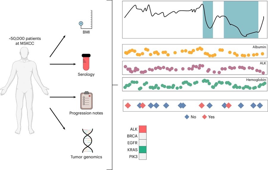
Multimodal Clinical Data Models Predict Cachexia Risk Across Cancers
Cancer cachexia is a severe wasting syndrome that affects roughly half of patients with cancer and cannot be reversed simply by increasing food intake. It can reduce treatment tolerance, impair quality... Read more

Hematology
view channel
New Myelofibrosis Guidelines Recommend Expanded Genetic Testing at Diagnosis
Myelofibrosis is a rare, life-threatening blood cancer affecting up to six per 100,000 people. The disease causes scarring in the bone marrow, limiting the production of healthy blood cells and potentially... Read more
Handheld Point-of-Care Test Aims to Enable Rapid Coagulation Assessment in Trauma
Severe blood loss and inadequate tissue oxygenation are leading causes of preventable death after traumatic injury. Blood products are essential in trauma care, but cold-chain storage, short shelf life,... Read more

Immunology
view channel
Immune Profiling Identifies Early Maternal Autoantibodies Linked to Preterm Birth Risk
A typical pregnancy lasts about 40 weeks, but labor begins prematurely in roughly one in 10 pregnancies. Preterm birth is the leading cause of newborn death worldwide, yet its underlying causes and which... Read more
Longitudinal Blood Testing Reveals Changes in Allergen Sensitization During Childhood
Food allergy is a major childhood health concern, and distinguishing persistent risk from transient sensitization can guide clinical decisions. Specific IgE antibodies may be detectable even when a child... Read more

Microbiology
view channel
Rapid Host-Response Test Aims to Predict Corticosteroid Benefit in Sepsis
Sepsis and severe infections are difficult to treat because patients with similar clinical presentations may have very different underlying immune responses. Corticosteroids are commonly used in severe... Read more
Oral and Lung Microbiome Changes Linked to Severe Long Covid Symptoms
Long Covid can involve persistent respiratory, cognitive, and gastrointestinal symptoms after acute infection, creating a difficult diagnostic and management challenge. Because COVID-19 initially affects... Read more

Pathology
view channel
Molecular Testing Refines Barrett’s Esophagus Progression Risk Beyond Biopsy Findings
Barrett’s esophagus is a precancerous condition associated with increased risk of esophageal adenocarcinoma. Routine endoscopic biopsy findings guide surveillance and therapy, but histopathology may not... Read more
Updated Digital Pathology Platform Expands AI Integration for Cancer Diagnosis
Digital pathology is reshaping how laboratories manage case review, image analysis, and collaboration. As diagnostic workflows become more complex, pathology teams need platforms that can support digital... Read more
Technology
view channel
Laser-Based Swab Analysis Shows Promise for Detecting Disease-Linked Odor Patterns
Disease-related changes in volatile organic compounds can alter body odor, producing measurable patterns in exhaled breath and bodily fluids. Current analytical methods can be complex, time-consuming,... Read more
Laser-Enhanced Assay Boosts Sensitivity for Colorectal Cancer Biomarker Detection
Colorectal cancer is the third most commonly diagnosed cancer and the second leading cause of cancer-related death worldwide. Early detection remains critical, but cancer biomarkers can produce only faint... Read more

Industry
view channel
Expanded Partnership Scales AI-Powered Digital Pathology for Oncology
Oncology diagnostics rely on pathology and biomarker information to guide personalized treatment. As computational pathology and artificial intelligence (AI) expand insights from tissue analysis, laboratories... Read more
Global TB Summit Connects Diagnostic Testing With Prevention and Clinical Care
Tuberculosis remains a major global infectious disease challenge despite declining deaths. In 2024, an estimated 10.7 million people worldwide developed the disease and nearly 1.2 million died from it.... Read more