Telomerase Reverse Transcriptase Protein Expression Evaluated for Melanomas
|
By LabMedica International staff writers Posted on 19 Jul 2021 |

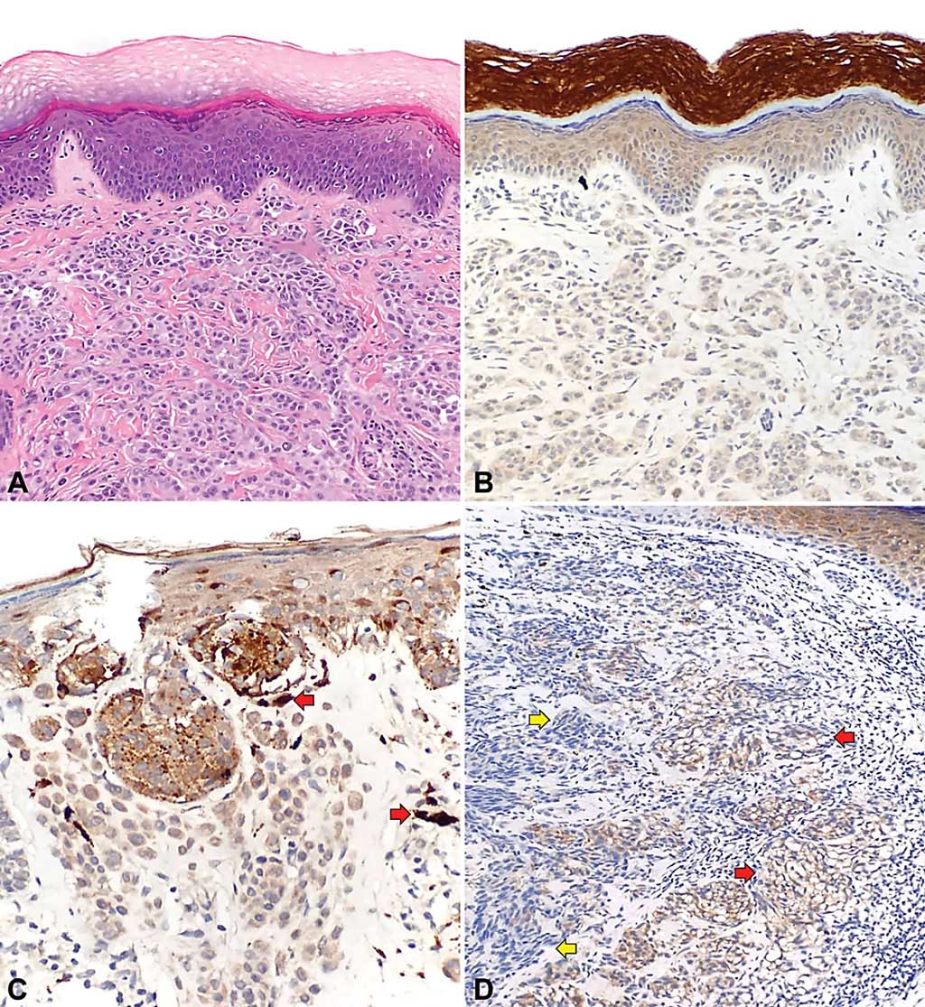
Image: Telomerase reverse transcriptase (TERT) expression in non-lentiginous acral melanoma (NLAM) and non-acral cutaneous melanoma (NACM): (A) exhibiting 1+ TERT staining intensity and (B) The intensity of TERT expression and proportion of TERT-positive cells could also vary in cutaneous melanomas (Photo courtesy of MD Anderson Cancer Center)
Telomeres are regions of repetitive nucleotide sequences located at the ends of chromosomes that play a key role in the maintenance of genomic integrity and stability in cells. In normal nonneoplastic somatic cells, telomeres progressively shorten with successive cell divisions.
Molecularly distinct from cutaneous melanomas arising from sun-exposed sites, acral lentiginous melanomas (ALMs) typically lack ultraviolet-signature mutations, such as telomerase reverse transcriptase (TERT) promoter mutations. Instead, ALMs show a high degree of copy number alterations, often with multiple amplifications of TERT, which are associated with adverse prognosis.
Pathologists at the University of Texas MD Anderson Cancer Center (Houston, TX, USA) identified a total of 57 cases of acral and non-acral melanocytic lesions, including 24 primary ALMs, six metastatic ALMs, 10 primary non-lentiginous acral melanomas (NLAMs), 12 primary NACMs, and five acral nevi (AN), diagnosed at their institution between 2003 and 2016. Demographic, clinical, and histopathologic parameters and follow-up data for the selected cases were retrieved through review of the final pathology reports and clinical charts.
Immunohistochemical (IHC) analysis of TERT protein expression was performed on a 5-μm–thick paraffin section was cut from each tissue block of selected cases. The paraffin sections were then tested for TERT protein expression by IHC using an anti-TERT monoclonal rabbit anti-human antibody (Abcam, Cambridge, MA, USA) performed on a Leica Bond autostainer (Leica Biosystems, Buffalo Grove, IL, USA) per routine laboratory protocols. The pattern of TERT protein expression was recorded as negative, cytoplasmic, nuclear, or Golgi/perinuclear.
The investigators reported that TERT expression was more frequent in ALMs than in non-lentiginous acral melanomas and non-acral cutaneous melanomas, and was absent in acral nevi. When present, TERT expression in ALMs was cytoplasmic and more intense than TERT expression in other melanocytic lesions (with a higher H-score). There was a trend toward decreased overall survival in patients with ALMs with TERT immunoreactivity, but it did not reach statistical significance. Furthermore, no correlation was found between TERT expression and disease-specific survival in patients with ALMs.
The authors concluded that their study demonstrated that unlike TERT promoter mutations, TERT protein expression was frequently detected in both primary and metastatic ALMs. In addition, the study was the first to demonstrate differences in TERT immunohistochemical expression between ALMs and NLAMs, which have never been separately studied but rather have been grouped together (i.e., as “acral melanomas”) without histologic distinction. Lastly, in their study, although TERT expression was more frequent and of stronger intensity in ALMs than in other types of melanocytic lesions, with higher overall H-scores, TERT immunoreactivity in ALMs did not correlate with survival. The study was published in the July 2021 issue of the journal Archives of Pathology and Laboratory Medicine.
Related Links:
University of Texas MD Anderson Cancer Center
Abcam
Leica Biosystems
Latest Pathology News
- Single Sample Classifier Predicts Cancer-Associated Fibroblast Subtypes in Patient Samples
- New AI-Driven Platform Standardizes Tuberculosis Smear Microscopy Workflow
- AI Tool Uses Blood Biomarkers to Predict Transplant Complications Before Symptoms Appear
- High-Resolution Cancer Virus Imaging Uncovers Potential Therapeutic Targets
- Research Consortium Harnesses AI and Spatial Biology to Advance Cancer Discovery
- AI Tool Helps See How Cells Work Together Inside Diseased Tissue
- AI-Powered Microscope Diagnoses Malaria in Blood Smears Within Minutes
- Engineered Yeast Cells Enable Rapid Testing of Cancer Immunotherapy
- First-Of-Its-Kind Test Identifies Autism Risk at Birth
- AI Algorithms Improve Genetic Mutation Detection in Cancer Diagnostics
- Skin Biopsy Offers New Diagnostic Method for Neurodegenerative Diseases
- Fast Label-Free Method Identifies Aggressive Cancer Cells
- New X-Ray Method Promises Advances in Histology
- Single-Cell Profiling Technique Could Guide Early Cancer Detection
- Intraoperative Tumor Histology to Improve Cancer Surgeries
- Rapid Stool Test Could Help Pinpoint IBD Diagnosis
Channels
Clinical Chemistry
view channel
Simple Blood Test Offers New Path to Alzheimer’s Assessment in Primary Care
Timely evaluation of cognitive symptoms in primary care is often limited by restricted access to specialized diagnostics and invasive confirmatory procedures. Clinicians need accessible tools to determine... Read more
Existing Hospital Analyzers Can Identify Fake Liquid Medical Products
Counterfeit and substandard medicines remain a serious global health threat, with World Health Organization estimates suggesting that 10.5% of medicines in low- and middle-income countries are either fake... Read moreMolecular Diagnostics
view channel
New Genome Sequencing Technique Measures Epstein-Barr Virus in Blood
The Epstein–Barr virus (EBV) infects up to 95% of adults worldwide and remains in the body for life. While usually kept under control, the virus is linked to cancers such as Hodgkin’s lymphoma and autoimmune... Read more
Blood Test Boosts Early Detection of Brain Cancer
Brain and central nervous system (CNS) tumors are often diagnosed at an advanced stage, when treatment options are limited, and survival rates remain low. Around 300,000 new cases are diagnosed each year... Read moreHematology
view channel
Rapid Cartridge-Based Test Aims to Expand Access to Hemoglobin Disorder Diagnosis
Sickle cell disease and beta thalassemia are hemoglobin disorders that often require referral to specialized laboratories for definitive diagnosis, delaying results for patients and clinicians.... Read more
New Guidelines Aim to Improve AL Amyloidosis Diagnosis
Light chain (AL) amyloidosis is a rare, life-threatening bone marrow disorder in which abnormal amyloid proteins accumulate in organs. Approximately 3,260 people in the United States are diagnosed... Read moreImmunology
view channel
New Biomarker Predicts Chemotherapy Response in Triple-Negative Breast Cancer
Triple-negative breast cancer is an aggressive form of breast cancer in which patients often show widely varying responses to chemotherapy. Predicting who will benefit from treatment remains challenging,... Read moreBlood Test Identifies Lung Cancer Patients Who Can Benefit from Immunotherapy Drug
Small cell lung cancer (SCLC) is an aggressive disease with limited treatment options, and even newly approved immunotherapies do not benefit all patients. While immunotherapy can extend survival for some,... Read more
Whole-Genome Sequencing Approach Identifies Cancer Patients Benefitting From PARP-Inhibitor Treatment
Targeted cancer therapies such as PARP inhibitors can be highly effective, but only for patients whose tumors carry specific DNA repair defects. Identifying these patients accurately remains challenging,... Read more
Ultrasensitive Liquid Biopsy Demonstrates Efficacy in Predicting Immunotherapy Response
Immunotherapy has transformed cancer treatment, but only a small proportion of patients experience lasting benefit, with response rates often remaining between 10% and 20%. Clinicians currently lack reliable... Read moreMicrobiology
view channel
Three-Test Panel Launched for Detection of Liver Fluke Infections
Parasitic liver fluke infections remain endemic in parts of Asia, where transmission commonly occurs through consumption of raw freshwater fish or aquatic plants. Chronic infection is a well-established... Read more
Rapid Test Promises Faster Answers for Drug-Resistant Infections
Drug-resistant pathogens continue to pose a growing threat in healthcare facilities, where delayed detection can impede outbreak control and increase mortality. Candida auris is notoriously difficult to... Read more
CRISPR-Based Technology Neutralizes Antibiotic-Resistant Bacteria
Antibiotic resistance has accelerated into a global health crisis, with projections estimating more than 10 million deaths per year by 2050 as drug-resistant “superbugs” continue to spread.... Read more
Comprehensive Review Identifies Gut Microbiome Signatures Associated With Alzheimer’s Disease
Alzheimer’s disease affects approximately 6.7 million people in the United States and nearly 50 million worldwide, yet early cognitive decline remains difficult to characterize. Increasing evidence suggests... Read moreTechnology
view channel
Blood Test “Clocks” Predict Start of Alzheimer’s Symptoms
More than 7 million Americans live with Alzheimer’s disease, and related health and long-term care costs are projected to reach nearly USD 400 billion in 2025. The disease has no cure, and symptoms often... Read more
AI-Powered Biomarker Predicts Liver Cancer Risk
Liver cancer, or hepatocellular carcinoma, causes more than 800,000 deaths worldwide each year and often goes undetected until late stages. Even after treatment, recurrence rates reach 70% to 80%, contributing... Read more
Robotic Technology Unveiled for Automated Diagnostic Blood Draws
Routine diagnostic blood collection is a high‑volume task that can strain staffing and introduce human‑dependent variability, with downstream implications for sample quality and patient experience.... Read more
ADLM Launches First-of-Its-Kind Data Science Program for Laboratory Medicine Professionals
Clinical laboratories generate billions of test results each year, creating a treasure trove of data with the potential to support more personalized testing, improve operational efficiency, and enhance patient care.... Read moreIndustry
view channel
QuidelOrtho Collaborates with Lifotronic to Expand Global Immunoassay Portfolio
QuidelOrtho (San Diego, CA, USA) has entered a long-term strategic supply agreement with Lifotronic Technology (Shenzhen, China) to expand its global immunoassay portfolio and accelerate customer access... Read more


