Novel CTC Culture Method Developed to Study Metastatic Cancer
|
By LabMedica International staff writers Posted on 26 Oct 2020 |


|
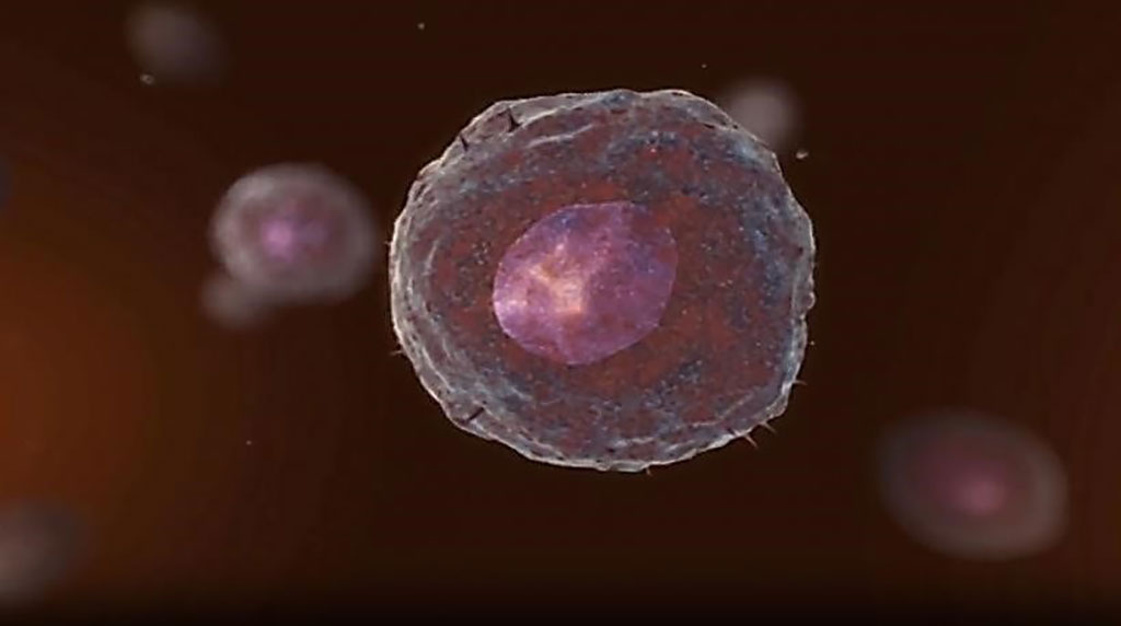
Image: Circulating tumor cells (CTCs) are cancer cells that are released and disseminated into the bloodstream and lymphatic system. CTC cultures were successfully propagated from breast epithelial cells (Photo courtesy of Menarini Silicon Biosystems).
Cancer metastasis is responsible for most cancer-associated death. During metastasis, cells that escape the primary tumor into the circulatory system are known as circulating tumor cells (CTCs).
Circulating tumor cells represent a unique population of cells that can be used to investigate the mechanistic underpinnings of metastasis. Unfortunately, current technologies designed for the isolation and capture of CTCs are inefficient. Existing literature for in vitro CTC cultures report low (6% to 20%) success rates.
Scientists from Georgetown University Medical Center (Washington, DC, USA) enrolled from 12 metastatic breast cancer patients representing all three major subtypes, HER2 positive, hormone receptor positive, and triple negative and samples from five healthy donors. After collecting a patient's blood sample, the team removed red blood cells (RBCs) and granulocytes using gradient centrifugation. They then harvest the rest of the cells, including CTCs, and put them into a custom cell culture growth medium for culture, followed by downstream analysis.
To see whether they had indeed cultured CTCs, the group then tested for the presence of epithelial, mesenchymal, and breast tissue markers to establish the cells' tissue of origin using reverse-transcriptase quantitative polymerase chain reaction (qRT-PCR). They selected cytokeratin 5 and 8 and mammaglobin and successfully identified all three biomarkers in all 12 cultures. The team then used RNA-seq on the six samples that yielded sufficient RNA and could be cultured for more than 30 days in order to characterize their gene expression. These six samples all contained CD45+ leukocytes, they noted, which have previously been shown to support CTC survival.
Overall, the investigators identified 7,234 genes that were significantly differentially expressed in the CTCs-containing samples compared to the healthy donors. Increased expression of multiple genes was correlated with a significant drop in overall patient survival, they found. Using multiple bioinformatics tools to confirm that the CTCs in the samples originated from the cancers, the team identified 52 significantly enriched key cancer pathways and 21 enriched genes that are important to breast cancer progression and metastasis. The group mainly found enriched CD8 T cells, neutrophils, and macrophages in the CTC cultures and noted that the isolation technique appeared to favor the survival of macrophages and neutrophils.
Seema Agarwal, PhD, the senior author and associate professor of pathology, said, “We could take DNA and RNA from cultured CTCs and do a detailed genomic analysis that can be done very quickly in a more meaningful way, as prior to expansion, there are very few CTCs in the patient's blood. We don't know whether the cultured cells represent the heterogeneity of the CTCs, but as we move forward, we hope to characterize the heterogeneity of the cells.”
The authors concluded that once profiled, CTCs can provide a significant amount of information based solely on their identity as an intermediary stage of metastasis. Comparison of CTCs with primary tumors would enable the identification of metastatic drivers and lead to the development of metastasis-preventing therapies. Thus, having a standardized method for the capture and culture of CTCs is a pressing need. The study was published on September 28, 2020 in the journal Cancers.
Related Links:
Georgetown University Medical Center






Circulating tumor cells represent a unique population of cells that can be used to investigate the mechanistic underpinnings of metastasis. Unfortunately, current technologies designed for the isolation and capture of CTCs are inefficient. Existing literature for in vitro CTC cultures report low (6% to 20%) success rates.
Scientists from Georgetown University Medical Center (Washington, DC, USA) enrolled from 12 metastatic breast cancer patients representing all three major subtypes, HER2 positive, hormone receptor positive, and triple negative and samples from five healthy donors. After collecting a patient's blood sample, the team removed red blood cells (RBCs) and granulocytes using gradient centrifugation. They then harvest the rest of the cells, including CTCs, and put them into a custom cell culture growth medium for culture, followed by downstream analysis.
To see whether they had indeed cultured CTCs, the group then tested for the presence of epithelial, mesenchymal, and breast tissue markers to establish the cells' tissue of origin using reverse-transcriptase quantitative polymerase chain reaction (qRT-PCR). They selected cytokeratin 5 and 8 and mammaglobin and successfully identified all three biomarkers in all 12 cultures. The team then used RNA-seq on the six samples that yielded sufficient RNA and could be cultured for more than 30 days in order to characterize their gene expression. These six samples all contained CD45+ leukocytes, they noted, which have previously been shown to support CTC survival.
Overall, the investigators identified 7,234 genes that were significantly differentially expressed in the CTCs-containing samples compared to the healthy donors. Increased expression of multiple genes was correlated with a significant drop in overall patient survival, they found. Using multiple bioinformatics tools to confirm that the CTCs in the samples originated from the cancers, the team identified 52 significantly enriched key cancer pathways and 21 enriched genes that are important to breast cancer progression and metastasis. The group mainly found enriched CD8 T cells, neutrophils, and macrophages in the CTC cultures and noted that the isolation technique appeared to favor the survival of macrophages and neutrophils.
Seema Agarwal, PhD, the senior author and associate professor of pathology, said, “We could take DNA and RNA from cultured CTCs and do a detailed genomic analysis that can be done very quickly in a more meaningful way, as prior to expansion, there are very few CTCs in the patient's blood. We don't know whether the cultured cells represent the heterogeneity of the CTCs, but as we move forward, we hope to characterize the heterogeneity of the cells.”
The authors concluded that once profiled, CTCs can provide a significant amount of information based solely on their identity as an intermediary stage of metastasis. Comparison of CTCs with primary tumors would enable the identification of metastatic drivers and lead to the development of metastasis-preventing therapies. Thus, having a standardized method for the capture and culture of CTCs is a pressing need. The study was published on September 28, 2020 in the journal Cancers.
Related Links:
Georgetown University Medical Center

Platinum Member
Automated Coagulation Analyzer
Hemolumi H6
Fully Automated Urinalysis System
DxU 1800 Fully Automated Urinalysis System
Gold Member
Aspiration System
VACUSAFE
Latest Pathology News
- AI Pre-Screening Helps Identify Lung Cancer Cases for Additional Biomarker Testing
- AI Pathology Platform Targets Workflow Delays and Molecular Test Failures
- Updated Digital Pathology Platform Expands AI Integration for Cancer Diagnosis
- Molecular Testing Refines Barrett’s Esophagus Progression Risk Beyond Biopsy Findings
- AI Histopathology Tool Distinguishes Ewing Sarcoma for Diagnosis
- Rapid Mass Spectrometry Test May Aid Glioma Margin Decisions
- Genomic Classifier Predicts Benefit From Adding Hormone Therapy to Salvage Prostate Radiation
- Multimodal AI Supports Prostate Cancer Risk Assessment Across Diverse Cohorts
- New AI Model Maps Where Glioblastoma Could Return After Surgery
- Simple Test Could Reduce the Need for Invasive Uterine Cancer Checks
- Tumor Blood Vessel Features May Help Predict Colorectal Cancer Survival
- Multimodal AI Improves Breast Cancer Recurrence Risk Prediction Beyond Standard Genomic Testing
- Machine Learning Cytology Tool Improves Cancer Cell Identification
- CAP Updates Cancer Protocols to Refine Breast Cancer Biomarker Reporting and GIST Molecular Testing
- Tumor Budding Grading May Predict Chemotherapy Benefit in Resected Lung Squamous Cancer
- AI Spatial Analysis of Routine Slides Helps Predict Pancreatic Cancer Recurrence
Channels
Clinical Chemistry
view channel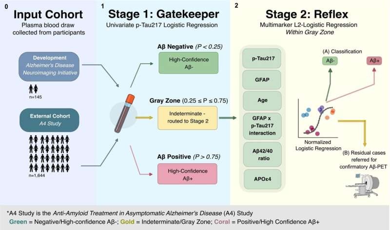
Two-Step Blood Testing Approach Reduces Uncertainty in Amyloid Assessment
Blood-based biomarkers offer a more accessible, less expensive way to detect Alzheimer’s-related brain changes than traditional methods such as amyloid-beta positron emission tomography (Aβ-PET).... Read more
Multimodal Platform to Combine Blood and Imaging Biomarkers for Lung Nodule Assessment
Lung nodules are commonly detected during chest imaging and require careful risk assessment to determine whether follow-up imaging, additional testing, or biopsy is needed. With millions of patients evaluated... Read more

Hematology
view channel
New Myelofibrosis Guidelines Recommend Expanded Genetic Testing at Diagnosis
Myelofibrosis is a rare, life-threatening blood cancer affecting up to six per 100,000 people. The disease causes scarring in the bone marrow, limiting the production of healthy blood cells and potentially... Read more
Handheld Point-of-Care Test Aims to Enable Rapid Coagulation Assessment in Trauma
Severe blood loss and inadequate tissue oxygenation are leading causes of preventable death after traumatic injury. Blood products are essential in trauma care, but cold-chain storage, short shelf life,... Read more

Immunology
view channel
Antibody Profiling Predicts Inflammatory Bowel Disease Years Before Diagnosis
Inflammatory bowel disease (IBD), which includes Crohn’s disease and ulcerative colitis, involves abnormal immune responses that cause persistent inflammation and damage in the digestive system.... Read more
Immune Profiling Initiative Aims to Improve Bone Marrow Analysis in Leukemia
Acute myeloid leukemia (AML) is an aggressive blood cancer in which assessing disease burden after therapy can help guide clinical evaluation. Patients with measurable residual disease (MRD) after treatment... Read more
Immune Profiling Identifies Early Maternal Autoantibodies Linked to Preterm Birth Risk
A typical pregnancy lasts about 40 weeks, but labor begins prematurely in roughly one in 10 pregnancies. Preterm birth is the leading cause of newborn death worldwide, yet its underlying causes and which... Read more
Longitudinal Blood Testing Reveals Changes in Allergen Sensitization During Childhood
Food allergy is a major childhood health concern, and distinguishing persistent risk from transient sensitization can guide clinical decisions. Specific IgE antibodies may be detectable even when a child... Read moreMicrobiology
view channel
Molecular Panel Gains WHO Emergency Use Listing to Support Ebola Outbreak Response
The ongoing Ebola Bundibugyo outbreak in the Democratic Republic of the Congo and neighboring countries has become one of the largest outbreaks of this Ebola virus species recorded to date.... Read more
Rapid Host-Response Test Aims to Predict Corticosteroid Benefit in Sepsis
Sepsis and severe infections are difficult to treat because patients with similar clinical presentations may have very different underlying immune responses. Corticosteroids are commonly used in severe... Read more
Pathology
view channel
AI Pre-Screening Helps Identify Lung Cancer Cases for Additional Biomarker Testing
Tempus AI (Chicago, IL, USA) will present findings from a study evaluating an artificial intelligence (AI)-enabled pre-screening panel for advanced non-small cell lung cancer (NSCLC) at the ESMO Congress... Read more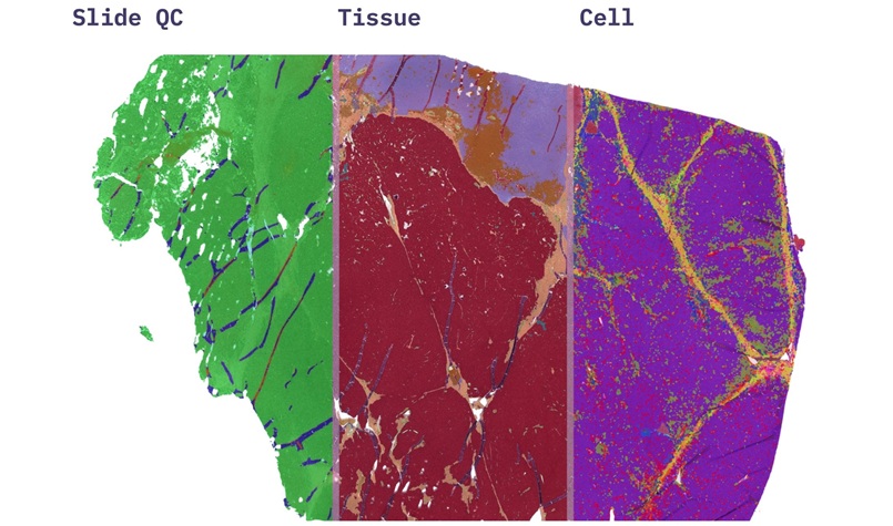
AI Pathology Platform Targets Workflow Delays and Molecular Test Failures
Pathology laboratories often rely on visual review of tissue slides to assess quality, identify tumors, count mitoses, and estimate tumor content. These tasks can be time-consuming and vary between readers,... Read more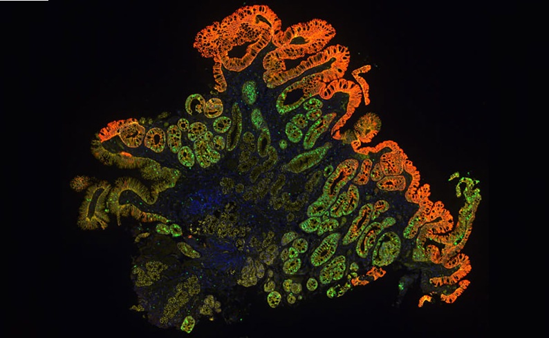
Molecular Testing Refines Barrett’s Esophagus Progression Risk Beyond Biopsy Findings
Barrett’s esophagus is a precancerous condition associated with increased risk of esophageal adenocarcinoma. Routine endoscopic biopsy findings guide surveillance and therapy, but histopathology may not... Read more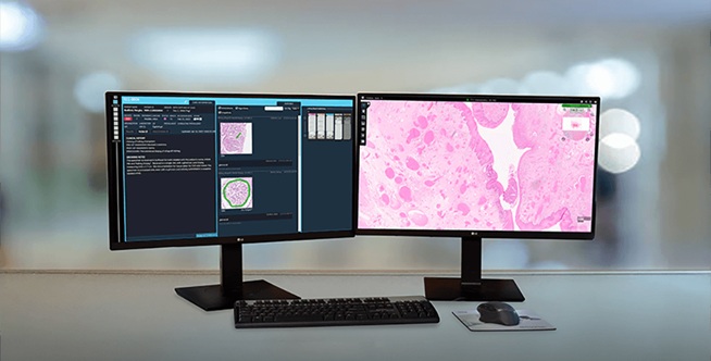
Technology
view channel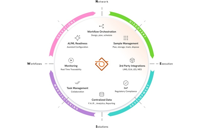
New AI-Powered System Aims to Streamline Laboratory and Manufacturing Workflows
Scientific laboratories supporting drug development, biotechnology, and materials research increasingly rely on coordinated digital workflows to move data, samples, and activities across teams.... Read more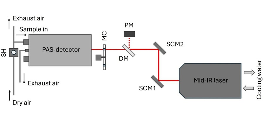
Laser-Based Swab Analysis Shows Promise for Detecting Disease-Linked Odor Patterns
Disease-related changes in volatile organic compounds can alter body odor, producing measurable patterns in exhaled breath and bodily fluids. Current analytical methods can be complex, time-consuming,... Read more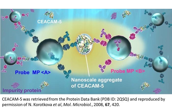
Industry
view channel
Hologic to Broaden Breast Cancer Testing Portfolio Through Reveal Genomics Acquisition
Hologic (Marlborough, MA, USA) has signed a definitive agreement to acquire Reveal Genomics (Barcelona, Spain), a precision oncology company developing genomic diagnostic tests to help personalize cancer treatment.... Read more