Automated Malaria Diagnosis Enhanced by Deep Neural Networks
|
By LabMedica International staff writers Posted on 14 Aug 2020 |

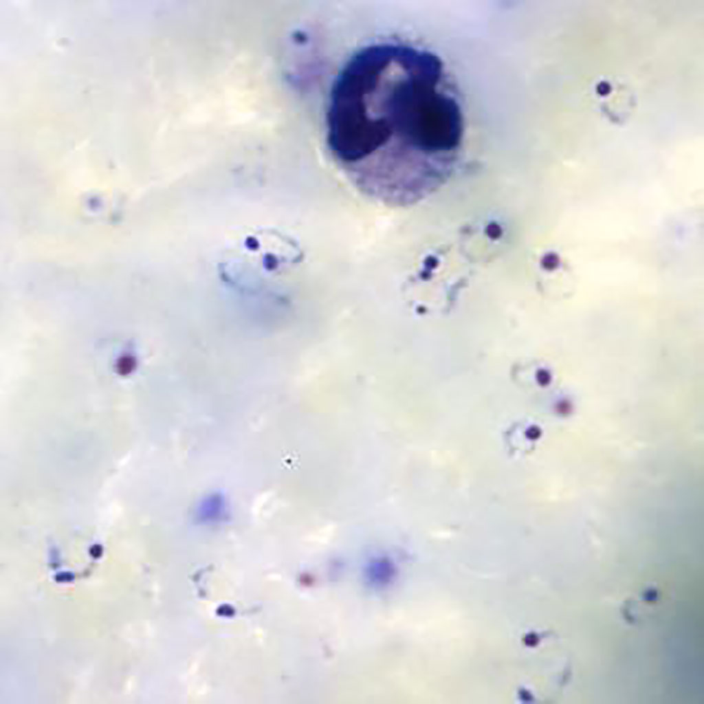
Ring-form trophozoites of Plasmodium falciparum and a white blood cell in a thick blood film (Photo courtesy of Medical Care Development International).
Plasmodium falciparum malaria remains one of the greatest global health burdens with over 228 million cases globally in 2018. In that year there were approximately 405,000 deaths due to malaria worldwide, with the African region accounting for 93% of these deaths, mostly among children.
Although there are a range of techniques that have been developed for the diagnosis of malaria, conventional light microscopy on Giemsa‐stained thick and thin blood films remains the gold standard. Techniques such as polymerase chain reaction, flow cytometric assay and fluorescence‐dye based approaches lack a universally standardized methodology, present high costs, and require quality control improvement.
A team of scientists from University College London (London, UK) leveraged routine clinical‐microscopy labels from their quality‐controlled malaria clinics, to train a Deep Malaria Convolutional Neural Network classifier (DeepMCNN) for automated malaria diagnosis. The DeepMCNN system also provides total Malaria Parasite (MP) and White Blood Cell (WBC) counts allowing parasitaemia estimation in MP/μL. Malaria parasites were detected and counted using human‐expert operated microscopy following Giemsa staining of thick and thin blood films. The criterion for declaring a participant to be malaria parasite‐free was no detectable parasites in 100 high‐power (100×) fields in thick films.
The investigators captured images using an upright bright-field BX63 microscope (Olympus, Tokyo, Japan) fitted with a 100×/1.4 NA objective lens, a motorized x‐y sample positioning stage (Prior Scientific, Cambridge, UK) and a color camera to capture images of Giemsa‐stained, thick blood smears. These smears prepared in their clinics tested the use of deep learning‐based object detection methods to identify both P. falciparum parasites and white‐blood‐cell (WBC) nuclei in the digitized extended depth of field (EDoF) thick blood films images.
The team reported that the prospective validation of the DeepMCNN achieved sensitivity/specificity of 0.92/0.90 against expert‐level malaria diagnosis. The PPV/NPV performance was 0.92/0.90, which is clinically usable in their holoendemic settings in a densely populated metropolis.
The authors concluded that their open data and easily deployable DeepMCNN provide a clinically relevant platform, where other healthcare providers could harness their readily available patient level diagnostic labels, to tailor and further improve the accuracy of the DeepMCNN classifier for their clinical pathway settings. The study was published in the August 2020 issue of the American Journal of Hematology.
Related Links:
University College London
Olympus
Prior Scientific
Latest Molecular Diagnostics News
- Ultra-Sensitive DNA Test Identifies Relapse Risk in Aggressive Leukemia
- Blood Test Could Help Detect Gallbladder Cancer Earlier
- New Blood Test Score Detects Hidden Alcohol-Related Liver Disease
- New Blood Test Predicts Who Will Most Likely Live Longer
- Genetic Test Predicts Radiation Therapy Risk for Prostate Cancer Patients
- Genetic Test Aids Early Detection and Improved Treatment for Cancers
- New Genome Sequencing Technique Measures Epstein-Barr Virus in Blood
- Blood Test Boosts Early Detection of Brain Cancer
- Molecular Monitoring Approach Helps Bladder Cancer Patients Avoid Surgery
- Genetic Tests to Speed Diagnosis of Lymphatic Disorders
- Changes In Lymphatic Vessels Can Aid Early Identification of Aggressive Oral Cancer
- New Extraction Kit Enables Consistent, Scalable cfDNA Isolation from Multiple Biofluids
- New CSF Liquid Biopsy Assay Reveals Genomic Insights for CNS Tumors
- AI-Powered Liquid Biopsy Classifies Pediatric Brain Tumors with High Accuracy
- Group A Strep Molecular Test Delivers Definitive Results at POC in 15 Minutes
- Rapid Molecular Test Identifies Sepsis Patients Most Likely to Have Positive Blood Cultures
Channels
Clinical Chemistry
view channelNew Blood Test Index Offers Earlier Detection of Liver Scarring
Metabolic fatty liver disease is highly prevalent and often silent, yet it can progress to fibrosis, cirrhosis, and liver failure. Current first-line blood test scores frequently return indeterminate results,... Read more
Electronic Nose Smells Early Signs of Ovarian Cancer in Blood
Ovarian cancer is often diagnosed at a late stage because its symptoms are vague and resemble those of more common conditions. Unlike breast cancer, there is currently no reliable screening method, and... Read moreHematology
view channel
Rapid Cartridge-Based Test Aims to Expand Access to Hemoglobin Disorder Diagnosis
Sickle cell disease and beta thalassemia are hemoglobin disorders that often require referral to specialized laboratories for definitive diagnosis, delaying results for patients and clinicians.... Read more
New Guidelines Aim to Improve AL Amyloidosis Diagnosis
Light chain (AL) amyloidosis is a rare, life-threatening bone marrow disorder in which abnormal amyloid proteins accumulate in organs. Approximately 3,260 people in the United States are diagnosed... Read moreImmunology
view channel
New Biomarker Predicts Chemotherapy Response in Triple-Negative Breast Cancer
Triple-negative breast cancer is an aggressive form of breast cancer in which patients often show widely varying responses to chemotherapy. Predicting who will benefit from treatment remains challenging,... Read moreBlood Test Identifies Lung Cancer Patients Who Can Benefit from Immunotherapy Drug
Small cell lung cancer (SCLC) is an aggressive disease with limited treatment options, and even newly approved immunotherapies do not benefit all patients. While immunotherapy can extend survival for some,... Read more
Whole-Genome Sequencing Approach Identifies Cancer Patients Benefitting From PARP-Inhibitor Treatment
Targeted cancer therapies such as PARP inhibitors can be highly effective, but only for patients whose tumors carry specific DNA repair defects. Identifying these patients accurately remains challenging,... Read more
Ultrasensitive Liquid Biopsy Demonstrates Efficacy in Predicting Immunotherapy Response
Immunotherapy has transformed cancer treatment, but only a small proportion of patients experience lasting benefit, with response rates often remaining between 10% and 20%. Clinicians currently lack reliable... Read moreMicrobiology
view channel
Hidden Gut Viruses Linked to Colorectal Cancer Risk
Colorectal cancer (CRC) remains a leading cause of cancer mortality in many Western countries, and existing risk-stratification approaches leave substantial room for improvement. Although age, diet, and... Read more
Three-Test Panel Launched for Detection of Liver Fluke Infections
Parasitic liver fluke infections remain endemic in parts of Asia, where transmission commonly occurs through consumption of raw freshwater fish or aquatic plants. Chronic infection is a well-established... Read morePathology
view channel
Molecular Imaging to Reduce Need for Melanoma Biopsies
Melanoma is the deadliest form of skin cancer and accounts for the vast majority of skin cancer-related deaths. Because early melanomas can closely resemble benign moles, clinicians often rely on visual... Read more
Urine Specimen Collection System Improves Diagnostic Accuracy and Efficiency
Urine testing is a critical, non-invasive diagnostic tool used to detect conditions such as pregnancy, urinary tract infections, metabolic disorders, cancer, and kidney disease. However, contaminated or... Read moreTechnology
view channel
Blood Test “Clocks” Predict Start of Alzheimer’s Symptoms
More than 7 million Americans live with Alzheimer’s disease, and related health and long-term care costs are projected to reach nearly USD 400 billion in 2025. The disease has no cure, and symptoms often... Read more
AI-Powered Biomarker Predicts Liver Cancer Risk
Liver cancer, or hepatocellular carcinoma, causes more than 800,000 deaths worldwide each year and often goes undetected until late stages. Even after treatment, recurrence rates reach 70% to 80%, contributing... Read more
Robotic Technology Unveiled for Automated Diagnostic Blood Draws
Routine diagnostic blood collection is a high‑volume task that can strain staffing and introduce human‑dependent variability, with downstream implications for sample quality and patient experience.... Read more
ADLM Launches First-of-Its-Kind Data Science Program for Laboratory Medicine Professionals
Clinical laboratories generate billions of test results each year, creating a treasure trove of data with the potential to support more personalized testing, improve operational efficiency, and enhance patient care.... Read moreIndustry
view channel
Cepheid Joins CDC Initiative to Strengthen U.S. Pandemic Testing Preparednesss
Cepheid (Sunnyvale, CA, USA) has been selected by the U.S. Centers for Disease Control and Prevention (CDC) as one of four national collaborators in a federal initiative to speed rapid diagnostic technologies... Read more
QuidelOrtho Collaborates with Lifotronic to Expand Global Immunoassay Portfolio
QuidelOrtho (San Diego, CA, USA) has entered a long-term strategic supply agreement with Lifotronic Technology (Shenzhen, China) to expand its global immunoassay portfolio and accelerate customer access... Read more


