Dual Role Discovered for Interleukin-17A in Autoimmune Uveitis
|
By LabMedica International staff writers Posted on 04 Aug 2020 |

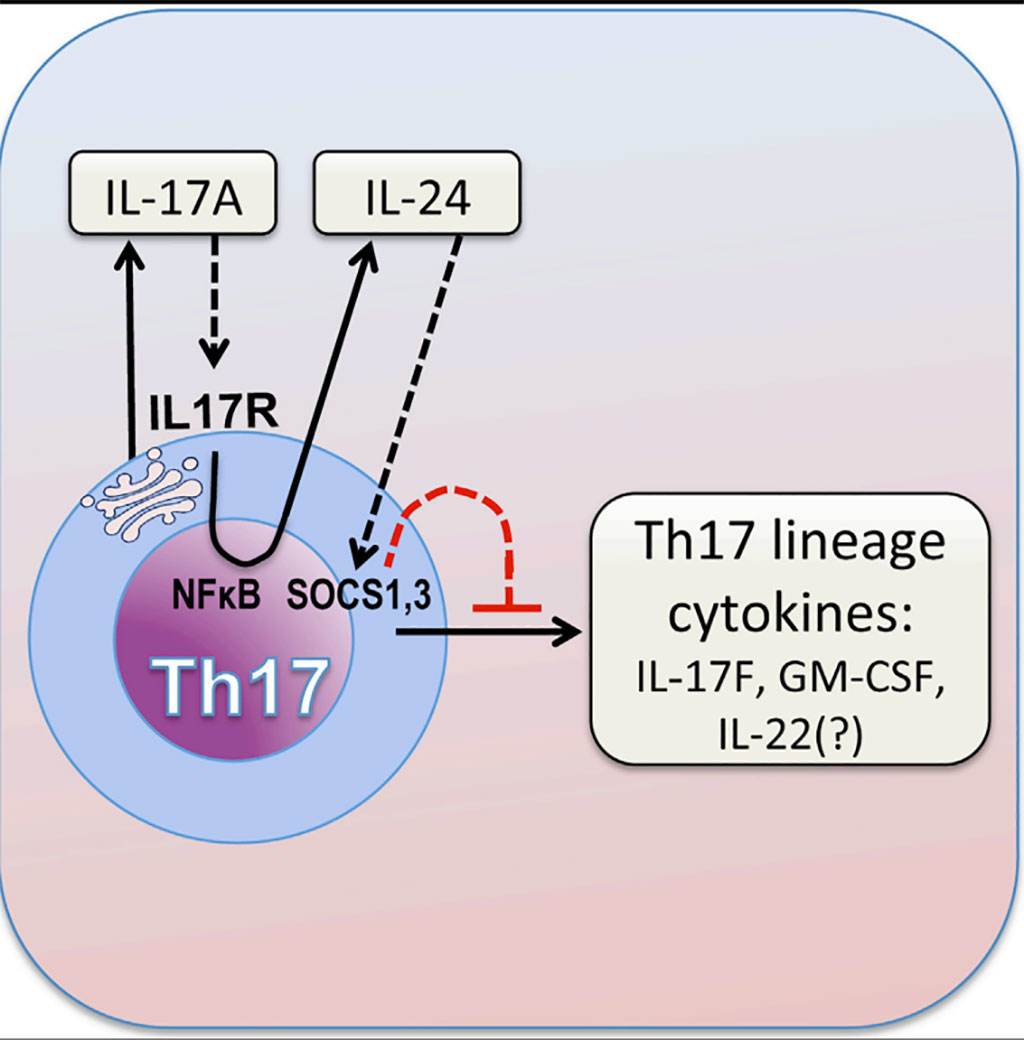
Image: Graphic diagram of how the cytokine IL-17A limits Th17 pathogenicity via a negative feedback loop driven by autocrine induction of IL-24 (Photo courtesy of the US National Eye Institute).
Autoimmune uveitis (AU) is an inflammatory process of the uveal components of the eye, due to an autoimmune reaction to self-antigens or caused by an innate inflammatory reaction secondary to an external stimulus. It can present as an isolated entity or associated with a systemic autoimmune or autoinflammatory disease.
In autoimmune uveitis, immune cells become abnormally activated and begin to destroy healthy cells, including light-sensing photoreceptors and neurons. A key immune cell involved in this response is the Th17 lymphocyte, which produces several pro-inflammatory molecules known as cytokines. A hallmark of Th17 cells is the ability to produce interleukin-17A (IL-17A), which attracts immune cells called neutrophils that can damage tissue.
Immunologists at the US National Eye Institute (Bethesda, MD, USA) and their international colleagues used mouse models and human peripheral blood samples collected from 14 healthy blood donors (seven males and seven females, ages ranging from 20 to 36 years) by venipuncture and peripheral blood mononuclear cells (PBMC) were isolated. Naive CD45RA+CD4+ T cells were isolated from the PBMCs using naive CD4+ T cell isolation kit II (Miltenyi Biotech, Bergisch Gladbach, Germany). Several other methods were used in the study, including immunofluorescence studies monitored with a Zeiss LSM 880 Confocal Microscope (Carl Zeiss, White Plains , NY, USA).
The team were able to selectively remove IL-17A from Th17 cells and examine the cells' behavior in models of both uveitis and multiple sclerosis (MS). Intriguingly, they found that these cells produce more IL-17F, granulocyte-macrophage colony-stimulating factor (GM-CSF), and possibly other inflammatory molecules. They concluded that these additional inflammatory cytokines compensate for the loss of IL-17A in driving inflammation. They found that when IL-17A binds to its receptor on Th17 cells, this triggers a signaling cascade that turns up the cells' production of an anti-inflammatory molecule, interleukin-24 (IL-24), which was not previously known to be produced by Th17 cells. IL-24 in turn suppresses the rest of the Th17 cells' inflammatory program, turning down the production of cytokines like IL-17F, GM-CSF and possibly IL-22. Thus, without IL-17A, this autocrine loop does not happen, causing the Th17 cells to overproduce the other inflammatory cytokines and thereby increase inflammation.
Rachel Caspi, PhD, chief of the Laboratory of Immunology and senior author of the study, said, “There are some diseases, like psoriasis, where anti-IL-17A therapy has been spectacularly successful. We expected that this would also apply to uveitis, but it turned out not to be the case. This study might explain why clinical trials targeting IL-17A to treat uveitis were not successful, and suggests that a combination approach involving both IL-17A and IL-24 may be more effective in treating autoimmune disorders of the nervous system.”
Related Links:
US National Eye Institute
Miltenyi Biotech
Carl Zeiss
Latest Immunology News
- Blood Test Identifies Lung Cancer Patients Who Can Benefit from Immunotherapy Drug
- Whole-Genome Sequencing Approach Identifies Cancer Patients Benefitting From PARP-Inhibitor Treatment
- Ultrasensitive Liquid Biopsy Demonstrates Efficacy in Predicting Immunotherapy Response
- Blood Test Could Identify Colon Cancer Patients to Benefit from NSAIDs
- Blood Test Could Detect Adverse Immunotherapy Effects
- Routine Blood Test Can Predict Who Benefits Most from CAR T-Cell Therapy
- New Test Distinguishes Vaccine-Induced False Positives from Active HIV Infection
- Gene Signature Test Predicts Response to Key Breast Cancer Treatment
- Chip Captures Cancer Cells from Blood to Help Select Right Breast Cancer Treatment
- Blood-Based Liquid Biopsy Model Analyzes Immunotherapy Effectiveness
- Signature Genes Predict T-Cell Expansion in Cancer Immunotherapy
- Molecular Microscope Diagnostic System Assesses Lung Transplant Rejection
- Blood Test Tracks Treatment Resistance in High-Grade Serous Ovarian Cancer
- Luminescent Probe Measures Immune Cell Activity in Real Time
- Blood-Based Immune Cell Signatures Could Guide Treatment Decisions for Critically Ill Patients
- Novel Tool Predicts Most Effective Multiple Sclerosis Medication for Patients
Channels
Clinical Chemistry
view channel
New PSA-Based Prognostic Model Improves Prostate Cancer Risk Assessment
Prostate cancer is the second-leading cause of cancer death among American men, and about one in eight will be diagnosed in their lifetime. Screening relies on blood levels of prostate-specific antigen... Read more
Extracellular Vesicles Linked to Heart Failure Risk in CKD Patients
Chronic kidney disease (CKD) affects more than 1 in 7 Americans and is strongly associated with cardiovascular complications, which account for more than half of deaths among people with CKD.... Read moreMolecular Diagnostics
view channel
Diagnostic Device Predicts Treatment Response for Brain Tumors Via Blood Test
Glioblastoma is one of the deadliest forms of brain cancer, largely because doctors have no reliable way to determine whether treatments are working in real time. Assessing therapeutic response currently... Read more
Blood Test Detects Early-Stage Cancers by Measuring Epigenetic Instability
Early-stage cancers are notoriously difficult to detect because molecular changes are subtle and often missed by existing screening tools. Many liquid biopsies rely on measuring absolute DNA methylation... Read more
“Lab-On-A-Disc” Device Paves Way for More Automated Liquid Biopsies
Extracellular vesicles (EVs) are tiny particles released by cells into the bloodstream that carry molecular information about a cell’s condition, including whether it is cancerous. However, EVs are highly... Read more
Blood Test Identifies Inflammatory Breast Cancer Patients at Increased Risk of Brain Metastasis
Brain metastasis is a frequent and devastating complication in patients with inflammatory breast cancer, an aggressive subtype with limited treatment options. Despite its high incidence, the biological... Read moreHematology
view channel
New Guidelines Aim to Improve AL Amyloidosis Diagnosis
Light chain (AL) amyloidosis is a rare, life-threatening bone marrow disorder in which abnormal amyloid proteins accumulate in organs. Approximately 3,260 people in the United States are diagnosed... Read more
Fast and Easy Test Could Revolutionize Blood Transfusions
Blood transfusions are a cornerstone of modern medicine, yet red blood cells can deteriorate quietly while sitting in cold storage for weeks. Although blood units have a fixed expiration date, cells from... Read more
Automated Hemostasis System Helps Labs of All Sizes Optimize Workflow
High-volume hemostasis sections must sustain rapid turnaround while managing reruns and reflex testing. Manual tube handling and preanalytical checks can strain staff time and increase opportunities for error.... Read more
High-Sensitivity Blood Test Improves Assessment of Clotting Risk in Heart Disease Patients
Blood clotting is essential for preventing bleeding, but even small imbalances can lead to serious conditions such as thrombosis or dangerous hemorrhage. In cardiovascular disease, clinicians often struggle... Read moreMicrobiology
view channel
Comprehensive Review Identifies Gut Microbiome Signatures Associated With Alzheimer’s Disease
Alzheimer’s disease affects approximately 6.7 million people in the United States and nearly 50 million worldwide, yet early cognitive decline remains difficult to characterize. Increasing evidence suggests... Read moreAI-Powered Platform Enables Rapid Detection of Drug-Resistant C. Auris Pathogens
Infections caused by the pathogenic yeast Candida auris pose a significant threat to hospitalized patients, particularly those with weakened immune systems or those who have invasive medical devices.... Read morePathology
view channel
Engineered Yeast Cells Enable Rapid Testing of Cancer Immunotherapy
Developing new cancer immunotherapies is a slow, costly, and high-risk process, particularly for CAR T cell treatments that must precisely recognize cancer-specific antigens. Small differences in tumor... Read more
First-Of-Its-Kind Test Identifies Autism Risk at Birth
Autism spectrum disorder is treatable, and extensive research shows that early intervention can significantly improve cognitive, social, and behavioral outcomes. Yet in the United States, the average age... Read moreTechnology
view channel
Robotic Technology Unveiled for Automated Diagnostic Blood Draws
Routine diagnostic blood collection is a high‑volume task that can strain staffing and introduce human‑dependent variability, with downstream implications for sample quality and patient experience.... Read more
ADLM Launches First-of-Its-Kind Data Science Program for Laboratory Medicine Professionals
Clinical laboratories generate billions of test results each year, creating a treasure trove of data with the potential to support more personalized testing, improve operational efficiency, and enhance patient care.... Read moreAptamer Biosensor Technology to Transform Virus Detection
Rapid and reliable virus detection is essential for controlling outbreaks, from seasonal influenza to global pandemics such as COVID-19. Conventional diagnostic methods, including cell culture, antigen... Read more
AI Models Could Predict Pre-Eclampsia and Anemia Earlier Using Routine Blood Tests
Pre-eclampsia and anemia are major contributors to maternal and child mortality worldwide, together accounting for more than half a million deaths each year and leaving millions with long-term health complications.... Read moreIndustry
view channelNew Collaboration Brings Automated Mass Spectrometry to Routine Laboratory Testing
Mass spectrometry is a powerful analytical technique that identifies and quantifies molecules based on their mass and electrical charge. Its high selectivity, sensitivity, and accuracy make it indispensable... Read more
AI-Powered Cervical Cancer Test Set for Major Rollout in Latin America
Noul Co., a Korean company specializing in AI-based blood and cancer diagnostics, announced it will supply its intelligence (AI)-based miLab CER cervical cancer diagnostic solution to Mexico under a multi‑year... Read more
Diasorin and Fisher Scientific Enter into US Distribution Agreement for Molecular POC Platform
Diasorin (Saluggia, Italy) has entered into an exclusive distribution agreement with Fisher Scientific, part of Thermo Fisher Scientific (Waltham, MA, USA), for the LIAISON NES molecular point-of-care... Read more


