Personal Lab Offers Rapid Detection of Food Allergens
|
By LabMedica International staff writers Posted on 06 Nov 2017 |


|
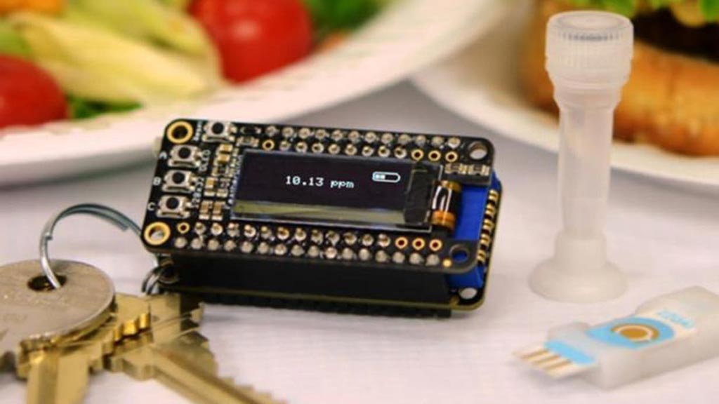
Image: The iEAT system for onsite antigen detection consists of a pocket-sized detector, an electrode chip, and a disposable kit for allergen extraction. The detector connects with a smartphone for system control and data upload to a cloud server (Photo courtesy of Lin, et al. ACS Nano, August 2017).
Researchers have developed a small, low cost device for individual use that can accurately detect food allergens in less than ten minutes.
Adverse food reactions, including food allergies, food sensitivities, and autoimmune reaction (e.g., celiac disease) affect 5-15% of the population of the USA and remain a considerable public health problem requiring stringent food avoidance and epinephrine availability for emergency events. Avoiding problematic foods is difficult in practical terms, given current reliance on prepared foods and out-of-home meals.
In response to the food allergy problem, investigators at Harvard Medical School (Boston, MA, USA) developed a portable, point-of-use detection technology, that they called "integrated exogenous antigen testing" (iEAT).
The iEAT device consists of three components: (1) a small plastic test tube, (2) a small electronic detection module, and (3) the electronic keychain reader. To perform the test, the user dissolves a small sample of the food in the plastic test tube and then adds magnetic beads that capture the food allergen of interest. A bit of this mixture is loaded onto electrode strips attached to a small module that is then inserted into the electronic keychain reader. The keychain reader has a small display that indicates whether the allergen is present, and if so, in what concentration.
The prototype iEAT system was optimized to detect five major food antigens in peanuts, hazelnuts, wheat, milk, and eggs. Antigen extraction and detection with iEAT required less than 10 minutes and achieved high-detection sensitivities (e.g., 0.1 milligram per kilogram for gluten, 200 times lower than regulatory limits of 20 milligram per kilogram).
The investigators also developed a dedicated cell phone application, which allows the user to compile and store the data collected by testing different foods for various allergens at different restaurants or in packaged foods. The application is set up to share this information online with both time and location stamps indicating when, where, and in what food or dish an allergen reading was taken.
“High accuracy built into a compact system was the key goals of the project,” said contributing author Dr. Ralph Weissleder, professor of radiology and systems biology at Harvard Medical School. “Users can be confident that even if they are sensitive to very low levels, iEAT will be able to give them exact concentrations. Armed with accurate concentration levels they will not have to completely avoid potentially problematic foods, but will know whether an allergen is at a dangerous level for them or a concentration that is safe for them to eat.”
The iEAT device was described in the August 2017 issue of the journal ACS Nano.
Related Links:
Harvard Medical School






Adverse food reactions, including food allergies, food sensitivities, and autoimmune reaction (e.g., celiac disease) affect 5-15% of the population of the USA and remain a considerable public health problem requiring stringent food avoidance and epinephrine availability for emergency events. Avoiding problematic foods is difficult in practical terms, given current reliance on prepared foods and out-of-home meals.
In response to the food allergy problem, investigators at Harvard Medical School (Boston, MA, USA) developed a portable, point-of-use detection technology, that they called "integrated exogenous antigen testing" (iEAT).
The iEAT device consists of three components: (1) a small plastic test tube, (2) a small electronic detection module, and (3) the electronic keychain reader. To perform the test, the user dissolves a small sample of the food in the plastic test tube and then adds magnetic beads that capture the food allergen of interest. A bit of this mixture is loaded onto electrode strips attached to a small module that is then inserted into the electronic keychain reader. The keychain reader has a small display that indicates whether the allergen is present, and if so, in what concentration.
The prototype iEAT system was optimized to detect five major food antigens in peanuts, hazelnuts, wheat, milk, and eggs. Antigen extraction and detection with iEAT required less than 10 minutes and achieved high-detection sensitivities (e.g., 0.1 milligram per kilogram for gluten, 200 times lower than regulatory limits of 20 milligram per kilogram).
The investigators also developed a dedicated cell phone application, which allows the user to compile and store the data collected by testing different foods for various allergens at different restaurants or in packaged foods. The application is set up to share this information online with both time and location stamps indicating when, where, and in what food or dish an allergen reading was taken.
“High accuracy built into a compact system was the key goals of the project,” said contributing author Dr. Ralph Weissleder, professor of radiology and systems biology at Harvard Medical School. “Users can be confident that even if they are sensitive to very low levels, iEAT will be able to give them exact concentrations. Armed with accurate concentration levels they will not have to completely avoid potentially problematic foods, but will know whether an allergen is at a dangerous level for them or a concentration that is safe for them to eat.”
The iEAT device was described in the August 2017 issue of the journal ACS Nano.
Related Links:
Harvard Medical School

Platinum Member
Automated Coagulation Analyzer
Hemolumi H6
Gold Member
Pre- Eclampsia Control
Acusera Pre-Eclampsia Control
Creatinine/eGFR Meter
StatSensor® Creatinine/eGFR Meter
New
MR-proADM Test
B•R•A•H•M•S MR-proADM KRYPTOR test
Latest Technology News
- Autonomous Robotic System Receives FDA Authorization for Blood Collection
- Interoperable Data Platform Standardizes Multi-Cancer Blood Test Results
- Training Device Improves Accuracy of Pooled Molecular Diagnostics
- New CE-Certified Software Advances Whole-Genome Cancer Testing
- National Rare Disease Registry Standardizes Genetic and Clinical Data for Coordinated Care
- AI Platform Links Biomarker Results to Cancer Clinical Trials and Guidelines
- Agentic AI Platform Supports Genomic Decision-Making in Oncology
- Algorithm Panel Aids Liver Fibrosis Assessment and Liver Cancer Surveillance
- Mailed Screening Kits Help Reduce Colorectal Cancer Screening Gaps
- AI-Enabled Assistant Unifies Molecular Workflow Planning and Support
- AI Tool Automates Validation of Laboratory Software Configuration Changes
- Point-of-Care Testing Enhances Health Literacy and Self-Management in Chronic Disease
- Fully Automated Sample-to-Insight Workflow Advances Latent TB Testing
- Tumor-on-a-Chip Platform Models Pancreatic Cancer Treatment Response
- New Platform Captures Extracellular Vesicles for Early Cancer Detection
- Microfluidic Single-Cell Assay Predicts Breast Cancer Risk
Channels
Clinical Chemistry
view channel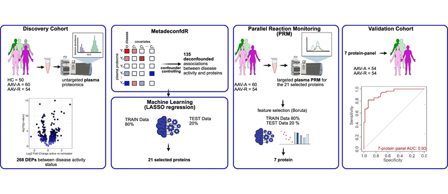
Multiprotein Plasma Panel Identifies Remission in Vasculitis
Vasculitis causes immune-mediated inflammation of blood vessel walls and can lead to kidney and lung damage. Clinicians rely on immunosuppressive drugs, but treatment carries significant side effects and... Read more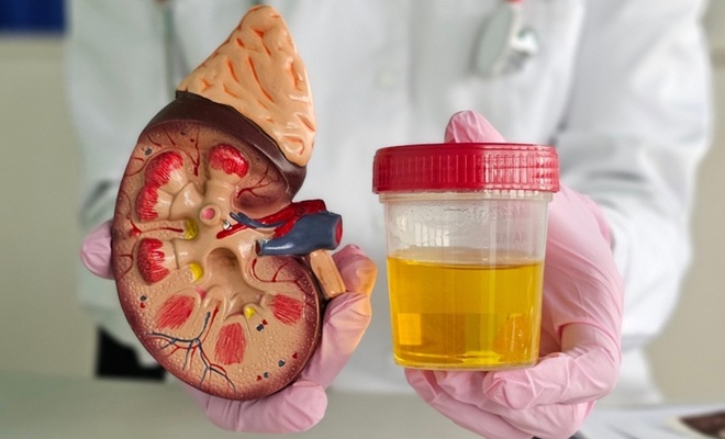
Rapid Urine Biomarker Panel Supports Better Kidney Transplant Decisions
Kidney transplantation remains constrained by organ scarcity and uncertainty about the quality of deceased-donor kidneys. In the United States, more than 92,000 people are awaiting a kidney transplant,... Read more
Hematology
view channel
New Genetic Findings Reveal Cause of Bone Marrow Failure Syndrome
Inherited bone marrow failure syndromes (IBMFS) impair the bone marrow’s ability to produce sufficient healthy blood cells and are associated with an increased risk of early-onset myelodysplastic syndromes (MDS).... Read more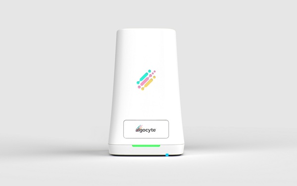
Ultra-Portable Device Enables Finger-Prick Blood Testing at Home
Patients who need frequent blood tests often face repeated clinic visits that burden services and disrupt care. In the UK, millions of tests are performed each year to diagnose disease, guide therapy,... Read more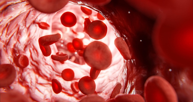
Immunology
view channel
Gut “Memory” May Explain Why IBD Flares Return
Inflammatory bowel diseases (IBD), including Crohn’s disease and ulcerative colitis, are characterized by recurring flare-ups that damage the intestinal mucosa and remain difficult to predict.... Read more
AI-Based Antibody Profiling Predicts Strength of COVID-19 Vaccine Response
Vaccine-induced protection can vary widely between individuals, creating uncertainty for clinicians, particularly in patients with immunosuppressive conditions. Demographic and health factors explain only... Read more
Microbiology
view channel
New Diagnostic Workflow Identifies Bloodstream Pathogens and Antibiotic Response in Hours
Sepsis is a life-threatening complication of infection that affects more than 1.5 million patients annually in the United States and contributes to roughly one in three in-hospital deaths.... Read more
Genetic Marker Identifies Emerging Resistance to Frontline Malaria Drugs
Artemisinin-based combination therapy remains central to controlling Plasmodium falciparum malaria, but emerging drug resistance is complicating treatment and surveillance. In Uganda and across sub-Saharan... Read more
Pathology
view channel
AI Matches Pathologists in Predicting Breast Cancer Prognosis from Immune Cells
Breast cancer is the most common cancer in Australian women, with more than 20,000 cases each year. Prognosis can be informed by counting tumor-infiltrating lymphocytes (TILs) on routine pathology slides,... Read more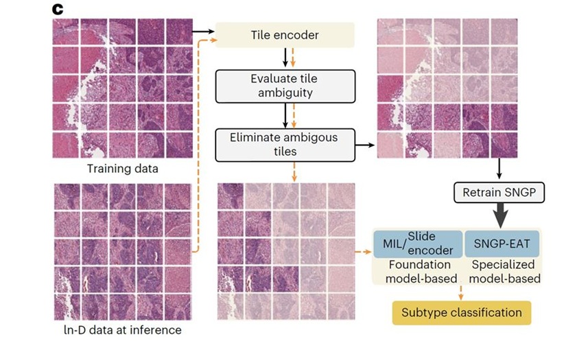
AI-Pathologist Framework Improves Accuracy and Reliability in Cancer Diagnosis
Ensuring reliable cancer diagnosis from digital pathology remains a critical challenge as clinical decisions rely on accurate slide interpretation. While artificial intelligence (AI) has accelerated whole-slide... Read more
New Review Highlights Intelligent Agents as Next Step for Digital Pathology
Digital pathology remains constrained by models that classify single images without mirroring how clinicians interrogate multiple slides, adjust magnification, and synthesize ancillary tests.... Read more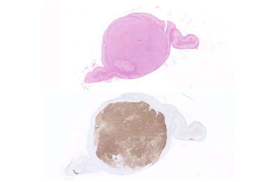
Simple Immunohistochemical Size Ratio Improves Classification of Primary Aldosteronism
Primary aldosteronism is a common but underdiagnosed cause of hypertension, in which excess aldosterone promotes salt retention and raises blood pressure. Identifying the dominant source of hormone overproduction... Read moreTechnology
view channel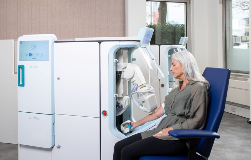
Autonomous Robotic System Receives FDA Authorization for Blood Collection
Venipuncture is central to many diagnostic pathways, but routine blood collection can be affected by staffing constraints and procedural variability that influence consistency and patient experience.... Read more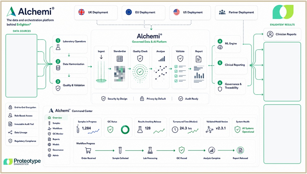
Interoperable Data Platform Standardizes Multi-Cancer Blood Test Results
Proteotype Diagnostics has introduced Alchemi, a proprietary data and clinical workflow platform being developed for Enlighten, the company’s investigational blood-based multi-cancer test.... Read more

Industry
view channel
Tumor-Informed MRD Assay Gains Medicare Coverage for Immunotherapy Monitoring
NeoGenomics’ RaDaR ST molecular residual disease (MRD) assay has received expanded coverage from the Centers for Medicare & Medicaid Services’ Molecular Diagnostic Services Program (MolDX) for monitoring... Read more