Changes in Eye Tissue May Enable Early Detection of Brain Diseases
|
By LabMedica International staff writers Posted on 11 Oct 2016 |

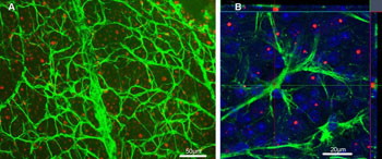
Image: An experiment examining retina tissue for mHtt deposition in GFAP-ir astrocytes in R6/2 mouse model of Huntington’s Disease. Green: glial fibrillary acidic protein (GFAP); Red: mutant huntingtin protein (mHtt). (A) A low magnification picture illustrates GFAP-ir astrocytes and mHtt deposits from the retinal wholemount of 12-week-old R6/2 (Huntington’s disease model) mouse. Scale bar = 50 µm. (B) A detailed confocal analysis of GFAP positivity, mHtt immunoreactivity, and DAPI counterstain (blue) revealed no colocalization of GFAP and mHtt. Scale bar = 20 µm (Image courtesy of PLoS One).
Research with mouse models has shown that at least some diseases of the central nervous system (CNS) manifest as pathological changes in the retina of the eye and that these changes may be detected earlier than brain changes. The findings suggest that eye examination could be used for minimally invasive screening for these diseases.
Retina tissue can be considered an integral part of the central nervous system (CNS). During fetal development, it matures from part of the brain and its innervation closely resembles that of the brain. Retinal structure and function can be readily examined with noninvasive or minimally invasive methods, whereas direct brain examination has numerous limitations. If, at least for some brain diseases, the health status of the brain could be indirectly assessed through the eyes, diagnostic screening could become more efficient.
In his PhD project at the University of Eastern Finland at Kuopio (Kuopio, Finland), Dr. Henri Leinonen and colleagues investigated functional abnormalities of the retina using mouse models of human CNS diseases. Electroretinography (ERG) and visual evoked potentials (VEP) were chosen as research techniques, since similar methodology can be applied in both laboratory animals and humans. ERG can precisely track the function of retina using corneal or skin electrodes, whereas VEP measures the function of visual cortex.
These methods were used to test different attributes of vision in 3 distinct genetically engineered mouse models of human CNS diseases. Also, basic life science methods were used to test the correlation between functional abnormalities and the anatomical status of the retina.
Day and color vision -associated retinal dysfunction was found in a mouse model of Huntington´s disease (HD) while the mouse was presymptomatic. Retinal structure remained relatively normal, even in an advanced disease state, although aggregation of toxic mutated huntingtin-protein was widespread in the diseased mouse retina. Although the retinopathy in mice is exaggerated compared to human HD patients, the finding is partly in line with patient data showing impaired color vision but no clear-cut anatomical retinopathy.
In a mouse model of Alzheimer´s disease (AD), the researchers observed abnormality in night vision -associated retinal function. Specifically, rod-mediated inner retinal responses to dim light flashes were faster in diseased mice than in their wild-type controls. The observation may be explained by impaired cholinergic neurotransmission that is also partly causative for the deterioration of memory in AD.
In a mouse model of late infantile neuronal ceroid lipofuscinosis (NCL), a pediatric neurological disease, the researchers described retinal degenerative changes that mimic the characteristic pathology of age-related macular degeneration (AMD). These included impaired function of retinal pigment epithelium and subsequent blindness due to photoreceptor atrophy and death. It has been postulated that the retinal degeneration in human patients progresses similarly.
Adding to the growing body of evidence, the results showed that functional changes of the retina occur in mouse models of three human CNS diseases whose phenotype, age of onset, and pathological mechanism clearly differ from each other. Visual impairment was the fastest progressive symptom in two models tested.
The findings support the idea of eye examinations as potential screening tools for CNS diseases. Development of efficient, safe, and economic screening is imperative since the diagnosis of these diseases is often obtained only in the advanced disease state, when as such satisfactory remedies are poorly effective. Since eye and vision research can be conducted noninvasively, advancement of trials from the preclinical to the clinical phase could be relatively fast.
Dr. Leinonen’s doctoral dissertation, entitled “Electrophysiology of visual pathways as a screening tool for neurodegenerative diseases: evidence from mouse disease models”, is available for download. The findings were published in PLoS One, the Journal of Alzheimer’s Disease, and most recently in the journal Human Molecular Genetics.
Related Links:
University of Eastern Finland
Latest Pathology News
- Engineered Yeast Cells Enable Rapid Testing of Cancer Immunotherapy
- First-Of-Its-Kind Test Identifies Autism Risk at Birth
- AI Algorithms Improve Genetic Mutation Detection in Cancer Diagnostics
- Skin Biopsy Offers New Diagnostic Method for Neurodegenerative Diseases
- Fast Label-Free Method Identifies Aggressive Cancer Cells
- New X-Ray Method Promises Advances in Histology
- Single-Cell Profiling Technique Could Guide Early Cancer Detection
- Intraoperative Tumor Histology to Improve Cancer Surgeries
- Rapid Stool Test Could Help Pinpoint IBD Diagnosis
- AI-Powered Label-Free Optical Imaging Accurately Identifies Thyroid Cancer During Surgery
- Deep Learning–Based Method Improves Cancer Diagnosis
- ADLM Updates Expert Guidance on Urine Drug Testing for Patients in Emergency Departments
- New Age-Based Blood Test Thresholds to Catch Ovarian Cancer Earlier
- Genetics and AI Improve Diagnosis of Aortic Stenosis
- AI Tool Simultaneously Identifies Genetic Mutations and Disease Type
- Rapid Low-Cost Tests Can Prevent Child Deaths from Contaminated Medicinal Syrups
Channels
Clinical Chemistry
view channel
New PSA-Based Prognostic Model Improves Prostate Cancer Risk Assessment
Prostate cancer is the second-leading cause of cancer death among American men, and about one in eight will be diagnosed in their lifetime. Screening relies on blood levels of prostate-specific antigen... Read more
Extracellular Vesicles Linked to Heart Failure Risk in CKD Patients
Chronic kidney disease (CKD) affects more than 1 in 7 Americans and is strongly associated with cardiovascular complications, which account for more than half of deaths among people with CKD.... Read moreMolecular Diagnostics
view channel
Diagnostic Device Predicts Treatment Response for Brain Tumors Via Blood Test
Glioblastoma is one of the deadliest forms of brain cancer, largely because doctors have no reliable way to determine whether treatments are working in real time. Assessing therapeutic response currently... Read more
Blood Test Detects Early-Stage Cancers by Measuring Epigenetic Instability
Early-stage cancers are notoriously difficult to detect because molecular changes are subtle and often missed by existing screening tools. Many liquid biopsies rely on measuring absolute DNA methylation... Read more
“Lab-On-A-Disc” Device Paves Way for More Automated Liquid Biopsies
Extracellular vesicles (EVs) are tiny particles released by cells into the bloodstream that carry molecular information about a cell’s condition, including whether it is cancerous. However, EVs are highly... Read more
Blood Test Identifies Inflammatory Breast Cancer Patients at Increased Risk of Brain Metastasis
Brain metastasis is a frequent and devastating complication in patients with inflammatory breast cancer, an aggressive subtype with limited treatment options. Despite its high incidence, the biological... Read moreHematology
view channel
New Guidelines Aim to Improve AL Amyloidosis Diagnosis
Light chain (AL) amyloidosis is a rare, life-threatening bone marrow disorder in which abnormal amyloid proteins accumulate in organs. Approximately 3,260 people in the United States are diagnosed... Read more
Fast and Easy Test Could Revolutionize Blood Transfusions
Blood transfusions are a cornerstone of modern medicine, yet red blood cells can deteriorate quietly while sitting in cold storage for weeks. Although blood units have a fixed expiration date, cells from... Read more
Automated Hemostasis System Helps Labs of All Sizes Optimize Workflow
High-volume hemostasis sections must sustain rapid turnaround while managing reruns and reflex testing. Manual tube handling and preanalytical checks can strain staff time and increase opportunities for error.... Read more
High-Sensitivity Blood Test Improves Assessment of Clotting Risk in Heart Disease Patients
Blood clotting is essential for preventing bleeding, but even small imbalances can lead to serious conditions such as thrombosis or dangerous hemorrhage. In cardiovascular disease, clinicians often struggle... Read moreImmunology
view channelBlood Test Identifies Lung Cancer Patients Who Can Benefit from Immunotherapy Drug
Small cell lung cancer (SCLC) is an aggressive disease with limited treatment options, and even newly approved immunotherapies do not benefit all patients. While immunotherapy can extend survival for some,... Read more
Whole-Genome Sequencing Approach Identifies Cancer Patients Benefitting From PARP-Inhibitor Treatment
Targeted cancer therapies such as PARP inhibitors can be highly effective, but only for patients whose tumors carry specific DNA repair defects. Identifying these patients accurately remains challenging,... Read more
Ultrasensitive Liquid Biopsy Demonstrates Efficacy in Predicting Immunotherapy Response
Immunotherapy has transformed cancer treatment, but only a small proportion of patients experience lasting benefit, with response rates often remaining between 10% and 20%. Clinicians currently lack reliable... Read moreMicrobiology
view channel
Comprehensive Review Identifies Gut Microbiome Signatures Associated With Alzheimer’s Disease
Alzheimer’s disease affects approximately 6.7 million people in the United States and nearly 50 million worldwide, yet early cognitive decline remains difficult to characterize. Increasing evidence suggests... Read moreAI-Powered Platform Enables Rapid Detection of Drug-Resistant C. Auris Pathogens
Infections caused by the pathogenic yeast Candida auris pose a significant threat to hospitalized patients, particularly those with weakened immune systems or those who have invasive medical devices.... Read moreTechnology
view channel
Robotic Technology Unveiled for Automated Diagnostic Blood Draws
Routine diagnostic blood collection is a high‑volume task that can strain staffing and introduce human‑dependent variability, with downstream implications for sample quality and patient experience.... Read more
ADLM Launches First-of-Its-Kind Data Science Program for Laboratory Medicine Professionals
Clinical laboratories generate billions of test results each year, creating a treasure trove of data with the potential to support more personalized testing, improve operational efficiency, and enhance patient care.... Read moreAptamer Biosensor Technology to Transform Virus Detection
Rapid and reliable virus detection is essential for controlling outbreaks, from seasonal influenza to global pandemics such as COVID-19. Conventional diagnostic methods, including cell culture, antigen... Read more
AI Models Could Predict Pre-Eclampsia and Anemia Earlier Using Routine Blood Tests
Pre-eclampsia and anemia are major contributors to maternal and child mortality worldwide, together accounting for more than half a million deaths each year and leaving millions with long-term health complications.... Read moreIndustry
view channelNew Collaboration Brings Automated Mass Spectrometry to Routine Laboratory Testing
Mass spectrometry is a powerful analytical technique that identifies and quantifies molecules based on their mass and electrical charge. Its high selectivity, sensitivity, and accuracy make it indispensable... Read more
AI-Powered Cervical Cancer Test Set for Major Rollout in Latin America
Noul Co., a Korean company specializing in AI-based blood and cancer diagnostics, announced it will supply its intelligence (AI)-based miLab CER cervical cancer diagnostic solution to Mexico under a multi‑year... Read more
Diasorin and Fisher Scientific Enter into US Distribution Agreement for Molecular POC Platform
Diasorin (Saluggia, Italy) has entered into an exclusive distribution agreement with Fisher Scientific, part of Thermo Fisher Scientific (Waltham, MA, USA), for the LIAISON NES molecular point-of-care... Read more


 Analyzer.jpg)


