Circulating Angiogenic Factor Levels Predict Hypertensive Disorders of Pregnancy
|
By LabMedica International staff writers Posted on 15 Nov 2022 |

Preeclampsia is the most common hypertensive disorder associated with pregnancy. The severe form of the disease can lead to dangerously high blood pressure, organ failure, vision loss or even stroke. It affects approximately 5% of pregnant women and is a leading cause of maternal and fetal death and serious illness.
Currently, the only cure for preeclampsia is delivery. A test indicating that a preterm patient, a woman who has completed less than 37 weeks of pregnancy, is likely to develop severe disease could help optimize care. Prior studies suggest that preeclampsia is characterized by angiogenic factor imbalance. Levels of soluble fms-like tyrosine kinase 1 (sFlt-1) and/or placental growth factor (PlGF), both angiogenic placental proteins, may be used to predict, diagnose, and prognosticate women with a suspicion of preeclampsia.
A large team of Medical Scientists led by those at Cedars-Sinai Medical Center (Los Angeles, CA, USA), prospectively measured across 18 USA centers, the ratio of serum soluble fms-like tyrosine kinase 1 (sFlt-1) to placental growth factor (PlGF) in pregnant women hospitalized between 23 and 35 weeks of gestation. The primary outcome was predicting preeclampsia with severe features (sPE), and secondary outcomes included predicting adverse outcomes within two weeks. The prognostic performance of the sFlt-1: PlGF ratio was assessed by using a derivation/validation design. Between 2019 and 2021, the enrolled 1,014 women at one of 18 USA hospitals with a singleton pregnancy and a hypertensive disorder of pregnancy.
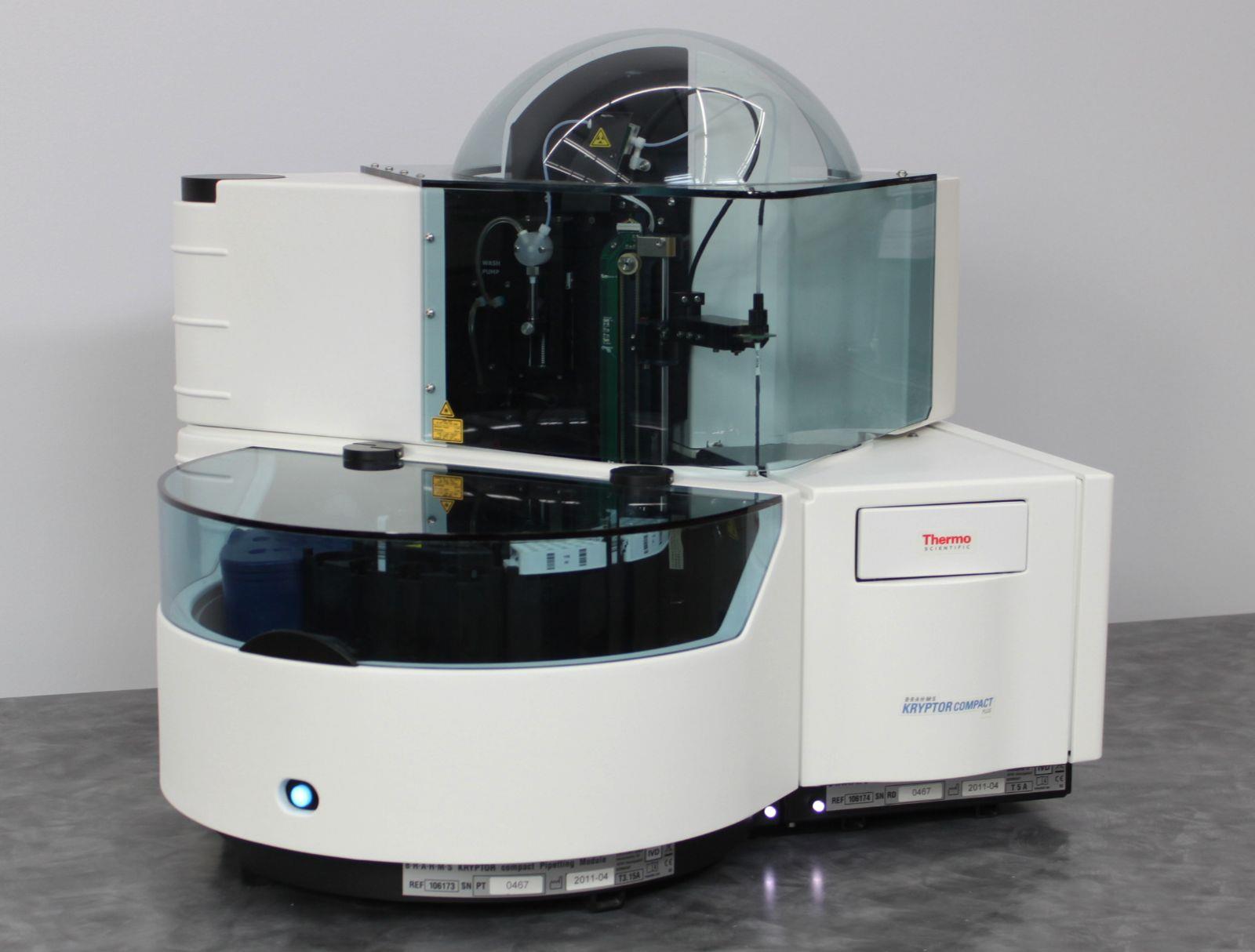
Serum samples were stored at –20 °C or lower within five hours of venipuncture. Specimens were routinely batch-shipped from sites to Cedars-Sinai Medical Center where sFlt-1 and PlGF levels (both in picograms per milliliter [hence the ratio is unitless]) were measured in a blinded fashion on an automated sandwich immunoassay platform: BRAHMS PlGF and sFlt-1 (KRYPTOR; Thermo Fisher Scientific, Henningsdorf, Germany). These assays are CE-marked and have been validated in pregnant populations.
The investigators reported that of the total of 1,014 pregnant women that were evaluated; 299 were included in the derivation cohort and 715 in the validation cohort. In the derivation cohort, the median sFlt-1: PlGF ratio was 200 (interquartile range, 53 to 458) among women who developed sPE compared with 6 (interquartile range, 3 to 26) in those who did not. The discriminatory ratio of ≥40 was then tested in the validation cohort and yielded a 65% positive and a 96% negative predictive value for the primary outcome. The ratio performed better than standard clinical measures (area under the receiver-operating characteristic curve, 0.92 versus <0.75 for standard-of-care tests). Compared with women with a ratio <40, women with a ratio ≥40 were at higher risk for adverse maternal outcomes (16.1% versus 2.8%; relative risk, 5.8).
Sarah Kilpatrick, MD, PhD, co-senior author of the study, said, “This test was significantly better than all the standard-of-care markers for preeclampsia with severe features. It predicted with over 90% accuracy whether the patient would develop preeclampsia with severe features or not, while the usual markers were accurate less than 75% of the time."
The authors concluded that in women with a hypertensive disorder of pregnancy presenting between 23 and 35 weeks of gestation, measurement of serum sFlt-1: PlGF provided stratification of the risk of progressing to sPE within the coming fortnight. The study was published on November 9, 2022 in the journal NEJM Evidence.
Related Links:
Cedars-Sinai Medical Center
Thermo Fisher Scientific
Latest Clinical Chem. News
- 3D Printed Point-Of-Care Mass Spectrometer Outperforms State-Of-The-Art Models
- POC Biomedical Test Spins Water Droplet Using Sound Waves for Cancer Detection
- Highly Reliable Cell-Based Assay Enables Accurate Diagnosis of Endocrine Diseases
- New Blood Testing Method Detects Potent Opioids in Under Three Minutes
- Wireless Hepatitis B Test Kit Completes Screening and Data Collection in One Step
- Pain-Free, Low-Cost, Sensitive, Radiation-Free Device Detects Breast Cancer in Urine
- Spit Test Detects Breast Cancer in Five Seconds
- Electrochemical Sensors with Next-Generation Coating Advances Precision Diagnostics at POC
- First-Of-Its-Kind Handheld Device Accurately Detects Fentanyl in Urine within Seconds
- New Fluorescent Sensor Array Lights up Alzheimer’s-Related Proteins for Earlier Detection
- Automated Mass Spectrometry-Based Clinical Analyzer Could Transform Lab Testing
- Highly Sensitive pH Sensor to Aid Detection of Cancers and Vector-Borne Viruses
- Non-Invasive Sensor Monitors Changes in Saliva Compositions to Rapidly Diagnose Diabetes
- Breakthrough Immunoassays to Aid in Risk Assessment of Preeclampsia
- Urine Test for Monitoring Changes in Kidney Health Markers Can Predict New-Onset Heart Failure
- AACC Releases Comprehensive Diabetes Testing Guidelines
Channels
Molecular Diagnostics
view channel
New Genetic Testing Procedure Combined With Ultrasound Detects High Cardiovascular Risk
A key interest area in cardiovascular research today is the impact of clonal hematopoiesis on cardiovascular diseases. Clonal hematopoiesis results from mutations in hematopoietic stem cells and may lead... Read more
Blood Samples Enhance B-Cell Lymphoma Diagnostics and Prognosis
B-cell lymphoma is the predominant form of cancer affecting the lymphatic system, with about 30% of patients with aggressive forms of this disease experiencing relapse. Currently, the disease’s risk assessment... Read moreHematology
view channel
Next Generation Instrument Screens for Hemoglobin Disorders in Newborns
Hemoglobinopathies, the most widespread inherited conditions globally, affect about 7% of the population as carriers, with 2.7% of newborns being born with these conditions. The spectrum of clinical manifestations... Read more
First 4-in-1 Nucleic Acid Test for Arbovirus Screening to Reduce Risk of Transfusion-Transmitted Infections
Arboviruses represent an emerging global health threat, exacerbated by climate change and increased international travel that is facilitating their spread across new regions. Chikungunya, dengue, West... Read more
POC Finger-Prick Blood Test Determines Risk of Neutropenic Sepsis in Patients Undergoing Chemotherapy
Neutropenia, a decrease in neutrophils (a type of white blood cell crucial for fighting infections), is a frequent side effect of certain cancer treatments. This condition elevates the risk of infections,... Read more
First Affordable and Rapid Test for Beta Thalassemia Demonstrates 99% Diagnostic Accuracy
Hemoglobin disorders rank as some of the most prevalent monogenic diseases globally. Among various hemoglobin disorders, beta thalassemia, a hereditary blood disorder, affects about 1.5% of the world's... Read moreImmunology
view channel
Diagnostic Blood Test for Cellular Rejection after Organ Transplant Could Replace Surgical Biopsies
Transplanted organs constantly face the risk of being rejected by the recipient's immune system which differentiates self from non-self using T cells and B cells. T cells are commonly associated with acute... Read more
AI Tool Precisely Matches Cancer Drugs to Patients Using Information from Each Tumor Cell
Current strategies for matching cancer patients with specific treatments often depend on bulk sequencing of tumor DNA and RNA, which provides an average profile from all cells within a tumor sample.... Read more
Genetic Testing Combined With Personalized Drug Screening On Tumor Samples to Revolutionize Cancer Treatment
Cancer treatment typically adheres to a standard of care—established, statistically validated regimens that are effective for the majority of patients. However, the disease’s inherent variability means... Read moreMicrobiology
view channel
Clinical Decision Support Software a Game-Changer in Antimicrobial Resistance Battle
Antimicrobial resistance (AMR) is a serious global public health concern that claims millions of lives every year. It primarily results from the inappropriate and excessive use of antibiotics, which reduces... Read more
New CE-Marked Hepatitis Assays to Help Diagnose Infections Earlier
According to the World Health Organization (WHO), an estimated 354 million individuals globally are afflicted with chronic hepatitis B or C. These viruses are the leading causes of liver cirrhosis, liver... Read more
1 Hour, Direct-From-Blood Multiplex PCR Test Identifies 95% of Sepsis-Causing Pathogens
Sepsis contributes to one in every three hospital deaths in the US, and globally, septic shock carries a mortality rate of 30-40%. Diagnosing sepsis early is challenging due to its non-specific symptoms... Read morePathology
view channel.jpg)
Use of DICOM Images for Pathology Diagnostics Marks Significant Step towards Standardization
Digital pathology is rapidly becoming a key aspect of modern healthcare, transforming the practice of pathology as laboratories worldwide adopt this advanced technology. Digital pathology systems allow... Read more
First of Its Kind Universal Tool to Revolutionize Sample Collection for Diagnostic Tests
The COVID pandemic has dramatically reshaped the perception of diagnostics. Post the pandemic, a groundbreaking device that combines sample collection and processing into a single, easy-to-use disposable... Read moreAI-Powered Digital Imaging System to Revolutionize Cancer Diagnosis
The process of biopsy is important for confirming the presence of cancer. In the conventional histopathology technique, tissue is excised, sliced, stained, mounted on slides, and examined under a microscope... Read more
New Mycobacterium Tuberculosis Panel to Support Real-Time Surveillance and Combat Antimicrobial Resistance
Tuberculosis (TB), the leading cause of death from an infectious disease globally, is a contagious bacterial infection that primarily spreads through the coughing of patients with active pulmonary TB.... Read moreTechnology
view channel
New Diagnostic System Achieves PCR Testing Accuracy
While PCR tests are the gold standard of accuracy for virology testing, they come with limitations such as complexity, the need for skilled lab operators, and longer result times. They also require complex... Read more
DNA Biosensor Enables Early Diagnosis of Cervical Cancer
Molybdenum disulfide (MoS2), recognized for its potential to form two-dimensional nanosheets like graphene, is a material that's increasingly catching the eye of the scientific community.... Read more
Self-Heating Microfluidic Devices Can Detect Diseases in Tiny Blood or Fluid Samples
Microfluidics, which are miniature devices that control the flow of liquids and facilitate chemical reactions, play a key role in disease detection from small samples of blood or other fluids.... Read more
Breakthrough in Diagnostic Technology Could Make On-The-Spot Testing Widely Accessible
Home testing gained significant importance during the COVID-19 pandemic, yet the availability of rapid tests is limited, and most of them can only drive one liquid across the strip, leading to continued... Read moreIndustry
view channel_1.jpg)
Thermo Fisher and Bio-Techne Enter Into Strategic Distribution Agreement for Europe
Thermo Fisher Scientific (Waltham, MA USA) has entered into a strategic distribution agreement with Bio-Techne Corporation (Minneapolis, MN, USA), resulting in a significant collaboration between two industry... Read more
ECCMID Congress Name Changes to ESCMID Global
Over the last few years, the European Society of Clinical Microbiology and Infectious Diseases (ESCMID, Basel, Switzerland) has evolved remarkably. The society is now stronger and broader than ever before... Read more


