AI-Enabled POC Device Detects Cancers Without Biopsies
|
By LabMedica International staff writers Posted on 15 Sep 2022 |


|
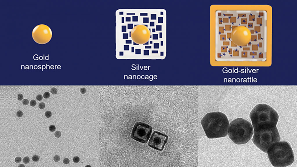
Researchers have developed a unique type of nanoparticle called a “nanorattle” that greatly enhances light emitted from within its outer shell. Loaded with light scattering dyes called Raman reporters commonly used to detect biomarkers of disease in organic samples, the approach can amplify and detect signals from separate types of nanoprobes without needing an expensive machine or medical professional to read the results. In a small proof-of-concept study, the nanorattles accurately identified head and neck cancers through an AI-enabled point-of-care device that could revolutionize how these cancers and other diseases are detected in low-resource areas to improve global health.
To make nanorattles, researchers at Duke University (Durham, NC, USA) start with a solid gold sphere about 20 nanometers wide. After growing a layer of silver around the gold core to make a larger sphere (or cube), they use a corrosion process called galvanic replacement that hollows out the silver, creating a cage-like shell around the core. The structure is then soaked in a solution containing positively charged Raman reporters, which are drawn into the outer cage by the negatively charged gold core. The outer hulls are then covered by an extremely thin layer of gold to lock the Raman reporters inside. The result is a nanosphere (or nanocube) about 60 nanometers wide with an architecture that resembles a rattle - a gold core trapped within a larger outer silver-gold shell. The gap between the two is only about a few nanometers, which is just large enough to fit the Raman reporters. Those tight tolerances are essential to controlling the Raman signal enhancement the nanorattles produce.
When a laser shines on the nanorattles, it travels through the extremely thin outer shell and hits the Raman reporters within, causing them to emit light of their own. Because of how close the surfaces of the gold core and the outer gold/silver shell are together, the laser also excites groups of electrons on the metallic structures, called plasmons. These groups of electrons create an extremely powerful electromagnetic field due to the plasmons’ interaction of the metallic core-shell architecture, a process called plasmonic coupling, which amplifies the light emitted by the Raman reporters millions of times over. The researchers applied the nanorattle technology to a lab-on-a-stick device capable of detecting head and neck cancers, which appear anywhere between the shoulders and the brain, typically in the mouth, nose and throat. Survival rate for these cancers have hovered between 40 and 60% for decades. While those statistics have improved in recent years in the U.S., they have gotten worse in low-resource settings, where risk factors such as smoking, drinking and betel nut chewing are much more prevalent.
The prototype device uses specific genetic sequences that act like Velcro for the biomarkers the researchers are looking for - in this case, a specific mRNA that is overly abundant in people with head and neck cancers. When the mRNA in question is present, it acts like a tether that binds nanorattles to magnetic beads. These beads are then concentrated and held in place by another magnet while everything else gets rinsed away. Researchers can then use a simple, inexpensive handheld device to look for light emitted from the nanorattles to see if any biomarkers were caught. In the experiments, the test determined whether or not 20 samples came from patients that had head and neck cancer with 100% accuracy. The experiments also showed that the nanorattle platform is capable of handling multiple types of nanoprobes, thanks to a machine learning algorithm that can tease apart the separate signals, meaning they can target multiple biomarkers at once.
“In low-resource settings, these cancers often present in advanced stages and result in poor outcomes due in part to limited examination equipment, lack of trained healthcare workers and essentially non-existent screening programs,” said Walter Lee, professor of head and neck surgery & communication sciences and radiation oncology at Duke, and a collaborator on the research. “Having the ability to detect these cancers early should lead to earlier treatment and improvement in outcomes, both in survival and quality of life,” Lee said. “This approach is exciting since it does not depend on a pathologist review and potentially could be used at the point of care.”
“Many mRNA biomarkers are overly abundant in multiple types of cancers, while other biomarkers can be used to evaluate patient risk and future treatment outcome,” said Tuan Vo-Dinh, the R. Eugene and Susie E. Goodson Distinguished Professor of Biomedical Engineering and professor of chemistry at Duke. “Detecting multiple biomarkers at once would help us differentiate between cancers, and also look for other prognostic markers such as Human Papillomavirus (HPV), and both positive and negative controls. Combining mRNA detection with novel nanorattle biosensing will result in a paradigm shift in achieving a diagnostic tool that could revolutionize how these cancers and other diseases are detected in low-resource areas”.
Related Links:
Duke University








Latest Molecular Diagnostics News
- Urine-Based RNA Improves Bladder Cancer Detection and Monitoring
- Blood RNA Panel Predicts Lung Cancer Risk Years Before Diagnosis
- Bile DNA Testing Detects Tumor Signals Before Conventional Cancer Diagnosis
- Blood-Based Test Broadens Colorectal Cancer Screening Options
- New Blood Test Could Help Predict Severe Scoliosis in Adolescents
- Single Assay Enables Simultaneous Newborn Screening for Three Severe Genetic Disorders
- Genetic Testing Identifies Inherited Predisposition in Children With Acute Leukemia
- Pooled Saliva PCR Screening Identifies Congenital CMV Missed by Targeted Testing
- Portable Test Differentiates Seven Mosquito-Borne Viruses at Point of Care
- Rapid Genetic Test Aims to Shorten Brain Tumor Diagnosis Time
- Long-Read Sequencing Test Identifies Genetic Causes in Inherited Muscle Disease
- Genetic Testing Program Helps Uncover Inherited Risk in Pediatric Cancer
- Genomic Test Guides Extended Endocrine Therapy in Early Breast Cancer
- Genomic Breast Cancer Study Adds Real-World Evidence Across Diverse Patient Populations
- New Oncology Assays Deliver Precise Molecular Counts with MRD-Grade Sensitivity
- Molecular Skin Test May Help Guide Treatment Selection in Atopic Dermatitis
Channels
Clinical Chemistry
view channel
Screening After Pneumococcal Disease May Reveal Undiagnosed Blood Cancer or Immune Disorders
Severe pneumococcal disease requiring hospitalization often presents as pneumonia, particularly in older adults and people with cancer or compromised immune function. Because M protein testing and antibody... Read more
Global Survey Underscores Need to Standardize Bone Biomarker Testing
Bone and mineral metabolism biomarkers can complement dual-energy X-ray absorptiometry (DXA) by providing information on disease activity and treatment response. For more than 20 years, standardization... Read more
Label-Free Platform Combines Microfluidics and AI for Circulating Tumor Cell Analysis
Liquid biopsy relies on detecting rare tumor-derived material in blood, but circulating tumor cells (CTCs) are especially difficult to capture because they are vastly outnumbered by normal blood cells.... Read more
Rapid D-Dimer Assay Supports Exclusion and Monitoring of Serious Clotting Conditions
Blood clots can rapidly become life‑threatening and affect approximately 10 million people worldwide each year. During time‑sensitive evaluations, clinicians use D‑dimer testing to help rule out deep vein... Read moreHematology
view channel
New Donor Genetic Marker May Help Predict Stem Cell Transplant Success
Donor selection for hematopoietic stem cell transplantation plays a major role in relapse risk and survival for patients with blood cancers and other blood disorders. Despite advances in genotyping, uncertainty... Read more
Updated Ferritin Thresholds Improve Detection of Iron Deficiency
Iron deficiency is one of the most common health conditions worldwide, yet its nonspecific symptoms can delay diagnosis for months. Variation in testing practices and ferritin thresholds may contribute... Read more
Immunology
view channel
Blood Test Differentiates Bacterial and Viral Infections in Febrile Infants
Fever in infants younger than 3 months is treated as a medical emergency because infections can become life-threatening while the immune system is still developing. Emergency department teams must quickly... Read more
New Autoantibody Test Targets Pre-Symptomatic Type 1 Diabetes Screening
Type 1 diabetes is often first recognized only after substantial beta-cell destruction, sometimes presenting as diabetic ketoacidosis. Population-scale screening could identify children at elevated risk... Read more
Microbiology
view channel
FDA-Cleared Multiplex PCR Test Detects 13 Respiratory Pathogens in a Single Sample
Respiratory tract infections can be difficult to distinguish at presentation because many cause overlapping, nonspecific symptoms and are initially grouped as influenza-like illnesses. Causes span a range... Read more
One-Hour Molecular Panel Expands Bloodstream Infection Testing for Gram-Negative Pathogens
Bloodstream infections can progress rapidly and lead to sepsis, organ failure, and death. In the United States, about 1.7 million adults develop sepsis each year, and at least 350,000 die during hospitalization... Read more
FDA Clears Rapid Phenotypic Antimicrobial Susceptibility System for Positive Blood Cultures
Bloodstream infections require prompt treatment, but antimicrobial susceptibility results often lag behind a positive blood culture. Conventional testing can take another 24 to 48 hours after a culture... Read more
New Urine Test Expands Mycotoxin Analysis to 31 Markers for Broader Exposure Assessment
Clinical evaluation of mold exposure increasingly relies on urinary mycotoxin testing, but limited marker coverage and metabolite masking can make results more difficult to interpret. Broader analysis... Read morePathology
view channel
Genomic Classifier Predicts Benefit From Adding Hormone Therapy to Salvage Prostate Radiation
Men who have undergone prostatectomy for prostate cancer may later develop a detectable or rising prostate-specific antigen, prompting salvage radiation therapy. A key challenge is determining who is most... Read more
New AI Model Maps Where Glioblastoma Could Return After Surgery
Glioblastoma is the most common malignant brain tumor in adults and the most lethal, with median survival of about 17 months after diagnosis. Even after surgeons remove all visible tumor and patients receive... Read more
Technology
view channel
Laser-Based Swab Analysis Shows Promise for Detecting Disease-Linked Odor Patterns
Disease-related changes in volatile organic compounds can alter body odor, producing measurable patterns in exhaled breath and bodily fluids. Current analytical methods can be complex, time-consuming,... Read more
Laser-Enhanced Assay Boosts Sensitivity for Colorectal Cancer Biomarker Detection
Colorectal cancer is the third most commonly diagnosed cancer and the second leading cause of cancer-related death worldwide. Early detection remains critical, but cancer biomarkers can produce only faint... Read more

Industry
view channel
Collaboration Combines AI Cognitive Assessment and RNA Blood Testing for Earlier Alzheimer’s Detection
Alzheimer’s disease is often identified only after substantial neurodegeneration, partly because current diagnostic pathways are fragmented and difficult to scale. As treatment shifts toward earlier intervention,... Read more
Mayo Clinic Laboratories and Pathology Asia Expand Genomic Testing Across Asia-Pacific
Mayo Clinic Laboratories and Pathology Asia Holdings (PAH), together with subsidiary LifeStrands Genomics, announced a strategic investment and collaboration focused on expanding access to advanced diagnostics... Read more