Simple Urine Test Detects Urothelial Cancers in Lynch Syndrome Patients
|
By LabMedica International staff writers Posted on 24 Nov 2021 |


|
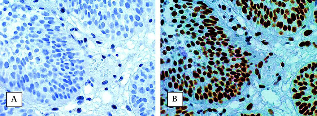
Image: Absence of nuclear immunohistochemical staining of MSH2 protein (A) and presence of MLH1 protein (B) in urothelial cell carcinoma of the urinary bladder of a patient carrying a germline MSH2 mutation. Observe the nuclear staining in stromal cells as an internal control (Photo courtesy of Radboud University Nijmegen Medical Centre)
Lynch Syndrome (LS) is an inherited genetic disorder that carries a high risk of cancer. LS is caused by mutations affecting MLH1, MSH2, MSH6 or PMS2 genes. More than one in 300 people have LS but most do not know it and that is equivalent to more than 2,300 new cases every year in the UK.
LS or hereditary nonpolyposis cancer patients are at high risk of bowel and endometrial cancers. The risk of urothelial cancer is less well recognized, with a lifetime risk of up to 28.5%, depending on which gene is involved. This means that those LS patients who have an underlying defect in the MSH2 gene are more than ten times as likely to get a potentially curable cancer in their urinary tract as the general population.
Medical Scientists at Newcastle University (Newcastle upon Tyne, UK) redesigned the Newcastle MSI-Plus Assay. This has recently become the standard test in North East England to find people with LS among those diagnosed with bowel cancer. MSI, which stands for microsatellite instability, shows that an important DNA repair system is not working. Most LS patients are in this group. The test is being evaluated for national rollout.
To see if the test could be used for urothelial cancers as well as bowel cancer, the investigators tested blood and urine samples in a LS patient who had been diagnosed with urothelial cancer in the upper urinary tract. They tested the urine before and after surgery to remove the tumour. In a review of 10,243 urothelial cancers in England in 2018, they found that only 43 patients (0.4%) had undergone testing for genetic variants that could identify whether or not they carried the variants for LS. They estimated that around 2% of urothelial cancers are in people with the LS genetic mutations and at least 5% should be tested to identify LS cases, so there is an urgent need to improve screening for this disease.
Sir John Burn, MB BS, MD, Professor of Clinical Genetics and the presenter of the study, said, “Dying cells within the human body shed small fragments of DNA into the surrounding tissues and circulation. These fragments are known as cell-free DNA and can be extracted from urine. LS is caused by inherited changes that affect the cells’ ability to repair DNA damage. As a result, almost all cancers that develop within individuals with LS have a characteristic pattern of errors called microsatellite instability. Microsatellites are regions of repetitive DNA that often change their length if not repaired correctly. Our test uses this ‘signature’ to identify DNA from tumour cells that are shedding into the urine.”
The author concluded their findings offer the potential to develop a cheap, easy and non-invasive way of screening LS patients for cancers of the bladder, kidney and ureter (the tube connecting the bladder with the kidneys). The study was presented on November 11, 2021 at the virtual National Cancer Research Institute Festival.
Related Links:
Newcastle University





LS or hereditary nonpolyposis cancer patients are at high risk of bowel and endometrial cancers. The risk of urothelial cancer is less well recognized, with a lifetime risk of up to 28.5%, depending on which gene is involved. This means that those LS patients who have an underlying defect in the MSH2 gene are more than ten times as likely to get a potentially curable cancer in their urinary tract as the general population.
Medical Scientists at Newcastle University (Newcastle upon Tyne, UK) redesigned the Newcastle MSI-Plus Assay. This has recently become the standard test in North East England to find people with LS among those diagnosed with bowel cancer. MSI, which stands for microsatellite instability, shows that an important DNA repair system is not working. Most LS patients are in this group. The test is being evaluated for national rollout.
To see if the test could be used for urothelial cancers as well as bowel cancer, the investigators tested blood and urine samples in a LS patient who had been diagnosed with urothelial cancer in the upper urinary tract. They tested the urine before and after surgery to remove the tumour. In a review of 10,243 urothelial cancers in England in 2018, they found that only 43 patients (0.4%) had undergone testing for genetic variants that could identify whether or not they carried the variants for LS. They estimated that around 2% of urothelial cancers are in people with the LS genetic mutations and at least 5% should be tested to identify LS cases, so there is an urgent need to improve screening for this disease.
Sir John Burn, MB BS, MD, Professor of Clinical Genetics and the presenter of the study, said, “Dying cells within the human body shed small fragments of DNA into the surrounding tissues and circulation. These fragments are known as cell-free DNA and can be extracted from urine. LS is caused by inherited changes that affect the cells’ ability to repair DNA damage. As a result, almost all cancers that develop within individuals with LS have a characteristic pattern of errors called microsatellite instability. Microsatellites are regions of repetitive DNA that often change their length if not repaired correctly. Our test uses this ‘signature’ to identify DNA from tumour cells that are shedding into the urine.”
The author concluded their findings offer the potential to develop a cheap, easy and non-invasive way of screening LS patients for cancers of the bladder, kidney and ureter (the tube connecting the bladder with the kidneys). The study was presented on November 11, 2021 at the virtual National Cancer Research Institute Festival.
Related Links:
Newcastle University

Platinum Member
Automated Coagulation Analyzer
Hemolumi H6
Gold Member
Fully-auto Specific Protein (Nephelometry) Analyzer
PA240
LAIR2 Antibody Pair Set
LAIR2 Antibody Pair [Biotin]
New
Microbiology Laboratory Automation Solution
BD Kiestra™ ReadA+BarcodA
Latest Molecular Diagnostics News
- Blood RNA Panel Predicts Lung Cancer Risk Years Before Diagnosis
- Bile DNA Testing Detects Tumor Signals Before Conventional Cancer Diagnosis
- Blood-Based Test Broadens Colorectal Cancer Screening Options
- New Blood Test Could Help Predict Severe Scoliosis in Adolescents
- Single Assay Enables Simultaneous Newborn Screening for Three Severe Genetic Disorders
- Genetic Testing Identifies Inherited Predisposition in Children With Acute Leukemia
- Pooled Saliva PCR Screening Identifies Congenital CMV Missed by Targeted Testing
- Portable Test Differentiates Seven Mosquito-Borne Viruses at Point of Care
- Rapid Genetic Test Aims to Shorten Brain Tumor Diagnosis Time
- Long-Read Sequencing Test Identifies Genetic Causes in Inherited Muscle Disease
- Genetic Testing Program Helps Uncover Inherited Risk in Pediatric Cancer
- Genomic Test Guides Extended Endocrine Therapy in Early Breast Cancer
- Genomic Breast Cancer Study Adds Real-World Evidence Across Diverse Patient Populations
- New Oncology Assays Deliver Precise Molecular Counts with MRD-Grade Sensitivity
- Molecular Skin Test May Help Guide Treatment Selection in Atopic Dermatitis
- CSF Liquid Biopsy Could Help Distinguish Cancer from Autoimmune Brain Inflammation
Channels
Clinical Chemistry
view channel
Screening After Pneumococcal Disease May Reveal Undiagnosed Blood Cancer or Immune Disorders
Severe pneumococcal disease requiring hospitalization often presents as pneumonia, particularly in older adults and people with cancer or compromised immune function. Because M protein testing and antibody... Read more
Global Survey Underscores Need to Standardize Bone Biomarker Testing
Bone and mineral metabolism biomarkers can complement dual-energy X-ray absorptiometry (DXA) by providing information on disease activity and treatment response. For more than 20 years, standardization... Read more
Label-Free Platform Combines Microfluidics and AI for Circulating Tumor Cell Analysis
Liquid biopsy relies on detecting rare tumor-derived material in blood, but circulating tumor cells (CTCs) are especially difficult to capture because they are vastly outnumbered by normal blood cells.... Read more
Rapid D-Dimer Assay Supports Exclusion and Monitoring of Serious Clotting Conditions
Blood clots can rapidly become life‑threatening and affect approximately 10 million people worldwide each year. During time‑sensitive evaluations, clinicians use D‑dimer testing to help rule out deep vein... Read moreHematology
view channel
New Donor Genetic Marker May Help Predict Stem Cell Transplant Success
Donor selection for hematopoietic stem cell transplantation plays a major role in relapse risk and survival for patients with blood cancers and other blood disorders. Despite advances in genotyping, uncertainty... Read more
Updated Ferritin Thresholds Improve Detection of Iron Deficiency
Iron deficiency is one of the most common health conditions worldwide, yet its nonspecific symptoms can delay diagnosis for months. Variation in testing practices and ferritin thresholds may contribute... Read more
Immunology
view channel
Blood Test Differentiates Bacterial and Viral Infections in Febrile Infants
Fever in infants younger than 3 months is treated as a medical emergency because infections can become life-threatening while the immune system is still developing. Emergency department teams must quickly... Read more
New Autoantibody Test Targets Pre-Symptomatic Type 1 Diabetes Screening
Type 1 diabetes is often first recognized only after substantial beta-cell destruction, sometimes presenting as diabetic ketoacidosis. Population-scale screening could identify children at elevated risk... Read more
Microbiology
view channel
FDA-Cleared Multiplex PCR Test Detects 13 Respiratory Pathogens in a Single Sample
Respiratory tract infections can be difficult to distinguish at presentation because many cause overlapping, nonspecific symptoms and are initially grouped as influenza-like illnesses. Causes span a range... Read more
One-Hour Molecular Panel Expands Bloodstream Infection Testing for Gram-Negative Pathogens
Bloodstream infections can progress rapidly and lead to sepsis, organ failure, and death. In the United States, about 1.7 million adults develop sepsis each year, and at least 350,000 die during hospitalization... Read more
FDA Clears Rapid Phenotypic Antimicrobial Susceptibility System for Positive Blood Cultures
Bloodstream infections require prompt treatment, but antimicrobial susceptibility results often lag behind a positive blood culture. Conventional testing can take another 24 to 48 hours after a culture... Read more
New Urine Test Expands Mycotoxin Analysis to 31 Markers for Broader Exposure Assessment
Clinical evaluation of mold exposure increasingly relies on urinary mycotoxin testing, but limited marker coverage and metabolite masking can make results more difficult to interpret. Broader analysis... Read morePathology
view channel
Genomic Classifier Predicts Benefit From Adding Hormone Therapy to Salvage Prostate Radiation
Men who have undergone prostatectomy for prostate cancer may later develop a detectable or rising prostate-specific antigen, prompting salvage radiation therapy. A key challenge is determining who is most... Read more
New AI Model Maps Where Glioblastoma Could Return After Surgery
Glioblastoma is the most common malignant brain tumor in adults and the most lethal, with median survival of about 17 months after diagnosis. Even after surgeons remove all visible tumor and patients receive... Read more
Technology
view channel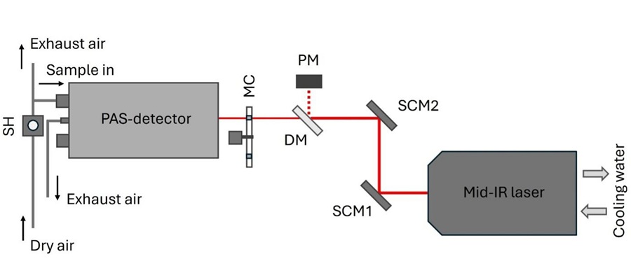
Laser-Based Swab Analysis Shows Promise for Detecting Disease-Linked Odor Patterns
Disease-related changes in volatile organic compounds can alter body odor, producing measurable patterns in exhaled breath and bodily fluids. Current analytical methods can be complex, time-consuming,... Read more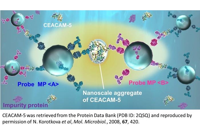
Laser-Enhanced Assay Boosts Sensitivity for Colorectal Cancer Biomarker Detection
Colorectal cancer is the third most commonly diagnosed cancer and the second leading cause of cancer-related death worldwide. Early detection remains critical, but cancer biomarkers can produce only faint... Read more

Industry
view channel
Collaboration Combines AI Cognitive Assessment and RNA Blood Testing for Earlier Alzheimer’s Detection
Alzheimer’s disease is often identified only after substantial neurodegeneration, partly because current diagnostic pathways are fragmented and difficult to scale. As treatment shifts toward earlier intervention,... Read more
Mayo Clinic Laboratories and Pathology Asia Expand Genomic Testing Across Asia-Pacific
Mayo Clinic Laboratories and Pathology Asia Holdings (PAH), together with subsidiary LifeStrands Genomics, announced a strategic investment and collaboration focused on expanding access to advanced diagnostics... Read more