Flow Cytometry Employed in Diagnosing Male Urethritis
|
By LabMedica International staff writers Posted on 17 Dec 2020 |

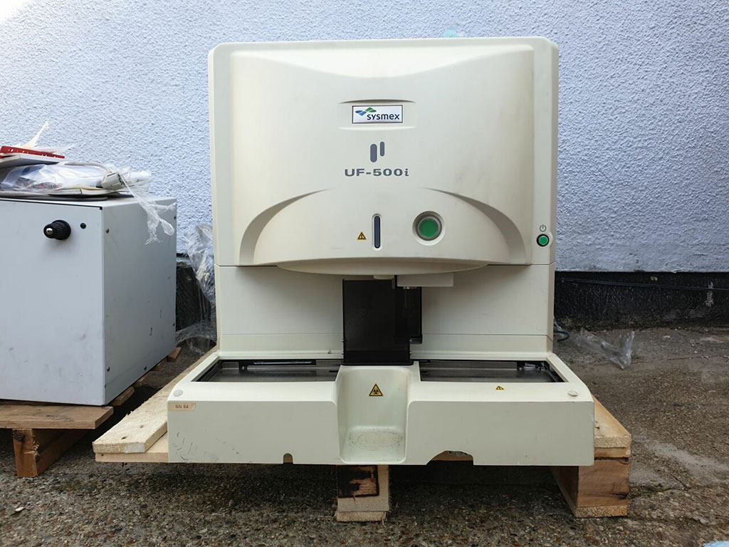
Image: The UF-500i fully automated urine particle analyzer (Photo courtesy of Sysmex Corporation).
According to the established guidelines, diagnosis of non-gonococcal urethritis should be confirmed by demonstrating polymorphonuclear leukocytes from the anterior urethra using a Gram-stained (GSS) or methylene blue-stained urethral smear.
There are several methods for detecting these infections. In recent years, new technologies have emerged in the field of urinalysis methodology, offering quick and standardized opportunities in everyday clinical practice. However, there is only limited information about how to use flow cytometry in diagnosing male urethritis.
Medical Andrologists and their colleagues at Tartu University Hospital (Tartu, Estonia) recruited 306 male patients with infectious urethritis caused by Chlamydia trachomatis, Neisseria gonorrhoeae, Mycoplasma genitalium and/or Trichomonas vaginalis. The control group consisted of 192 patients without uro-genital complaints, negative tests for C. trachomatis, N. gonorrhoeae, M. genitalium and T. vaginalis from first-voided urine and no inflammation in first-voided urine, mid-stream urine and urine after prostate massage.
The concentration and total count of white blood cells and bacteria in urine were analyzed using urine flow cytometry. The analyses were performed using fully automated urine particle analyzer Sysmex UF-500i (Sysmex Corporation, Kobe, Japan). Urethritis-associated sexually transmitted infections (STIs were detected from the first-voided urine using a PCR method: C. trachomatis and N. gonorrhoeae DNA by cobas 4800 CT/NG Test (Roche Diagnostics, Risch-Rotkreuz, Switzerland); M. genitalium DNA /Mycoplasma genitalium Real-TM; T. vaginalis DNA by Trichomonas vaginalis Real-TM (Sacace Biotehnologies, Como, Italy).
The investigators reported that the most prevalent infection was chlamydia (64.1%), followed by Mycoplasma genitalium (20.9%), gonorrhoea (7.8%) and trichomoniasis (1.6%). Gonorrhoea caused the highest flow-cytometric leucocyte/bacteria count, followed by chlamydia and Mycoplasma genitalium. Trichomonas vaginalis showed nearly absent inflammation in first-voided urine. Using an empiric flow-cytometry diagnostic threshold for urethritis in first-voided urine (leucocytes ≥ 15/μL and bacteria ≥ 20/μL), the total calculated sensitivity was over 90%. However, when applying such criteria for deciding whether to perform first-voided urine PCR for C. trachomatis, N. gonorrhoeae, M. genitalium and T. vaginalis or not, the team could miss 23 cases with infectious urethritis that makes up 7.5% of all proven cases.
The authors concluded that C. trachomatis (CT) is the most prevalent urethritis-associated STI among the men consulting an andrologist, followed by M. genitalium (MG). A strong inflammatory reaction accompanied by high bacterial concentration in first-voided urine as revealed by flow cytometry is highly predictive of NG infection (sensitivity >95%), while the sensitivity of this method remains slightly lower for CT and MG (>92%) and very low for infrequently occurring T. vaginalis. The study was published on December 2 2020 in the journal PLOS ONE.
Related Links:
Tartu University Hospital
Sysmex Corporation
Roche Diagnostics
Sacace Biotehnologies
Latest Microbiology News
- Comprehensive Review Identifies Gut Microbiome Signatures Associated With Alzheimer’s Disease
- AI-Powered Platform Enables Rapid Detection of Drug-Resistant C. Auris Pathogens
- New Test Measures How Effectively Antibiotics Kill Bacteria
- New Antimicrobial Stewardship Standards for TB Care to Optimize Diagnostics
- New UTI Diagnosis Method Delivers Antibiotic Resistance Results 24 Hours Earlier
- Breakthroughs in Microbial Analysis to Enhance Disease Prediction
- Blood-Based Diagnostic Method Could Identify Pediatric LRTIs
- Rapid Diagnostic Test Matches Gold Standard for Sepsis Detection
- Rapid POC Tuberculosis Test Provides Results Within 15 Minutes
- Rapid Assay Identifies Bloodstream Infection Pathogens Directly from Patient Samples
- Blood-Based Molecular Signatures to Enable Rapid EPTB Diagnosis
- 15-Minute Blood Test Diagnoses Life-Threatening Infections in Children
- High-Throughput Enteric Panels Detect Multiple GI Bacterial Infections from Single Stool Swab Sample
- Fast Noninvasive Bedside Test Uses Sugar Fingerprint to Detect Fungal Infections
- Rapid Sepsis Diagnostic Device to Enable Personalized Critical Care for ICU Patients
- Microfluidic Platform Assesses Neutrophil Function in Sepsis Patients
Channels
Clinical Chemistry
view channel
New PSA-Based Prognostic Model Improves Prostate Cancer Risk Assessment
Prostate cancer is the second-leading cause of cancer death among American men, and about one in eight will be diagnosed in their lifetime. Screening relies on blood levels of prostate-specific antigen... Read more
Extracellular Vesicles Linked to Heart Failure Risk in CKD Patients
Chronic kidney disease (CKD) affects more than 1 in 7 Americans and is strongly associated with cardiovascular complications, which account for more than half of deaths among people with CKD.... Read moreMolecular Diagnostics
view channel
Diagnostic Device Predicts Treatment Response for Brain Tumors Via Blood Test
Glioblastoma is one of the deadliest forms of brain cancer, largely because doctors have no reliable way to determine whether treatments are working in real time. Assessing therapeutic response currently... Read more
Blood Test Detects Early-Stage Cancers by Measuring Epigenetic Instability
Early-stage cancers are notoriously difficult to detect because molecular changes are subtle and often missed by existing screening tools. Many liquid biopsies rely on measuring absolute DNA methylation... Read more
“Lab-On-A-Disc” Device Paves Way for More Automated Liquid Biopsies
Extracellular vesicles (EVs) are tiny particles released by cells into the bloodstream that carry molecular information about a cell’s condition, including whether it is cancerous. However, EVs are highly... Read more
Blood Test Identifies Inflammatory Breast Cancer Patients at Increased Risk of Brain Metastasis
Brain metastasis is a frequent and devastating complication in patients with inflammatory breast cancer, an aggressive subtype with limited treatment options. Despite its high incidence, the biological... Read moreHematology
view channel
New Guidelines Aim to Improve AL Amyloidosis Diagnosis
Light chain (AL) amyloidosis is a rare, life-threatening bone marrow disorder in which abnormal amyloid proteins accumulate in organs. Approximately 3,260 people in the United States are diagnosed... Read more
Fast and Easy Test Could Revolutionize Blood Transfusions
Blood transfusions are a cornerstone of modern medicine, yet red blood cells can deteriorate quietly while sitting in cold storage for weeks. Although blood units have a fixed expiration date, cells from... Read more
Automated Hemostasis System Helps Labs of All Sizes Optimize Workflow
High-volume hemostasis sections must sustain rapid turnaround while managing reruns and reflex testing. Manual tube handling and preanalytical checks can strain staff time and increase opportunities for error.... Read more
High-Sensitivity Blood Test Improves Assessment of Clotting Risk in Heart Disease Patients
Blood clotting is essential for preventing bleeding, but even small imbalances can lead to serious conditions such as thrombosis or dangerous hemorrhage. In cardiovascular disease, clinicians often struggle... Read moreImmunology
view channelBlood Test Identifies Lung Cancer Patients Who Can Benefit from Immunotherapy Drug
Small cell lung cancer (SCLC) is an aggressive disease with limited treatment options, and even newly approved immunotherapies do not benefit all patients. While immunotherapy can extend survival for some,... Read more
Whole-Genome Sequencing Approach Identifies Cancer Patients Benefitting From PARP-Inhibitor Treatment
Targeted cancer therapies such as PARP inhibitors can be highly effective, but only for patients whose tumors carry specific DNA repair defects. Identifying these patients accurately remains challenging,... Read more
Ultrasensitive Liquid Biopsy Demonstrates Efficacy in Predicting Immunotherapy Response
Immunotherapy has transformed cancer treatment, but only a small proportion of patients experience lasting benefit, with response rates often remaining between 10% and 20%. Clinicians currently lack reliable... Read morePathology
view channel
Engineered Yeast Cells Enable Rapid Testing of Cancer Immunotherapy
Developing new cancer immunotherapies is a slow, costly, and high-risk process, particularly for CAR T cell treatments that must precisely recognize cancer-specific antigens. Small differences in tumor... Read more
First-Of-Its-Kind Test Identifies Autism Risk at Birth
Autism spectrum disorder is treatable, and extensive research shows that early intervention can significantly improve cognitive, social, and behavioral outcomes. Yet in the United States, the average age... Read moreTechnology
view channel
Robotic Technology Unveiled for Automated Diagnostic Blood Draws
Routine diagnostic blood collection is a high‑volume task that can strain staffing and introduce human‑dependent variability, with downstream implications for sample quality and patient experience.... Read more
ADLM Launches First-of-Its-Kind Data Science Program for Laboratory Medicine Professionals
Clinical laboratories generate billions of test results each year, creating a treasure trove of data with the potential to support more personalized testing, improve operational efficiency, and enhance patient care.... Read moreAptamer Biosensor Technology to Transform Virus Detection
Rapid and reliable virus detection is essential for controlling outbreaks, from seasonal influenza to global pandemics such as COVID-19. Conventional diagnostic methods, including cell culture, antigen... Read more
AI Models Could Predict Pre-Eclampsia and Anemia Earlier Using Routine Blood Tests
Pre-eclampsia and anemia are major contributors to maternal and child mortality worldwide, together accounting for more than half a million deaths each year and leaving millions with long-term health complications.... Read moreIndustry
view channelNew Collaboration Brings Automated Mass Spectrometry to Routine Laboratory Testing
Mass spectrometry is a powerful analytical technique that identifies and quantifies molecules based on their mass and electrical charge. Its high selectivity, sensitivity, and accuracy make it indispensable... Read more
AI-Powered Cervical Cancer Test Set for Major Rollout in Latin America
Noul Co., a Korean company specializing in AI-based blood and cancer diagnostics, announced it will supply its intelligence (AI)-based miLab CER cervical cancer diagnostic solution to Mexico under a multi‑year... Read more
Diasorin and Fisher Scientific Enter into US Distribution Agreement for Molecular POC Platform
Diasorin (Saluggia, Italy) has entered into an exclusive distribution agreement with Fisher Scientific, part of Thermo Fisher Scientific (Waltham, MA, USA), for the LIAISON NES molecular point-of-care... Read more


