Genetic Alterations Associated with Sporadic Congenital Hydrocephalus
|
By LabMedica International staff writers Posted on 02 Nov 2020 |


|
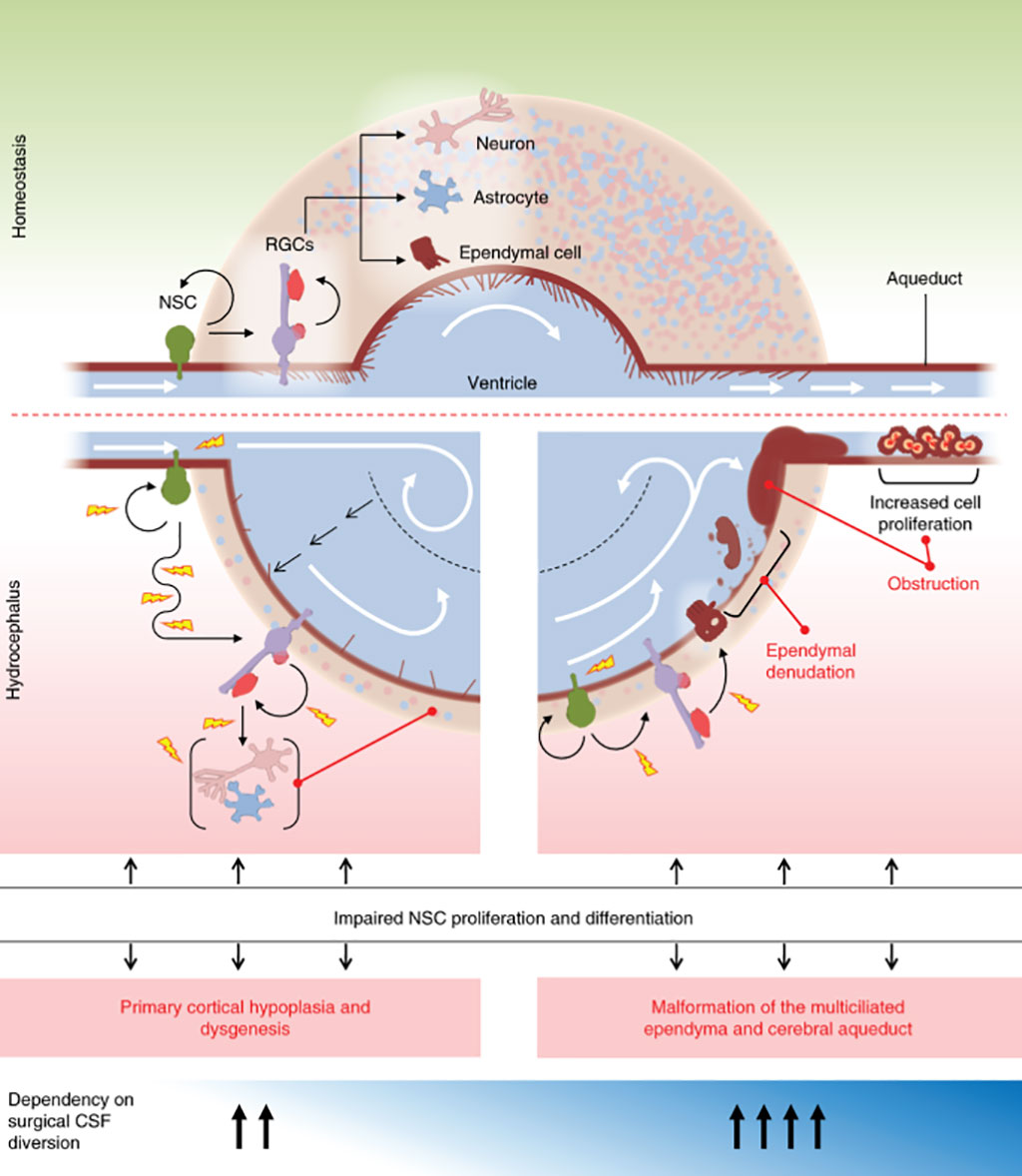
Image: A neural stem cell model of sporadic congenital hydrocephalus (Photo courtesy of Yale University School of Medicine).
Congenital hydrocephalus (CH), characterized by enlarged brain ventricles, is considered a disease of excessive cerebrospinal fluid (CSF) accumulation and thereby treated with neurosurgical CSF diversion with high morbidity and failure rates.
Congenital hydrocephalus is present in the infant prior to birth, meaning the fetus developed hydrocephalus in utero during fetal development. The most common cause of congenital hydrocephalus is aqueductal stenosis, which occurs when the narrow passage between the third and fourth ventricles in the brain is blocked or too narrow to allow sufficient CSF to drain.
A multidisciplinary team at Yale University School of Medicine (New Haven, CT, USA) performed whole-exome sequencing of 381 individuals with sporadic congenital hydrocephalus who were treated with neurosurgery, a cohort that included 232 parent-offspring trios. Another 1,798 trios of unaffected siblings and parents of individuals with autism spectrum disorder were analyzed in parallel as a control group.
The team uncovered 12 genes with two or more de novo mutations each that were predicted to be protein damaging. Five of these genes: TRIM71, SMARCC1, PTEN, PIK3CA, and FOXJ1, had significantly more mutations than expected. The investigators further estimated that about 22% of sporadic congenital hydrocephalus are due to rare, damaging mutations. The teams’ analysis additionally implicated a number of genes in the PI3K signaling pathway in congenital hydrocephalus. These genes regulate cell growth, proliferation, and differentiation in numerous tissues, including developing neural stem cells. In particular, they uncovered three de novo mutations in PI3KCA, three in PTEN, and two in MTOR.
Individuals with mutations in TRIM71, which maintains stem cell pluripotency, are more likely to have cranial nerve defect, non-obstructive inter-hemispheric cysts, and hearing loss. Meanwhile, individuals with mutations in SMARCC1, which regulates gene expression needed for neural stem cell proliferation, differentiation, and survival during telencephalon development, are more likely to have aqueductal stenosis and cardiac and skeletal abnormalities.
All together, the known, high-confidence, and probable risk genes for congenital hydrocephalus converge in gene co-expression networks of the mid-gestational human cortex, In particular, the congenital hydrocephalus risk genes converge on a network previously linked to autism spectrum disorder that is enriched for neuronal differentiation and RNA processing gene ontology terms.
Kristopher T. Kahle, MD, PhD, an assistant professor of neurosurgery and a senior author of the study, said, “It is possible that some of the neurological problems that many of these congenital hydrocephalus patients have may not be due to inadequately working shunts, but are in fact neurodevelopmental sequelae of a genetic condition that alters neuronal function.”
The authors concluded that that genetic disruptions affecting early brain development may drive sporadic congenital hydrocephalus. Their data implicate genetically encoded neural stem cell dysregulation and an associated impairment of fetal neurogliogenesis as primary pathophysiological events in a significant number of congenital hydrocephalus cases. The study was published on October 19, 2020 in the journal Nature Medicine.
Related Links:
Yale University School of Medicine








Congenital hydrocephalus is present in the infant prior to birth, meaning the fetus developed hydrocephalus in utero during fetal development. The most common cause of congenital hydrocephalus is aqueductal stenosis, which occurs when the narrow passage between the third and fourth ventricles in the brain is blocked or too narrow to allow sufficient CSF to drain.
A multidisciplinary team at Yale University School of Medicine (New Haven, CT, USA) performed whole-exome sequencing of 381 individuals with sporadic congenital hydrocephalus who were treated with neurosurgery, a cohort that included 232 parent-offspring trios. Another 1,798 trios of unaffected siblings and parents of individuals with autism spectrum disorder were analyzed in parallel as a control group.
The team uncovered 12 genes with two or more de novo mutations each that were predicted to be protein damaging. Five of these genes: TRIM71, SMARCC1, PTEN, PIK3CA, and FOXJ1, had significantly more mutations than expected. The investigators further estimated that about 22% of sporadic congenital hydrocephalus are due to rare, damaging mutations. The teams’ analysis additionally implicated a number of genes in the PI3K signaling pathway in congenital hydrocephalus. These genes regulate cell growth, proliferation, and differentiation in numerous tissues, including developing neural stem cells. In particular, they uncovered three de novo mutations in PI3KCA, three in PTEN, and two in MTOR.
Individuals with mutations in TRIM71, which maintains stem cell pluripotency, are more likely to have cranial nerve defect, non-obstructive inter-hemispheric cysts, and hearing loss. Meanwhile, individuals with mutations in SMARCC1, which regulates gene expression needed for neural stem cell proliferation, differentiation, and survival during telencephalon development, are more likely to have aqueductal stenosis and cardiac and skeletal abnormalities.
All together, the known, high-confidence, and probable risk genes for congenital hydrocephalus converge in gene co-expression networks of the mid-gestational human cortex, In particular, the congenital hydrocephalus risk genes converge on a network previously linked to autism spectrum disorder that is enriched for neuronal differentiation and RNA processing gene ontology terms.
Kristopher T. Kahle, MD, PhD, an assistant professor of neurosurgery and a senior author of the study, said, “It is possible that some of the neurological problems that many of these congenital hydrocephalus patients have may not be due to inadequately working shunts, but are in fact neurodevelopmental sequelae of a genetic condition that alters neuronal function.”
The authors concluded that that genetic disruptions affecting early brain development may drive sporadic congenital hydrocephalus. Their data implicate genetically encoded neural stem cell dysregulation and an associated impairment of fetal neurogliogenesis as primary pathophysiological events in a significant number of congenital hydrocephalus cases. The study was published on October 19, 2020 in the journal Nature Medicine.
Related Links:
Yale University School of Medicine

Platinum Member
Automated Coagulation Analyzer
Hemolumi H6
Gold Member
Blood-Based Protein Biomarker Solution for Alzheimer's Disease
BG-DTi2000.
Microbiology Laboratory Automation Solution
BD Kiestra™ ReadA+BarcodA
Urine Analyzer
respons® UDS100
Latest Pathology News
- Updated Digital Pathology Platform Expands AI Integration for Cancer Diagnosis
- Molecular Testing Refines Barrett’s Esophagus Progression Risk Beyond Biopsy Findings
- AI Histopathology Tool Distinguishes Ewing Sarcoma for Diagnosis
- Rapid Mass Spectrometry Test May Aid Glioma Margin Decisions
- Genomic Classifier Predicts Benefit From Adding Hormone Therapy to Salvage Prostate Radiation
- Multimodal AI Supports Prostate Cancer Risk Assessment Across Diverse Cohorts
- New AI Model Maps Where Glioblastoma Could Return After Surgery
- Simple Test Could Reduce the Need for Invasive Uterine Cancer Checks
- Tumor Blood Vessel Features May Help Predict Colorectal Cancer Survival
- Multimodal AI Improves Breast Cancer Recurrence Risk Prediction Beyond Standard Genomic Testing
- Machine Learning Cytology Tool Improves Cancer Cell Identification
- CAP Updates Cancer Protocols to Refine Breast Cancer Biomarker Reporting and GIST Molecular Testing
- Tumor Budding Grading May Predict Chemotherapy Benefit in Resected Lung Squamous Cancer
- AI Spatial Analysis of Routine Slides Helps Predict Pancreatic Cancer Recurrence
- Hybrid Computational Imaging Method Improves Digital Pathology Resolution
- FDA Clearance Expands Digital Pathology Options for Clinical Laboratories
Channels
Clinical Chemistry
view channel
Multimodal Clinical Data Models Predict Cachexia Risk Across Cancers
Cancer cachexia is a severe wasting syndrome that affects roughly half of patients with cancer and cannot be reversed simply by increasing food intake. It can reduce treatment tolerance, impair quality... Read more
Study Evaluates Alzheimer’s Blood Biomarker Across Racial and Ethnic Groups
Alzheimer’s disease trials increasingly depend on identifying cognitively unimpaired people who already have elevated brain amyloid. Blood biomarkers may help reduce reliance on positron emission tomography... Read more
Hematology
view channel
New Myelofibrosis Guidelines Recommend Expanded Genetic Testing at Diagnosis
Myelofibrosis is a rare, life-threatening blood cancer affecting up to six per 100,000 people. The disease causes scarring in the bone marrow, limiting the production of healthy blood cells and potentially... Read more
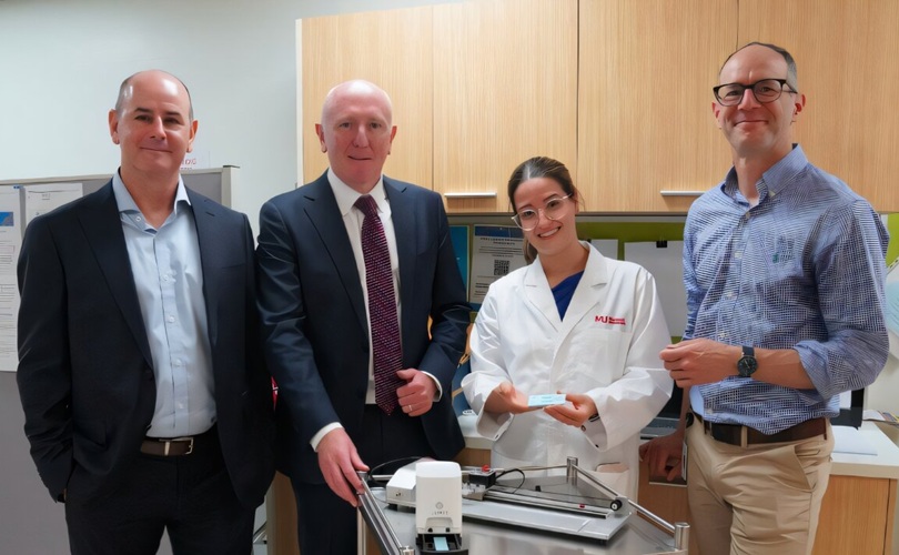
Handheld Point-of-Care Test Aims to Enable Rapid Coagulation Assessment in Trauma
Severe blood loss and inadequate tissue oxygenation are leading causes of preventable death after traumatic injury. Blood products are essential in trauma care, but cold-chain storage, short shelf life,... Read more

Immunology
view channel
FDA Approval Adds Automated Testing Option for Parvovirus B19 Serology
Human Parvovirus B19 is a highly contagious erythrovirus transmitted mainly through respiratory droplets and close contact. Diagnosis relies on IgM antibodies to detect active infection and IgG antibodies... Read more
Blood-Based Immune Aging Map Could Help Spot Disease Risk Earlier
Chronological age does not always reflect immune health or future disease risk. Two adults of the same age can have immune systems that age very differently, with some remaining resilient while others... Read more
Microbiology
view channel
Rapid Host-Response Test Aims to Predict Corticosteroid Benefit in Sepsis
Sepsis and severe infections are difficult to treat because patients with similar clinical presentations may have very different underlying immune responses. Corticosteroids are commonly used in severe... Read more
Oral and Lung Microbiome Changes Linked to Severe Long Covid Symptoms
Long Covid can involve persistent respiratory, cognitive, and gastrointestinal symptoms after acute infection, creating a difficult diagnostic and management challenge. Because COVID-19 initially affects... Read more

Pathology
view channel
Molecular Testing Refines Barrett’s Esophagus Progression Risk Beyond Biopsy Findings
Barrett’s esophagus is a precancerous condition associated with increased risk of esophageal adenocarcinoma. Routine endoscopic biopsy findings guide surveillance and therapy, but histopathology may not... Read more
Updated Digital Pathology Platform Expands AI Integration for Cancer Diagnosis
Digital pathology is reshaping how laboratories manage case review, image analysis, and collaboration. As diagnostic workflows become more complex, pathology teams need platforms that can support digital... Read more
Technology
view channel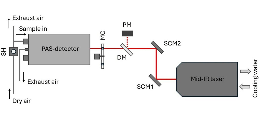
Laser-Based Swab Analysis Shows Promise for Detecting Disease-Linked Odor Patterns
Disease-related changes in volatile organic compounds can alter body odor, producing measurable patterns in exhaled breath and bodily fluids. Current analytical methods can be complex, time-consuming,... Read more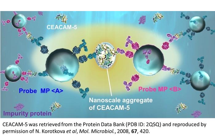
Laser-Enhanced Assay Boosts Sensitivity for Colorectal Cancer Biomarker Detection
Colorectal cancer is the third most commonly diagnosed cancer and the second leading cause of cancer-related death worldwide. Early detection remains critical, but cancer biomarkers can produce only faint... Read more

Industry
view channel
Global TB Summit Connects Diagnostic Testing With Prevention and Clinical Care
Tuberculosis remains a major global infectious disease challenge despite declining deaths. In 2024, an estimated 10.7 million people worldwide developed the disease and nearly 1.2 million died from it.... Read more