Mycobacterium Infection Found in Gastric Patients’ Stomachs
|
By LabMedica International staff writers Posted on 26 Nov 2019 |

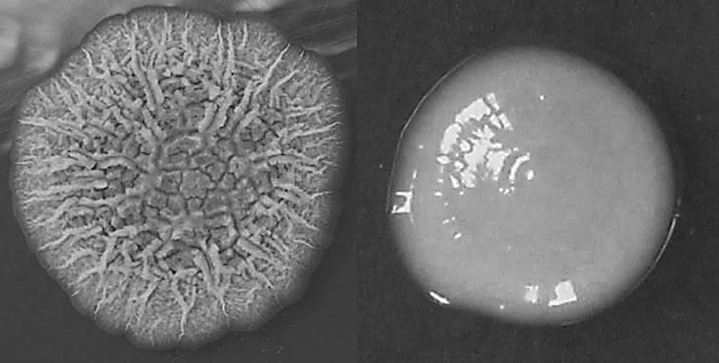
Image: Growth characteristics of rough and smooth phenotypes of Mycobacterium abscessus on 7H11 agar cultured at 37 °C: representative single rough (left) and smooth (right) colonies (Photo courtesy of Hannover Medical School)
Development of gastric diseases such as gastritis, peptic ulcer and gastric cancer is often associated with several biotic and abiotic factors. Helicobacter pylori infection is such a well-known biotic factor. However, not all H. pylori-infected individuals develop gastric diseases and not all individuals with gastric diseases are infected with H. pylori.
H. pylori is not the only bacterium that can colonize human stomach. Culture independent metagenomic sequence analyses have shown that human stomach carries a unique microbiota. The dominant phyla that are present in human stomach are Proteobacteria, Firmicutes, Actinobacteria and Fusobacterium. Interestingly, however, most of these bacteria cannot be cultured using traditional techniques.
Microbiologists at the Rajiv Gandhi Centre for Biotechnology (Thiruvananthapuram, India) recruited patients aged between 20 and 70 with various gastric and esophageal symptoms ranging from mild dyspepsia, gastro-esophageal reflux disorder to severe gastric diseases like gastric cancer and who were recommended to have upper gastrointestinal endoscopy. Three gastric biopsy specimens were collected from each patient for this study. The aim of this study was to isolate prevalent gastric bacteria under microaerobic condition and identify them by 16S rRNA gene sequence analysis.
The team employed various technologies including gastric bacteria culture, bacterial DNA isolation, extraction of intracellular bacterial DNA from biopsy tissues, molecular characterization of the bacteria isolated from stomach. The purified DNA fragments were sequenced by a 3730XL DNA analyzer (Thermo Fisher Scientific, Waltham, Massachusetts, USA). The team also performed Hematoxylin and Eosin (H&E) and Ziehl-Neelsen Acid-fast staining on tissue biopsies, and immunohistochemistry.
Analysis of gastric biopsies showed infection of Mycobacterium abscessus (phylum Actinobacteria) to be highly prevalent in the stomachs of subjects included. The data showed that of 129 (67 male and 62 female) patients with gastric symptoms, 96 (51 male and 45 female) showed the presence of M. abscessus in stomach tissues. Infection of M. abscessus in gastric epithelium was further confirmed by imaging with acid fast staining, immunohistochemistry and immunofluorescence. Surprisingly, the subjects studied, the prevalence of M. abscessus infection in stomach is even higher than the prevalence of H. pylori infection.
The authors concluded that their study on 129 individuals with gastric diseases shows that the prevalence of gastric M. abscessus is higher in the local population as compared to the prevalence of H. pylori. The route of transmission is not known at present, but water could be a source. Significance of this infection is also presently unknown, but it may have a significant role in the formation or progression of gastric disease. The study was published on November 4, 2019 in the journal PLOS Neglected Tropical Diseases.
Related Links:
Rajiv Gandhi Centre for Biotechnology
Thermo Fisher Scientific
Latest Molecular Diagnostics News
- New Genome Sequencing Technique Measures Epstein-Barr Virus in Blood
- Blood Test Boosts Early Detection of Brain Cancer
- Molecular Monitoring Approach Helps Bladder Cancer Patients Avoid Surgery
- Genetic Tests to Speed Diagnosis of Lymphatic Disorders
- Changes In Lymphatic Vessels Can Aid Early Identification of Aggressive Oral Cancer
- New Extraction Kit Enables Consistent, Scalable cfDNA Isolation from Multiple Biofluids
- New CSF Liquid Biopsy Assay Reveals Genomic Insights for CNS Tumors
- AI-Powered Liquid Biopsy Classifies Pediatric Brain Tumors with High Accuracy
- Group A Strep Molecular Test Delivers Definitive Results at POC in 15 Minutes
- Rapid Molecular Test Identifies Sepsis Patients Most Likely to Have Positive Blood Cultures
- Light-Based Sensor Detects Early Molecular Signs of Cancer in Blood
- New Testing Method Predicts Trauma Patient Recovery Days in Advance
- Simple Method Predicts Risk of Brain Tumor Recurrence
- Genetic Test Could Improve Early Detection of Prostate Cancer
- Bone Molecular Maps to Transform Early Osteoarthritis Detection
- POC Testing for Hepatitis B DNA as Effective as Traditional Laboratory Testing
Channels
Clinical Chemistry
view channel
Simple Blood Test Offers New Path to Alzheimer’s Assessment in Primary Care
Timely evaluation of cognitive symptoms in primary care is often limited by restricted access to specialized diagnostics and invasive confirmatory procedures. Clinicians need accessible tools to determine... Read more
Existing Hospital Analyzers Can Identify Fake Liquid Medical Products
Counterfeit and substandard medicines remain a serious global health threat, with World Health Organization estimates suggesting that 10.5% of medicines in low- and middle-income countries are either fake... Read moreMolecular Diagnostics
view channel
New Genome Sequencing Technique Measures Epstein-Barr Virus in Blood
The Epstein–Barr virus (EBV) infects up to 95% of adults worldwide and remains in the body for life. While usually kept under control, the virus is linked to cancers such as Hodgkin’s lymphoma and autoimmune... Read more
Blood Test Boosts Early Detection of Brain Cancer
Brain and central nervous system (CNS) tumors are often diagnosed at an advanced stage, when treatment options are limited, and survival rates remain low. Around 300,000 new cases are diagnosed each year... Read moreHematology
view channel
Rapid Cartridge-Based Test Aims to Expand Access to Hemoglobin Disorder Diagnosis
Sickle cell disease and beta thalassemia are hemoglobin disorders that often require referral to specialized laboratories for definitive diagnosis, delaying results for patients and clinicians.... Read more
New Guidelines Aim to Improve AL Amyloidosis Diagnosis
Light chain (AL) amyloidosis is a rare, life-threatening bone marrow disorder in which abnormal amyloid proteins accumulate in organs. Approximately 3,260 people in the United States are diagnosed... Read moreImmunology
view channel
New Biomarker Predicts Chemotherapy Response in Triple-Negative Breast Cancer
Triple-negative breast cancer is an aggressive form of breast cancer in which patients often show widely varying responses to chemotherapy. Predicting who will benefit from treatment remains challenging,... Read moreBlood Test Identifies Lung Cancer Patients Who Can Benefit from Immunotherapy Drug
Small cell lung cancer (SCLC) is an aggressive disease with limited treatment options, and even newly approved immunotherapies do not benefit all patients. While immunotherapy can extend survival for some,... Read more
Whole-Genome Sequencing Approach Identifies Cancer Patients Benefitting From PARP-Inhibitor Treatment
Targeted cancer therapies such as PARP inhibitors can be highly effective, but only for patients whose tumors carry specific DNA repair defects. Identifying these patients accurately remains challenging,... Read more
Ultrasensitive Liquid Biopsy Demonstrates Efficacy in Predicting Immunotherapy Response
Immunotherapy has transformed cancer treatment, but only a small proportion of patients experience lasting benefit, with response rates often remaining between 10% and 20%. Clinicians currently lack reliable... Read morePathology
view channel
Single Sample Classifier Predicts Cancer-Associated Fibroblast Subtypes in Patient Samples
Pancreatic ductal adenocarcinoma (PDAC) remains one of the deadliest cancers, in part because of its dense tumor microenvironment that influences how tumors grow and respond to treatment.... Read more
New AI-Driven Platform Standardizes Tuberculosis Smear Microscopy Workflow
Sputum smear microscopy remains central to tuberculosis treatment monitoring and follow-up, particularly in high‑burden settings where serial testing is routine. Yet consistent, repeatable bacillary assessment... Read more
AI Tool Uses Blood Biomarkers to Predict Transplant Complications Before Symptoms Appear
Stem cell and bone marrow transplants can be lifesaving, but serious complications may arise months after patients leave the hospital. One of the most dangerous is chronic graft-versus-host disease, in... Read moreTechnology
view channel
Blood Test “Clocks” Predict Start of Alzheimer’s Symptoms
More than 7 million Americans live with Alzheimer’s disease, and related health and long-term care costs are projected to reach nearly USD 400 billion in 2025. The disease has no cure, and symptoms often... Read more
AI-Powered Biomarker Predicts Liver Cancer Risk
Liver cancer, or hepatocellular carcinoma, causes more than 800,000 deaths worldwide each year and often goes undetected until late stages. Even after treatment, recurrence rates reach 70% to 80%, contributing... Read more
Robotic Technology Unveiled for Automated Diagnostic Blood Draws
Routine diagnostic blood collection is a high‑volume task that can strain staffing and introduce human‑dependent variability, with downstream implications for sample quality and patient experience.... Read more
ADLM Launches First-of-Its-Kind Data Science Program for Laboratory Medicine Professionals
Clinical laboratories generate billions of test results each year, creating a treasure trove of data with the potential to support more personalized testing, improve operational efficiency, and enhance patient care.... Read moreIndustry
view channel
QuidelOrtho Collaborates with Lifotronic to Expand Global Immunoassay Portfolio
QuidelOrtho (San Diego, CA, USA) has entered a long-term strategic supply agreement with Lifotronic Technology (Shenzhen, China) to expand its global immunoassay portfolio and accelerate customer access... Read more


