Novel Nanosensing Technologies Developed for Exosome Detection and Profiling
|
By LabMedica International staff writers Posted on 20 Nov 2019 |

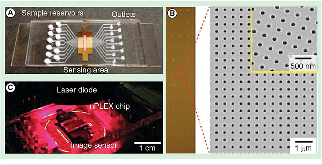
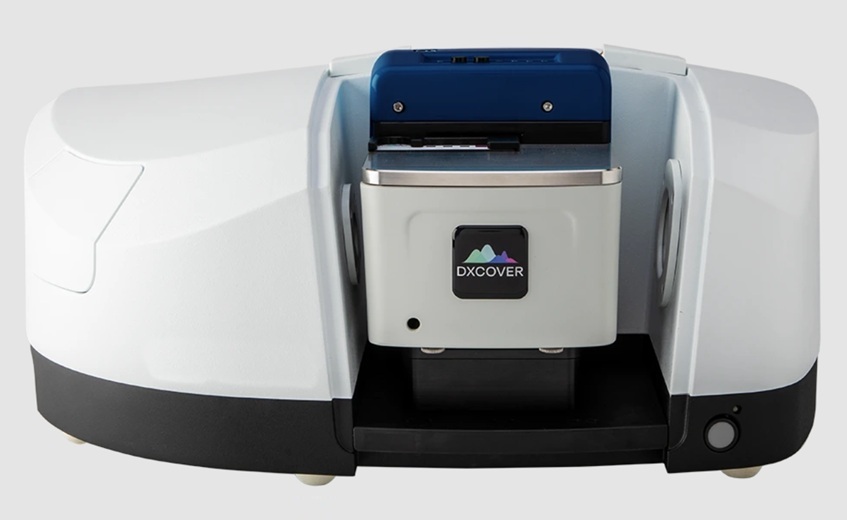
Image: The nano-plasmonic exosome (nPLEX) sensor (Photo courtesy of Massachusetts General Hospital).
Exosomes are extracellular vesicles (EVs) that are produced in the endosomal compartment of most eukaryotic cells. Liquid biopsy technologies have been developed to do high-throughput exosome protein profiling and point-of-care (POC) exosome analysis.
The aim is to develop multiplexed assay systems to streamline the analysis of exosomes and evaluate their clinical utility for cancer management, both as diagnostic tools and to predict disease recurrence. Nanoplasmonic sensors are fabricated to accommodate at most one exosome and individually imaged in real time, enabling the label-free recording of digital responses in a highly multiplexed geometry.
Scientists at the Massachusetts General Hospital (MGH, Boston, MA, USA) and their colleagues developed the nano-plasmonic exosome (nPLEX) sensor, in routine use at MGH, which detects exosome protein levels based on “spectral shifts” (intensity changes) of light through thousands of optimally spaced 200-nanometer (nm) holes. Antibodies against cancer biomarkers get immobilized on the nanopores for capture.
Nanoholes on the sensor are gold and exosomes are labeled by gold nanoparticles for signal amplification. Gold has proven to have the best chemical stability for this type of assay. The device is being manufactured on a glass-based substrate, as silicon wafers proved to be too fragile. The iMEX (integrated magnetic-electrochemical exosome) device, meanwhile, integrates vesicle isolation and detection in a single platform. Target-specific exosomes first get enriched through immunomagnetic selection, for high detection sensitivity. The sensors can be miniaturized and expanded for parallel measurements.
The nPLEX has demonstrated good accuracy and 100 times greater sensitivity than the commonly used enzyme-linked immunosorbent assay (ELISA) and able to detect as few as 3,000 exosomes. The team has also come up with a protein signature for pancreatic ductal adenocarcinoma (PDAC) that is powerful enough for diagnostic purposes, a combination of EGFR (epidermal growth factor receptor), EpCAM, MUC1, GPC1 and WNT2. As demonstrated in a prospective study involving more than 100 patients and age-matched controls, as the tumors shrink the biomarker combination gets lower and lower.
The scientists have also used nPLEX technology to show how exosome protein expression of cancer cells changes with drug treatment. Discovery of these unique, drug-dependent protein signatures suggests the exosome screening assay will be a potentially powerful molecular screening tool. Hyungsoon Im, PhD, an assistant professor of radiology and a senior author of the study, said, “The screening assay could be incorporated into point–of–care (POC) instruments for more rigorous testing of different drugs in different settings. The study was presented at the 2019 Next Generation Dx Summit held August 20-22, 2019 in Washington, DC, USA.
Related Links:
Massachusetts General Hospital
Latest Molecular Diagnostics News
- Genetic Test Aids Early Detection and Improved Treatment for Cancers
- New Genome Sequencing Technique Measures Epstein-Barr Virus in Blood
- Blood Test Boosts Early Detection of Brain Cancer
- Molecular Monitoring Approach Helps Bladder Cancer Patients Avoid Surgery
- Genetic Tests to Speed Diagnosis of Lymphatic Disorders
- Changes In Lymphatic Vessels Can Aid Early Identification of Aggressive Oral Cancer
- New Extraction Kit Enables Consistent, Scalable cfDNA Isolation from Multiple Biofluids
- New CSF Liquid Biopsy Assay Reveals Genomic Insights for CNS Tumors
- AI-Powered Liquid Biopsy Classifies Pediatric Brain Tumors with High Accuracy
- Group A Strep Molecular Test Delivers Definitive Results at POC in 15 Minutes
- Rapid Molecular Test Identifies Sepsis Patients Most Likely to Have Positive Blood Cultures
- Light-Based Sensor Detects Early Molecular Signs of Cancer in Blood
- New Testing Method Predicts Trauma Patient Recovery Days in Advance
- Simple Method Predicts Risk of Brain Tumor Recurrence
- Genetic Test Could Improve Early Detection of Prostate Cancer
- Bone Molecular Maps to Transform Early Osteoarthritis Detection
Channels
Clinical Chemistry
view channel
Simple Blood Test Offers New Path to Alzheimer’s Assessment in Primary Care
Timely evaluation of cognitive symptoms in primary care is often limited by restricted access to specialized diagnostics and invasive confirmatory procedures. Clinicians need accessible tools to determine... Read more
Existing Hospital Analyzers Can Identify Fake Liquid Medical Products
Counterfeit and substandard medicines remain a serious global health threat, with World Health Organization estimates suggesting that 10.5% of medicines in low- and middle-income countries are either fake... Read moreMolecular Diagnostics
view channel
Genetic Test Aids Early Detection and Improved Treatment for Cancers
Lynch syndrome is a hereditary genetic condition that significantly increases the risk of several cancers, including those of the bowel and urinary tract. Urinary tract cancers—affecting the kidney, bladder,... Read more
New Genome Sequencing Technique Measures Epstein-Barr Virus in Blood
The Epstein–Barr virus (EBV) infects up to 95% of adults worldwide and remains in the body for life. While usually kept under control, the virus is linked to cancers such as Hodgkin’s lymphoma and autoimmune... Read moreHematology
view channel
Rapid Cartridge-Based Test Aims to Expand Access to Hemoglobin Disorder Diagnosis
Sickle cell disease and beta thalassemia are hemoglobin disorders that often require referral to specialized laboratories for definitive diagnosis, delaying results for patients and clinicians.... Read more
New Guidelines Aim to Improve AL Amyloidosis Diagnosis
Light chain (AL) amyloidosis is a rare, life-threatening bone marrow disorder in which abnormal amyloid proteins accumulate in organs. Approximately 3,260 people in the United States are diagnosed... Read moreImmunology
view channel
New Biomarker Predicts Chemotherapy Response in Triple-Negative Breast Cancer
Triple-negative breast cancer is an aggressive form of breast cancer in which patients often show widely varying responses to chemotherapy. Predicting who will benefit from treatment remains challenging,... Read moreBlood Test Identifies Lung Cancer Patients Who Can Benefit from Immunotherapy Drug
Small cell lung cancer (SCLC) is an aggressive disease with limited treatment options, and even newly approved immunotherapies do not benefit all patients. While immunotherapy can extend survival for some,... Read more
Whole-Genome Sequencing Approach Identifies Cancer Patients Benefitting From PARP-Inhibitor Treatment
Targeted cancer therapies such as PARP inhibitors can be highly effective, but only for patients whose tumors carry specific DNA repair defects. Identifying these patients accurately remains challenging,... Read more
Ultrasensitive Liquid Biopsy Demonstrates Efficacy in Predicting Immunotherapy Response
Immunotherapy has transformed cancer treatment, but only a small proportion of patients experience lasting benefit, with response rates often remaining between 10% and 20%. Clinicians currently lack reliable... Read moreMicrobiology
view channel
Three-Test Panel Launched for Detection of Liver Fluke Infections
Parasitic liver fluke infections remain endemic in parts of Asia, where transmission commonly occurs through consumption of raw freshwater fish or aquatic plants. Chronic infection is a well-established... Read more
Rapid Test Promises Faster Answers for Drug-Resistant Infections
Drug-resistant pathogens continue to pose a growing threat in healthcare facilities, where delayed detection can impede outbreak control and increase mortality. Candida auris is notoriously difficult to... Read more
CRISPR-Based Technology Neutralizes Antibiotic-Resistant Bacteria
Antibiotic resistance has accelerated into a global health crisis, with projections estimating more than 10 million deaths per year by 2050 as drug-resistant “superbugs” continue to spread.... Read more
Comprehensive Review Identifies Gut Microbiome Signatures Associated With Alzheimer’s Disease
Alzheimer’s disease affects approximately 6.7 million people in the United States and nearly 50 million worldwide, yet early cognitive decline remains difficult to characterize. Increasing evidence suggests... Read morePathology
view channel
Single Sample Classifier Predicts Cancer-Associated Fibroblast Subtypes in Patient Samples
Pancreatic ductal adenocarcinoma (PDAC) remains one of the deadliest cancers, in part because of its dense tumor microenvironment that influences how tumors grow and respond to treatment.... Read more
New AI-Driven Platform Standardizes Tuberculosis Smear Microscopy Workflow
Sputum smear microscopy remains central to tuberculosis treatment monitoring and follow-up, particularly in high‑burden settings where serial testing is routine. Yet consistent, repeatable bacillary assessment... Read more
AI Tool Uses Blood Biomarkers to Predict Transplant Complications Before Symptoms Appear
Stem cell and bone marrow transplants can be lifesaving, but serious complications may arise months after patients leave the hospital. One of the most dangerous is chronic graft-versus-host disease, in... Read moreIndustry
view channel
QuidelOrtho Collaborates with Lifotronic to Expand Global Immunoassay Portfolio
QuidelOrtho (San Diego, CA, USA) has entered a long-term strategic supply agreement with Lifotronic Technology (Shenzhen, China) to expand its global immunoassay portfolio and accelerate customer access... Read more


