Genotyping Performed by FRET-PCR Without DNA Extraction
|
By LabMedica International staff writers Posted on 07 Jul 2014 |

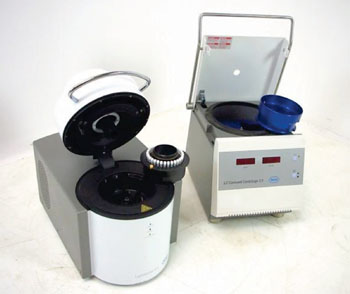
Image: The LightCycler 2.0 real-time polymerase chain reaction analyzer and centrifuge (Photo courtesy of Roche).”

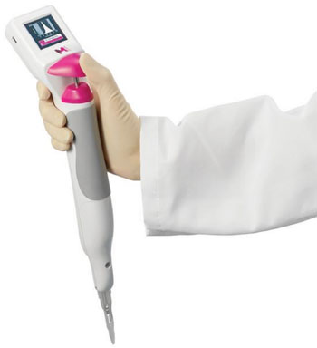
Image: The Scepter 2.0 Automated Cell Counter (Photo courtesy of Merck Millipore).
Blood samples are extensively used for the molecular diagnosis of many hematological diseases using a variety of techniques, based on the amplification of nucleic acids.
Current methods for polymerase chain reaction (PCR) use purified genomic DNA, mostly isolated from total peripheral blood cells or white blood cells (WBC), which can be improved by a real-time fluorescence resonance energy transfer-based method for genotyping directly from blood cells.
Hematologists at the Hospital Universitari Son Espases (Palma de Mallorca, Spain) studied peripheral blood from 34 patients collected into tubes containing ethylenediaminetetraacetic acid (EDTA). Among the samples, they included a mixture of mutant alleles for patients suffering from thrombosis or hereditary hemochromatosis. Red blood cells (RBCs) were lysed and white blood cells (WBCs) isolated. A real-time PCR was then performed followed by a melting curve analysis for different genes including methylenetetrahydrofolate reductase (MTHFR), hemochromatosis (HFE), coagulation factor V Leiden (F5), prothrombin factor two (F2) and coagulation factor XII (F12).
The real time PCR was performed on the LightCycler 2.0 Instrument (Roche Diagnostics Corporation, Indianapolis, IN, USA). In order to standardize the samples for the real-time PCR reaction, cells were counted in a Scepter 2.0 Automated Cell Counter (Merck Millipore, Billerica, MA, USA) and adjusted to 5×10⁶ cells/mL. After testing 34 samples comparing the real-time crossing point (CP) values between 5×10⁶ WBC/mL and 20 ng/µL of purified DNA, the results for F5 Leiden were as follows: CP mean value for WBC was 29.26 ± 0.57 versus purified DNA 24.79 ± 0.56. There was an observed delay of about four cycles when PCR was performed from WBC instead of DNA.
The authors concluded that their protocol obviates the DNA purification stage, thereby saving time and resources. Furthermore, since the manipulation performed on the sample is minimal, it may decrease the risk of contamination. As they reported the results from a variety of genes, they contend that their protocol will be suitable for the genotyping of almost any inherited polymorphism. The study was published on June 25, 2014, in the Journal of Blood Medicine.
Related Links:
Hospital Universitari Son Espases
Roche Diagnostics Corporation
Merck Millipore
Latest Molecular Diagnostics News
- Diagnostic Device Predicts Treatment Response for Brain Tumors Via Blood Test
- Blood Test Detects Early-Stage Cancers by Measuring Epigenetic Instability
- Two-in-One DNA Analysis Improves Diagnostic Accuracy While Saving Time and Costs
- “Lab-On-A-Disc” Device Paves Way for More Automated Liquid Biopsies
- New Tool Maps Chromosome Shifts in Cancer Cells to Predict Tumor Evolution
- Blood Test Identifies Inflammatory Breast Cancer Patients at Increased Risk of Brain Metastasis
- Newly-Identified Parkinson’s Biomarkers to Enable Early Diagnosis Via Blood Tests
- New Blood Test Could Detect Pancreatic Cancer at More Treatable Stage
- Liquid Biopsy Could Replace Surgical Biopsy for Diagnosing Primary Central Nervous Lymphoma
- New Tool Reveals Hidden Metabolic Weakness in Blood Cancers
- World's First Blood Test Distinguishes Between Benign and Cancerous Lung Nodules
- Rapid Test Uses Mobile Phone to Identify Severe Imported Malaria Within Minutes
- Gut Microbiome Signatures Predict Long-Term Outcomes in Acute Pancreatitis
- Blood Test Promises Faster Answers for Deadly Fungal Infections
- Blood Test Could Detect Infection Exposure History
- Urine-Based MRD Test Tracks Response to Bladder Cancer Surgery
Channels
Clinical Chemistry
view channel
New PSA-Based Prognostic Model Improves Prostate Cancer Risk Assessment
Prostate cancer is the second-leading cause of cancer death among American men, and about one in eight will be diagnosed in their lifetime. Screening relies on blood levels of prostate-specific antigen... Read more
Extracellular Vesicles Linked to Heart Failure Risk in CKD Patients
Chronic kidney disease (CKD) affects more than 1 in 7 Americans and is strongly associated with cardiovascular complications, which account for more than half of deaths among people with CKD.... Read moreMolecular Diagnostics
view channel
Diagnostic Device Predicts Treatment Response for Brain Tumors Via Blood Test
Glioblastoma is one of the deadliest forms of brain cancer, largely because doctors have no reliable way to determine whether treatments are working in real time. Assessing therapeutic response currently... Read more
Blood Test Detects Early-Stage Cancers by Measuring Epigenetic Instability
Early-stage cancers are notoriously difficult to detect because molecular changes are subtle and often missed by existing screening tools. Many liquid biopsies rely on measuring absolute DNA methylation... Read more
“Lab-On-A-Disc” Device Paves Way for More Automated Liquid Biopsies
Extracellular vesicles (EVs) are tiny particles released by cells into the bloodstream that carry molecular information about a cell’s condition, including whether it is cancerous. However, EVs are highly... Read more
Blood Test Identifies Inflammatory Breast Cancer Patients at Increased Risk of Brain Metastasis
Brain metastasis is a frequent and devastating complication in patients with inflammatory breast cancer, an aggressive subtype with limited treatment options. Despite its high incidence, the biological... Read moreImmunology
view channelBlood Test Identifies Lung Cancer Patients Who Can Benefit from Immunotherapy Drug
Small cell lung cancer (SCLC) is an aggressive disease with limited treatment options, and even newly approved immunotherapies do not benefit all patients. While immunotherapy can extend survival for some,... Read more
Whole-Genome Sequencing Approach Identifies Cancer Patients Benefitting From PARP-Inhibitor Treatment
Targeted cancer therapies such as PARP inhibitors can be highly effective, but only for patients whose tumors carry specific DNA repair defects. Identifying these patients accurately remains challenging,... Read more
Ultrasensitive Liquid Biopsy Demonstrates Efficacy in Predicting Immunotherapy Response
Immunotherapy has transformed cancer treatment, but only a small proportion of patients experience lasting benefit, with response rates often remaining between 10% and 20%. Clinicians currently lack reliable... Read moreMicrobiology
view channel
Comprehensive Review Identifies Gut Microbiome Signatures Associated With Alzheimer’s Disease
Alzheimer’s disease affects approximately 6.7 million people in the United States and nearly 50 million worldwide, yet early cognitive decline remains difficult to characterize. Increasing evidence suggests... Read moreAI-Powered Platform Enables Rapid Detection of Drug-Resistant C. Auris Pathogens
Infections caused by the pathogenic yeast Candida auris pose a significant threat to hospitalized patients, particularly those with weakened immune systems or those who have invasive medical devices.... Read morePathology
view channel
Engineered Yeast Cells Enable Rapid Testing of Cancer Immunotherapy
Developing new cancer immunotherapies is a slow, costly, and high-risk process, particularly for CAR T cell treatments that must precisely recognize cancer-specific antigens. Small differences in tumor... Read more
First-Of-Its-Kind Test Identifies Autism Risk at Birth
Autism spectrum disorder is treatable, and extensive research shows that early intervention can significantly improve cognitive, social, and behavioral outcomes. Yet in the United States, the average age... Read moreTechnology
view channel
Robotic Technology Unveiled for Automated Diagnostic Blood Draws
Routine diagnostic blood collection is a high‑volume task that can strain staffing and introduce human‑dependent variability, with downstream implications for sample quality and patient experience.... Read more
ADLM Launches First-of-Its-Kind Data Science Program for Laboratory Medicine Professionals
Clinical laboratories generate billions of test results each year, creating a treasure trove of data with the potential to support more personalized testing, improve operational efficiency, and enhance patient care.... Read moreAptamer Biosensor Technology to Transform Virus Detection
Rapid and reliable virus detection is essential for controlling outbreaks, from seasonal influenza to global pandemics such as COVID-19. Conventional diagnostic methods, including cell culture, antigen... Read more
AI Models Could Predict Pre-Eclampsia and Anemia Earlier Using Routine Blood Tests
Pre-eclampsia and anemia are major contributors to maternal and child mortality worldwide, together accounting for more than half a million deaths each year and leaving millions with long-term health complications.... Read moreIndustry
view channelNew Collaboration Brings Automated Mass Spectrometry to Routine Laboratory Testing
Mass spectrometry is a powerful analytical technique that identifies and quantifies molecules based on their mass and electrical charge. Its high selectivity, sensitivity, and accuracy make it indispensable... Read more
AI-Powered Cervical Cancer Test Set for Major Rollout in Latin America
Noul Co., a Korean company specializing in AI-based blood and cancer diagnostics, announced it will supply its intelligence (AI)-based miLab CER cervical cancer diagnostic solution to Mexico under a multi‑year... Read more
Diasorin and Fisher Scientific Enter into US Distribution Agreement for Molecular POC Platform
Diasorin (Saluggia, Italy) has entered into an exclusive distribution agreement with Fisher Scientific, part of Thermo Fisher Scientific (Waltham, MA, USA), for the LIAISON NES molecular point-of-care... Read more


