Leptomeningeal Disease Diagnosed Using CSF Cell-Free DNA
|
By LabMedica International staff writers Posted on 26 Aug 2021 |
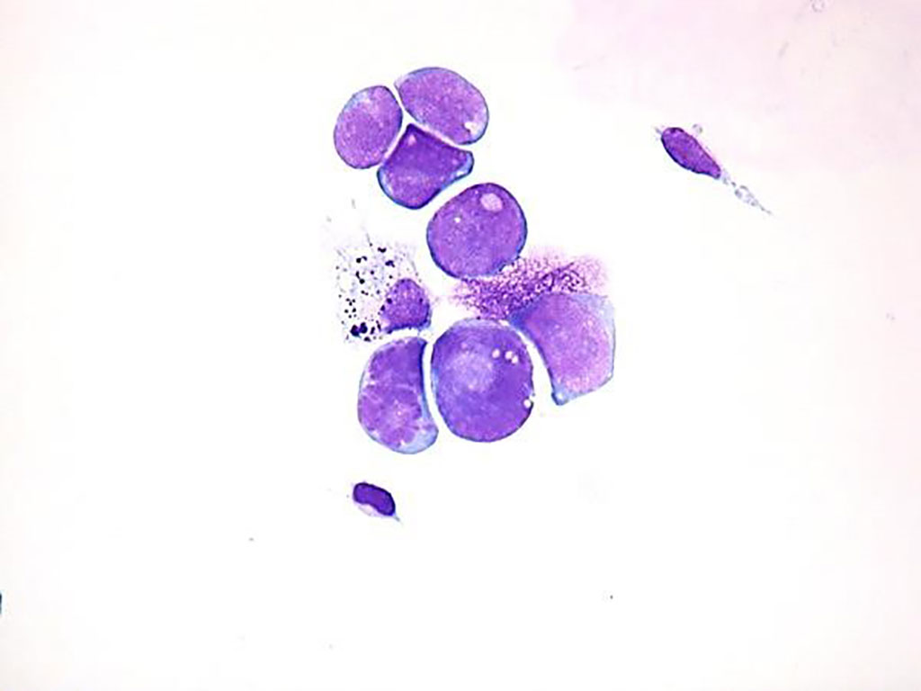
Image: Small round blue tumor cells found in the CSF of a patient with leptomeningeal disease from rhabdomyosarcoma (Photo courtesy of Pediatric Oncology)
Leptomeningeal disease (LMD) is a devastating complication of solid and liquid systemic and central nervous system malignant neoplasms. LMD, in which cancer metastasizes to the cerebrospinal fluid, occurs in 4% to 15% of cancer patients and is associated with poor survival, with untreated patients dying within four to six weeks.
Leptomeningeal disease is typically diagnosed via identification of malignant cells in cerebrospinal fluid (CSF), most commonly obtained via lumbar puncture or ventriculoperitoneal (VP) shunt access. CSF cytological analysis. The currently most reliable diagnostic method, has a sensitivity of approximately only 75% and is known to produce persistently negative results in approximately 10% of patients.
A large team of Neuro-oncologists at the Massachusetts General Hospital (Boston, MA, USA) and their colleagues conducted a diagnostic study to assess the use of genomic sequencing of CSF samples obtained from 30 patients with suspected or confirmed LMD from 2015 through 2018 to identify tumor-derived cfDNA. This study consisted of two patient populations: 22 patients with cytologically confirmed LMD without parenchymal tumors abutting their CSF and eight patients with parenchymal brain metastases with no evidence of LMD.
Cell-free DNA was isolated from blood plasma and CSF using a QIASymphony instrument with the QIASymphony DSP Circulating DNA Kit (Qiagen, Hilden, Germany). A subset of libraries was prepared with unique molecular identifier adapters (Integrated DNA Technologies, Coralville, IA, USA). Prepared libraries were sequenced on HiSeq X, HiSeq 2500, or HiSeq 4000 instruments (Illumina, San Diego, CA, USA) to a targeted mean depth of 0.1X. Samples were aligned to hg19 using bwa-mem, version 0.7.7-r441.
The scientists reported that in total, 30 patients (23 women [77%]; median age, 51 years [range, 28-81 years]), primarily presenting with metastatic solid malignant neoplasms, participated in this study. For 48 follow-up samples from patients previously diagnosed via cytological analysis as having LMD with no parenchymal tumor abutting CSF, cfDNA findings were accurate in the assessment of LMD in 45 samples, whereas cytological analysis was accurate in only 36 samples. Of 43 LMD-positive samples, CSF cfDNA analysis was sensitive to LMD in 40 samples and cytological analysis was sensitive to LMD in 31 samples, a significant difference. For three patients with parenchymal brain metastases abutting the CSF and no suspicion of LMD, cytological findings were negative for LMD in all three patients, whereas cfDNA findings were positive in all three patients.
Priscilla K. Brastianos, MD, Assistant Professor of Medicine and a co-senior author of the study, said, “If we are able to confidently diagnose LMD using cell-free DNA earlier and with fewer invasive procedures, then we can institute treatment sooner and enroll patients in clinical trials for new LMD treatments. The ultimate hope is that we can improve patient survival with earlier diagnosis and treatment for this deadly disease.”
The authors concluded that the diagnostic study found improved sensitivity and accuracy of cfDNA CSF testing versus cytological assessment for diagnosing LMD with the exception of parenchymal tumors abutting CSF, suggesting improved ability to diagnosis LMD. The study was published on August 9, 2021 in the journal JAMA Network Open.
Related Links:
Massachusetts General Hospital
Qiagen
Integrated DNA Technologies
Illumina
Latest Pathology News
- AI Integrated With Optical Imaging Technology Enables Rapid Intraoperative Diagnosis
- HPV Self-Collection Solution Improves Access to Cervical Cancer Testing
- Hyperspectral Dark-Field Microscopy Enables Rapid and Accurate Identification of Cancerous Tissues
- AI Advancements Enable Leap into 3D Pathology
- New Blood Test Device Modeled on Leeches to Help Diagnose Malaria
- Robotic Blood Drawing Device to Revolutionize Sample Collection for Diagnostic Testing
- Use of DICOM Images for Pathology Diagnostics Marks Significant Step towards Standardization
- First of Its Kind Universal Tool to Revolutionize Sample Collection for Diagnostic Tests
- AI-Powered Digital Imaging System to Revolutionize Cancer Diagnosis
- New Mycobacterium Tuberculosis Panel to Support Real-Time Surveillance and Combat Antimicrobial Resistance
- New Method Offers Sustainable Approach to Universal Metabolic Cancer Diagnosis
- Spatial Tissue Analysis Identifies Patterns Associated With Ovarian Cancer Relapse
- Unique Hand-Warming Technology Supports High-Quality Fingertip Blood Sample Collection
- Image-Based AI Shows Promise for Parasite Detection in Digitized Stool Samples
- Deep Learning Powered AI Algorithms Improve Skin Cancer Diagnostic Accuracy
- Microfluidic Device for Cancer Detection Precisely Separates Tumor Entities
Channels
Clinical Chemistry
view channel
3D Printed Point-Of-Care Mass Spectrometer Outperforms State-Of-The-Art Models
Mass spectrometry is a precise technique for identifying the chemical components of a sample and has significant potential for monitoring chronic illness health states, such as measuring hormone levels... Read more.jpg)
POC Biomedical Test Spins Water Droplet Using Sound Waves for Cancer Detection
Exosomes, tiny cellular bioparticles carrying a specific set of proteins, lipids, and genetic materials, play a crucial role in cell communication and hold promise for non-invasive diagnostics.... Read more
Highly Reliable Cell-Based Assay Enables Accurate Diagnosis of Endocrine Diseases
The conventional methods for measuring free cortisol, the body's stress hormone, from blood or saliva are quite demanding and require sample processing. The most common method, therefore, involves collecting... Read moreHematology
view channel
Next Generation Instrument Screens for Hemoglobin Disorders in Newborns
Hemoglobinopathies, the most widespread inherited conditions globally, affect about 7% of the population as carriers, with 2.7% of newborns being born with these conditions. The spectrum of clinical manifestations... Read more
First 4-in-1 Nucleic Acid Test for Arbovirus Screening to Reduce Risk of Transfusion-Transmitted Infections
Arboviruses represent an emerging global health threat, exacerbated by climate change and increased international travel that is facilitating their spread across new regions. Chikungunya, dengue, West... Read more
POC Finger-Prick Blood Test Determines Risk of Neutropenic Sepsis in Patients Undergoing Chemotherapy
Neutropenia, a decrease in neutrophils (a type of white blood cell crucial for fighting infections), is a frequent side effect of certain cancer treatments. This condition elevates the risk of infections,... Read more
First Affordable and Rapid Test for Beta Thalassemia Demonstrates 99% Diagnostic Accuracy
Hemoglobin disorders rank as some of the most prevalent monogenic diseases globally. Among various hemoglobin disorders, beta thalassemia, a hereditary blood disorder, affects about 1.5% of the world's... Read moreImmunology
view channel.jpg)
AI Predicts Tumor-Killing Cells with High Accuracy
Cellular immunotherapy involves extracting immune cells from a patient's tumor, potentially enhancing their cancer-fighting capabilities through engineering, and then expanding and reintroducing them into the body.... Read more
Diagnostic Blood Test for Cellular Rejection after Organ Transplant Could Replace Surgical Biopsies
Transplanted organs constantly face the risk of being rejected by the recipient's immune system which differentiates self from non-self using T cells and B cells. T cells are commonly associated with acute... Read more
AI Tool Precisely Matches Cancer Drugs to Patients Using Information from Each Tumor Cell
Current strategies for matching cancer patients with specific treatments often depend on bulk sequencing of tumor DNA and RNA, which provides an average profile from all cells within a tumor sample.... Read more
Genetic Testing Combined With Personalized Drug Screening On Tumor Samples to Revolutionize Cancer Treatment
Cancer treatment typically adheres to a standard of care—established, statistically validated regimens that are effective for the majority of patients. However, the disease’s inherent variability means... Read moreMicrobiology
view channel
Integrated Solution Ushers New Era of Automated Tuberculosis Testing
Tuberculosis (TB) is responsible for 1.3 million deaths every year, positioning it as one of the top killers globally due to a single infectious agent. In 2022, around 10.6 million people were diagnosed... Read more
Automated Sepsis Test System Enables Rapid Diagnosis for Patients with Severe Bloodstream Infections
Sepsis affects up to 50 million people globally each year, with bacteraemia, formerly known as blood poisoning, being a major cause. In the United States alone, approximately two million individuals are... Read moreEnhanced Rapid Syndromic Molecular Diagnostic Solution Detects Broad Range of Infectious Diseases
GenMark Diagnostics (Carlsbad, CA, USA), a member of the Roche Group (Basel, Switzerland), has rebranded its ePlex® system as the cobas eplex system. This rebranding under the globally renowned cobas name... Read more
Clinical Decision Support Software a Game-Changer in Antimicrobial Resistance Battle
Antimicrobial resistance (AMR) is a serious global public health concern that claims millions of lives every year. It primarily results from the inappropriate and excessive use of antibiotics, which reduces... Read morePathology
view channel
AI Integrated With Optical Imaging Technology Enables Rapid Intraoperative Diagnosis
Rapid and accurate intraoperative diagnosis is essential for tumor surgery as it guides surgical decisions with precision. Traditional intraoperative assessments, such as frozen sections based on H&E... Read more
HPV Self-Collection Solution Improves Access to Cervical Cancer Testing
Annually, over 604,000 women across the world are diagnosed with cervical cancer, and about 342,000 die from this disease, which is preventable and primarily caused by the Human Papillomavirus (HPV).... Read moreHyperspectral Dark-Field Microscopy Enables Rapid and Accurate Identification of Cancerous Tissues
Breast cancer remains a major cause of cancer-related mortality among women. Breast-conserving surgery (BCS), also known as lumpectomy, is the removal of the cancerous lump and a small margin of surrounding tissue.... Read moreTechnology
view channel
New Diagnostic System Achieves PCR Testing Accuracy
While PCR tests are the gold standard of accuracy for virology testing, they come with limitations such as complexity, the need for skilled lab operators, and longer result times. They also require complex... Read more
DNA Biosensor Enables Early Diagnosis of Cervical Cancer
Molybdenum disulfide (MoS2), recognized for its potential to form two-dimensional nanosheets like graphene, is a material that's increasingly catching the eye of the scientific community.... Read more
Self-Heating Microfluidic Devices Can Detect Diseases in Tiny Blood or Fluid Samples
Microfluidics, which are miniature devices that control the flow of liquids and facilitate chemical reactions, play a key role in disease detection from small samples of blood or other fluids.... Read more
Breakthrough in Diagnostic Technology Could Make On-The-Spot Testing Widely Accessible
Home testing gained significant importance during the COVID-19 pandemic, yet the availability of rapid tests is limited, and most of them can only drive one liquid across the strip, leading to continued... Read moreIndustry
view channel
Danaher and Johns Hopkins University Collaborate to Improve Neurological Diagnosis
Unlike severe traumatic brain injury (TBI), mild TBI often does not show clear correlations with abnormalities detected through head computed tomography (CT) scans. Consequently, there is a pressing need... Read more
Beckman Coulter and MeMed Expand Host Immune Response Diagnostics Partnership
Beckman Coulter Diagnostics (Brea, CA, USA) and MeMed BV (Haifa, Israel) have expanded their host immune response diagnostics partnership. Beckman Coulter is now an authorized distributor of the MeMed... Read more_1.jpg)


